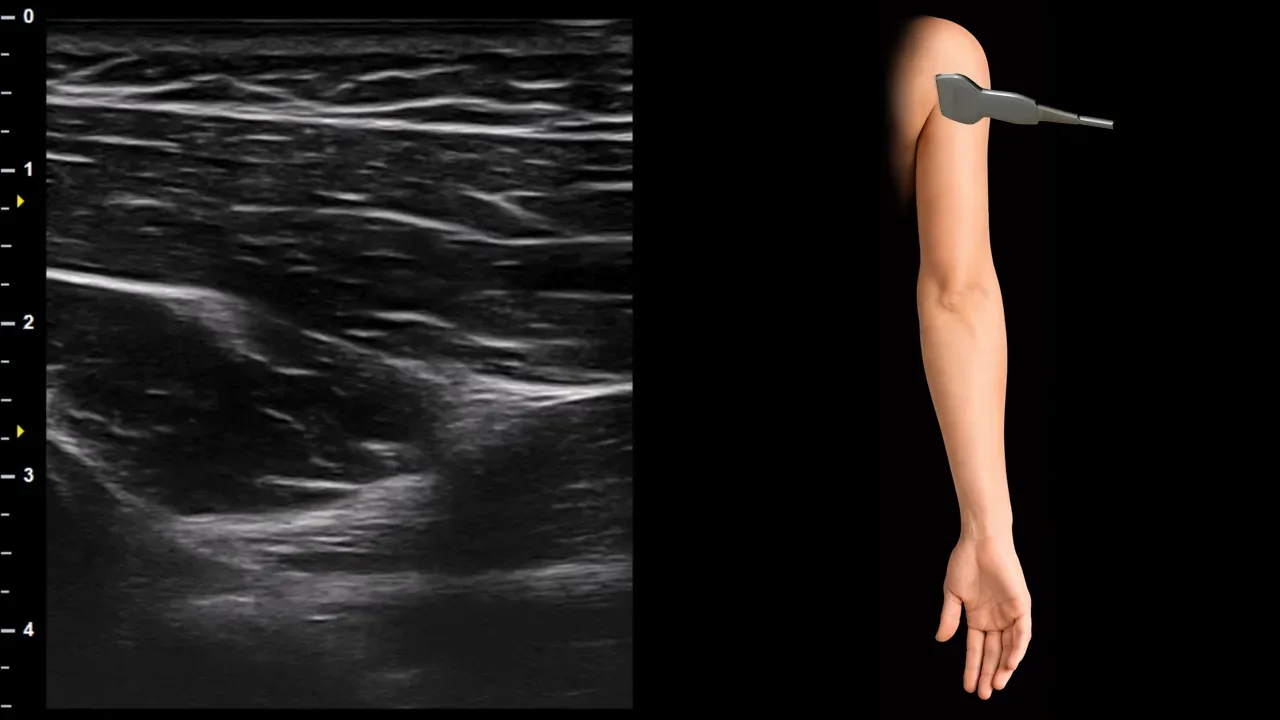
Figure 1. Posterior shoulder / quadrangular space, sagittal plane. A: n. axillaris
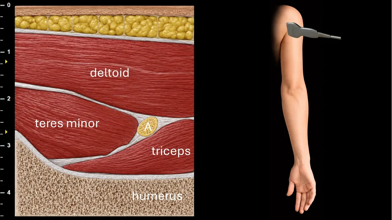
Sagittal ultrasound section of the posterior shoulder focused on visualization of the n. axillaris (A). The nerve is seen as an oval fascicular structure located in the intermuscular space between the m. teres minor and the m. triceps brachii, deep to the m. deltoideus. In this projection, the n. axillaris courses through the quadrangular space together with the posterior circumflex humeral vessels, close to the posterior aspect of the humerus.
The image also demonstrates the surrounding muscular landmarks, particularly the m. deltoideus superficially, the m. teres minor posteriorly and cranially, and the m. triceps brachii caudally. The humerus forms the main deep bony landmark. This level is useful for identifying the n. axillaris as it passes around the surgical neck of the humerus before entering the deep surface of the deltoid muscle.