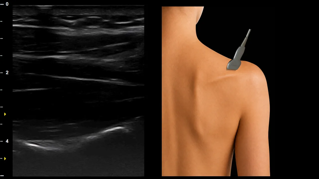
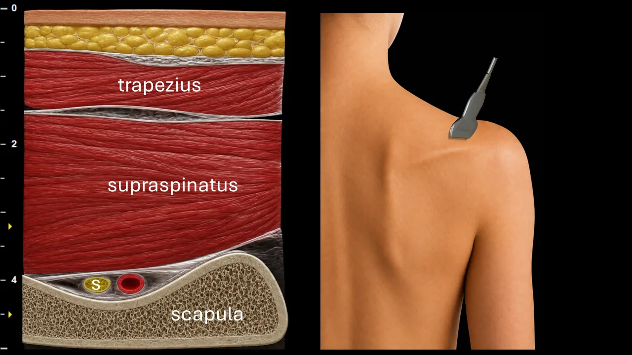
Figure 1. Suprascapular region / supraspinous fossa, transverse plane. S: n. suprascapularis
Transverse ultrasound section of the suprascapular region focused on visualization of the n. suprascapularis (S). The nerve is seen as a small oval fascicular structure located deep to the m. supraspinatus, in close relationship to the bony contour of the scapula. In this projection, the n. suprascapularis lies within the supraspinous fossa, accompanied by the suprascapular vessels.
The image also demonstrates the surrounding muscular landmarks, particularly the m. trapezius superficially and the m. supraspinatus lying directly above the scapula. The scapula forms the main deep bony landmark. This level is useful for identifying the n. suprascapularis as it courses through the suprascapular region before supplying the supraspinatus muscle and continuing toward the spinoglenoid notch.
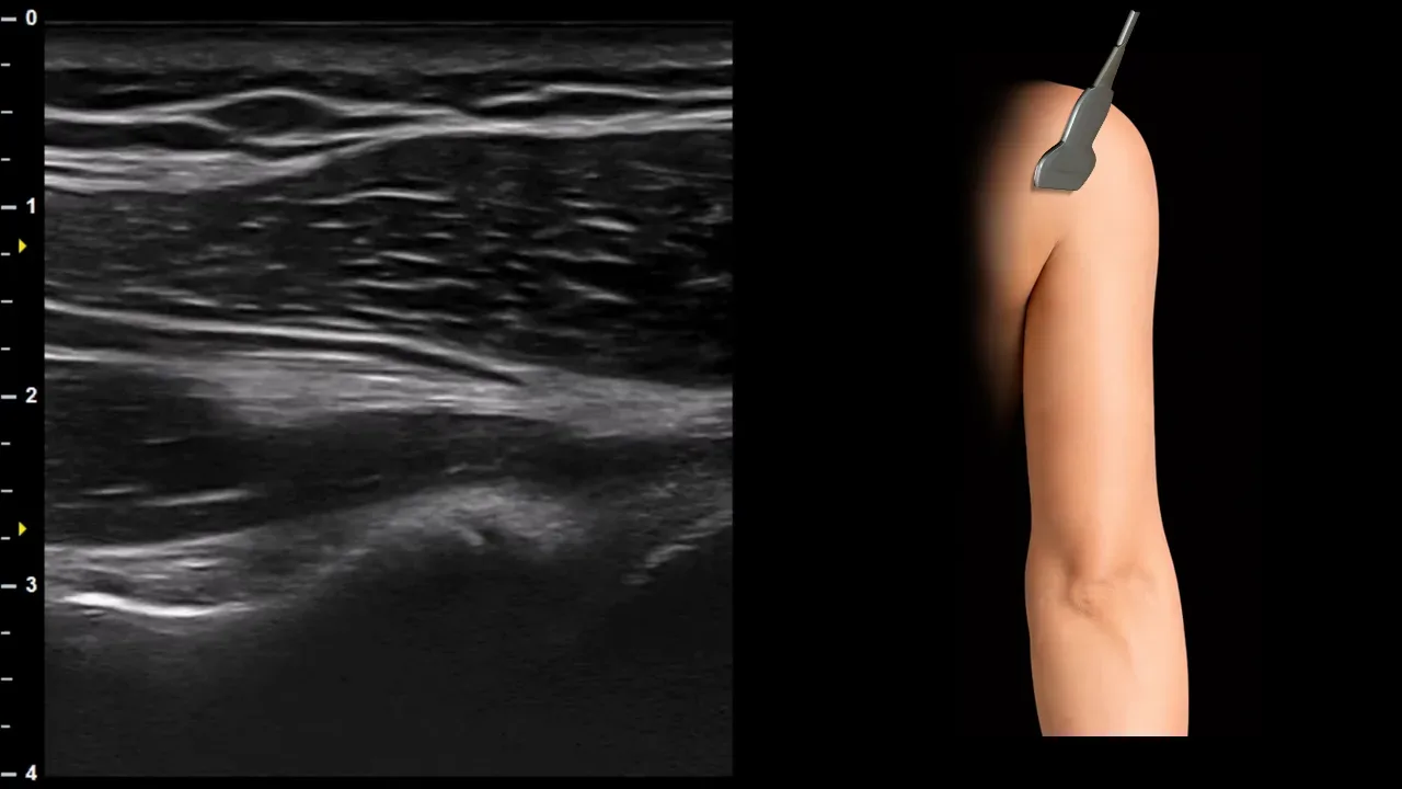
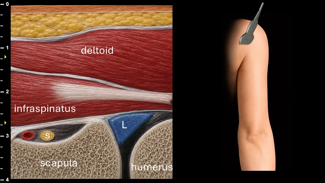
Figure 2. Spinoglenoid region / posterior shoulder, transverse plane. S: n. suprascapularis, L: labrum
Transverse ultrasound section of the posterior shoulder focused on visualization of the n. suprascapularis (S) in the spinoglenoid region. The nerve is seen as a small oval fascicular structure located deep to the m. infraspinatus, close to the posterior contour of the scapula. In this projection, the n. suprascapularis courses through the spinoglenoid notch region, accompanied by the suprascapular vessels.
The image also demonstrates the surrounding anatomical landmarks, particularly the m. deltoideus superficially and the m. infraspinatus lying directly above the scapula. Laterally, the humerus and the posterior glenohumeral region are visible, with the labrum (L) shown between the scapular and humeral contours. This level is useful for identifying the n. suprascapularis after it has passed from the supraspinous fossa toward the infraspinous fossa, before supplying the m. infraspinatus.