Lateral Ligaments Masterclass
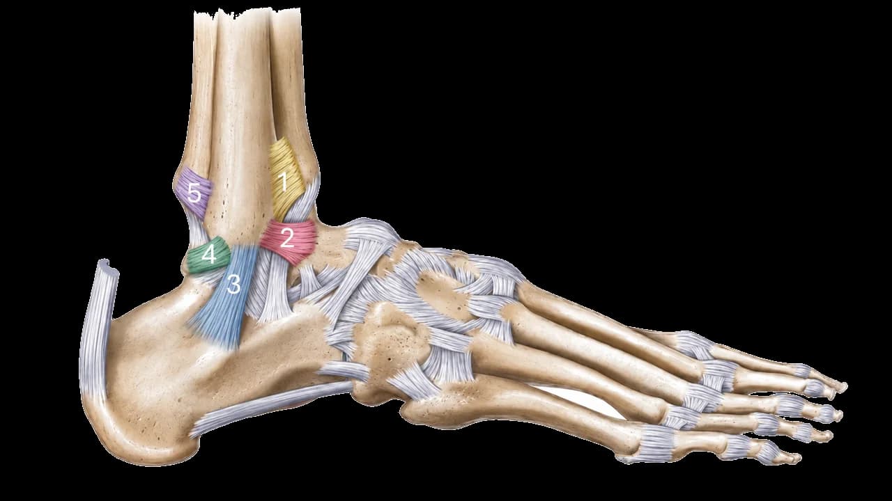
Figure 1. Lateral Ankle Ligaments. 1: anterior inferior tibiofibular ligament, 2: anterior talofibular ligament, 3: calcaneofibular ligament, 4: posterior talofibular ligament, 5: pposterior inferior tibiofibular ligament
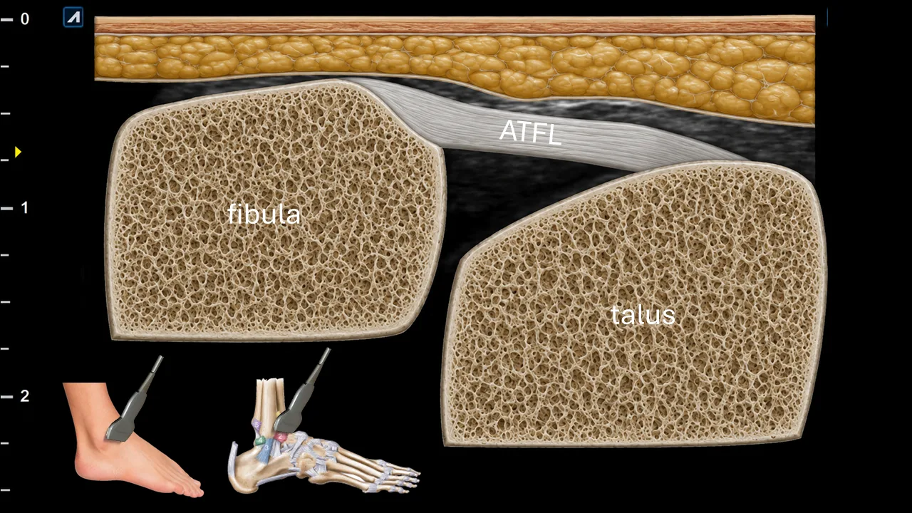
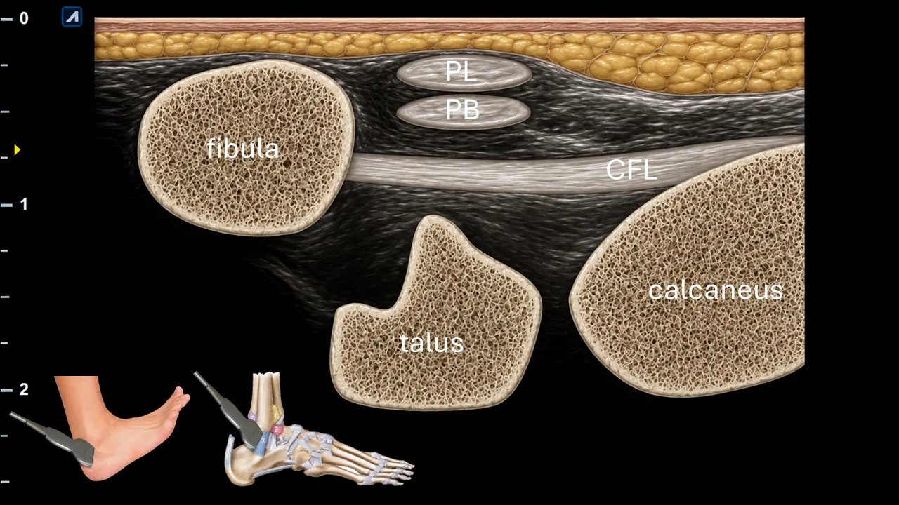
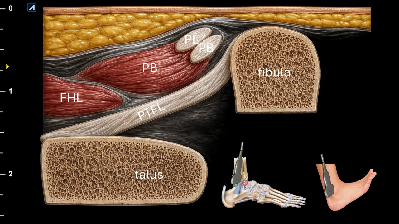
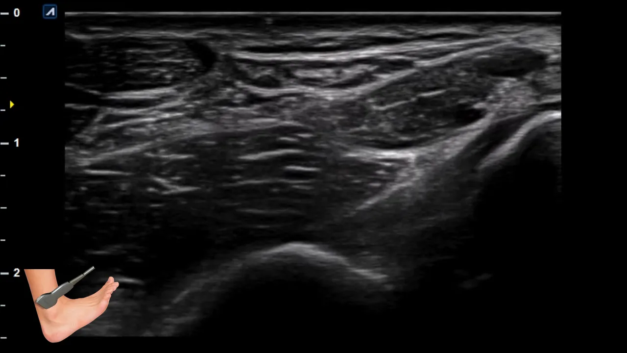
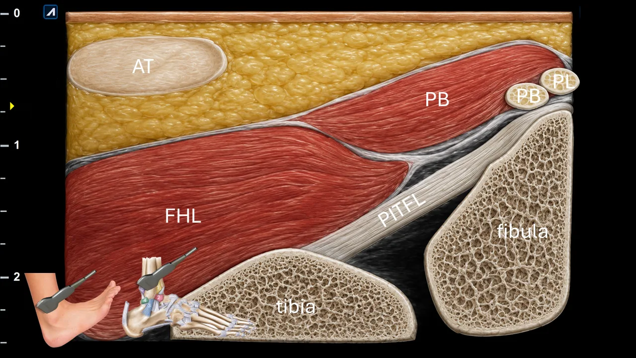
Ultrasound examination of the lateral ankle ligaments is focused on the lateral collateral ligament complex and the distal tibiofibular syndesmosis. The anterior inferior tibiofibular ligament, anterior talofibular ligament, calcaneofibular ligament, posterior talofibular ligament, and posterior inferior tibiofibular ligament should be assessed systematically in both short- and long-axis views.
A key principle of the examination is that each ligament should be evaluated under appropriate tension. Correct positioning of the foot is therefore essential, because a relaxed ligament may appear wavy, poorly defined, or falsely thickened. Depending on the ligament examined, the foot should be gently positioned to place the ligament on stretch, allowing better visualization of its fibrillar structure, thickness, continuity, and possible injury.
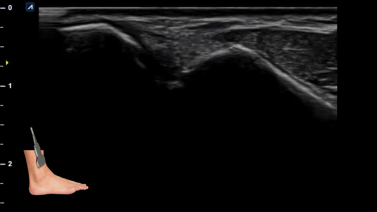
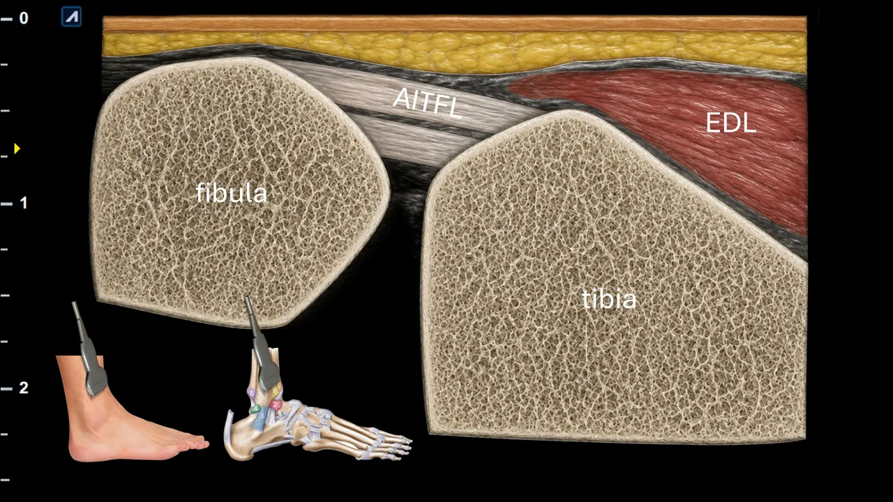
1. Anterior inferior tibiofibular ligament - AITFL: neutral position or slight plantar flexion with mild internal rotation of the foot; the syndesmosis should be gently placed under tension.
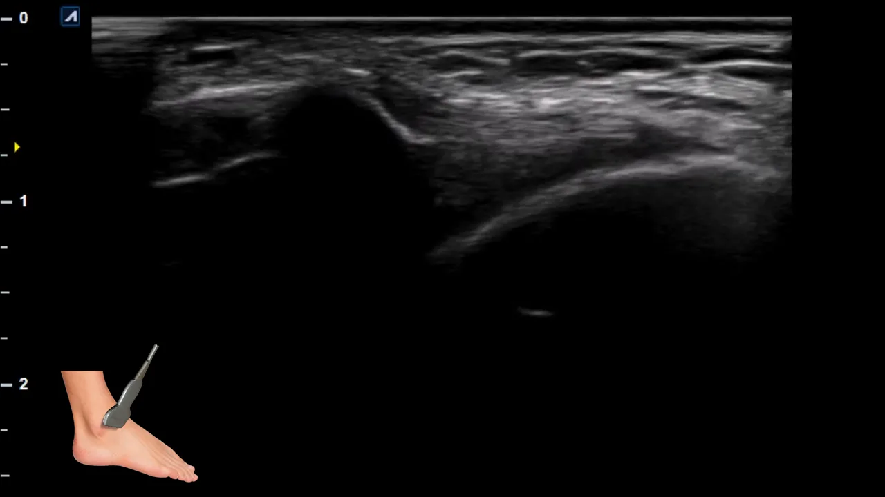
2. Anterior talofibular ligament - ATFL: plantar flexion with slight inversion of the foot. This position places the ATFL under tension and improves visualization of its fibers between the lateral malleolus and talus.
3. Calcaneofibular ligament - CFL: dorsiflexion with inversion of the hindfoot. This position helps tension the CFL as it courses from the fibula to the calcaneus, deep to the peroneal tendons.

4. Posterior talofibular ligament - PTFL: dorsiflexion with slight external rotation or neutral position of the foot. The posterior part of the lateral ligament complex should be examined with the posterior fibers gently tensioned.
5. Posterior inferior tibiofibular ligament - PITFL: neutral to dorsiflexed position, with gentle external rotation stress if needed to assess the posterior syndesmotic fibers.
Schalten Sie die vollständige Health Library frei
Voller Zugriff auf Scan-Protokolle, Anatomie und klinische Referenzen. Jederzeit kündbar.
- Alle Protokolle und Anatomiereferenzen
- Originale Ultraschall-Illustrationen und Video-Demonstrationen
- Synchronisierung zwischen Mobilgerät und Web