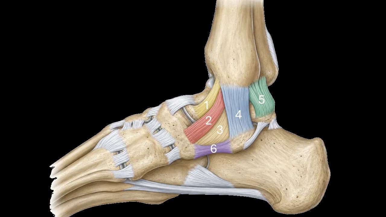
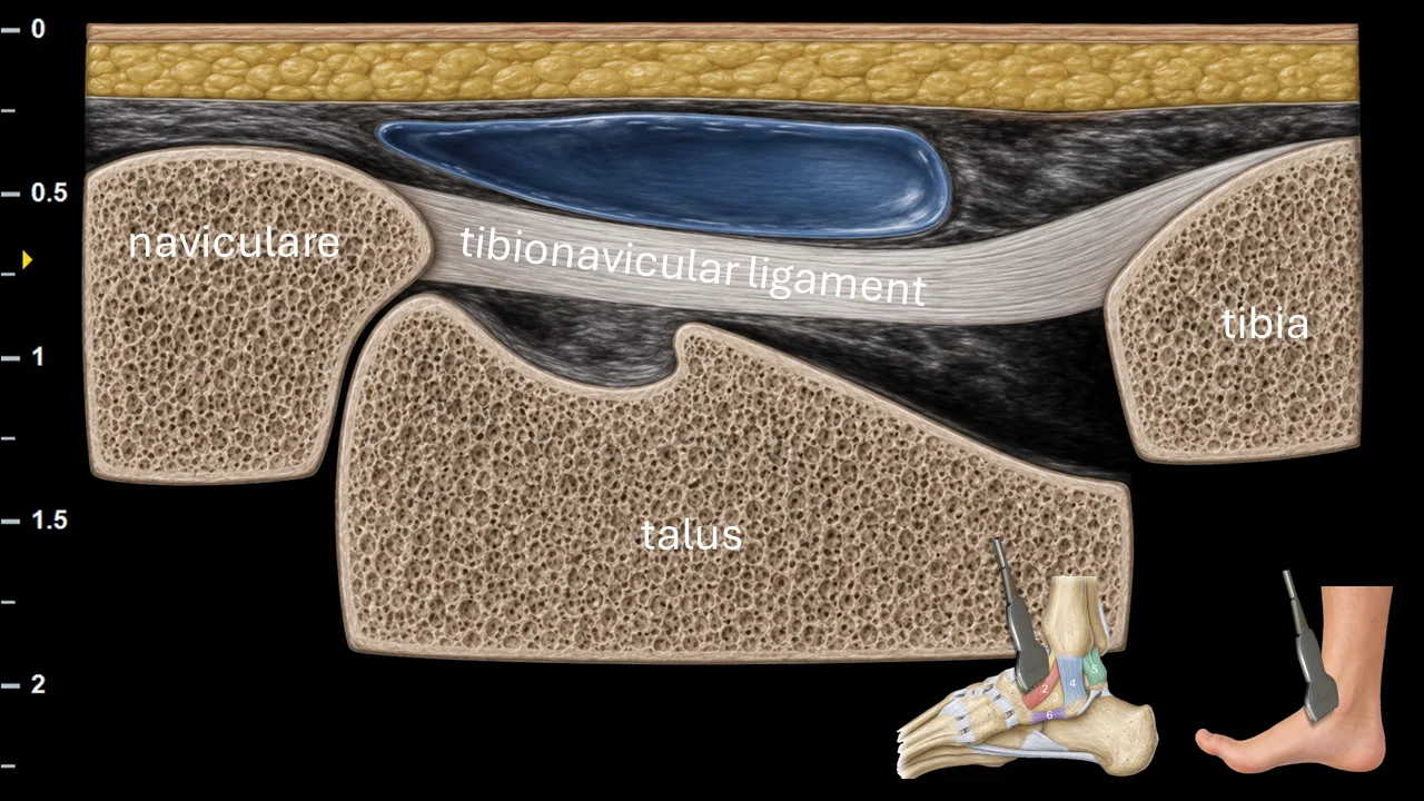
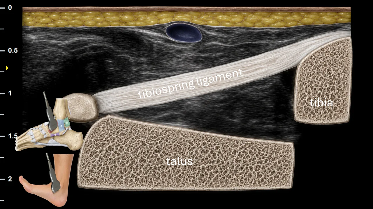
Figure 1. Medial Ankle Ligaments. 1: anterior tibiotalar ligament, 2: tibionavicular ligament, 3: tibiospring ligament, 4: tibiocalcaneal ligament, 5: posterior tibiotalar ligament, 6: spring ligament
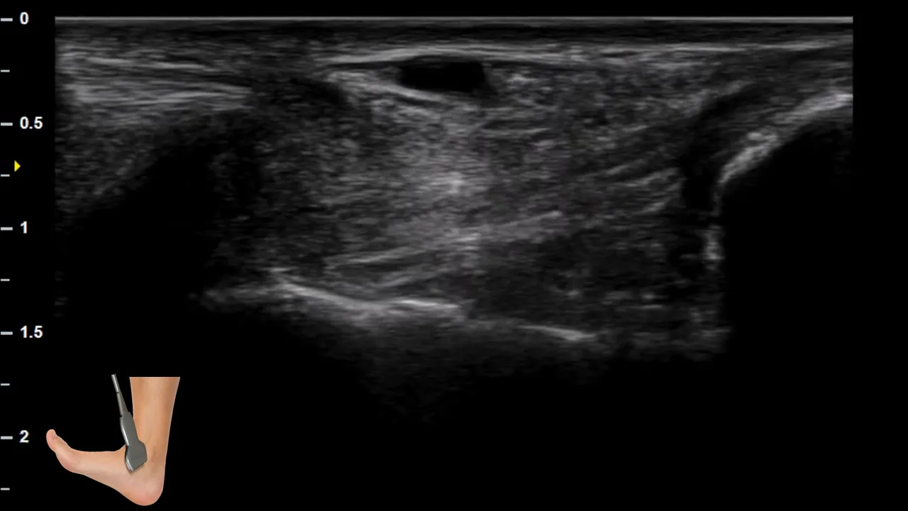
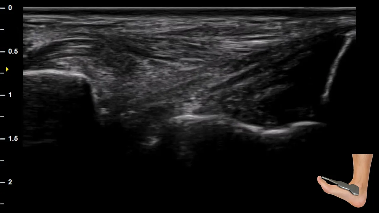
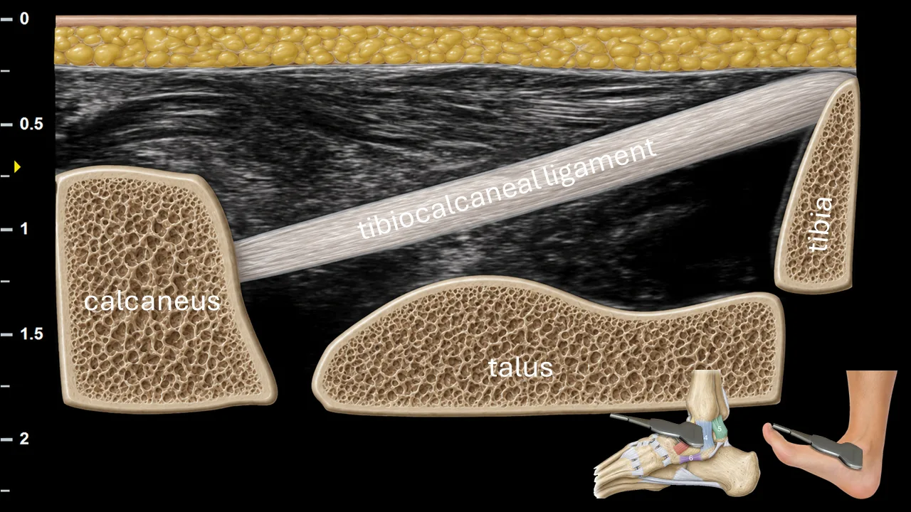
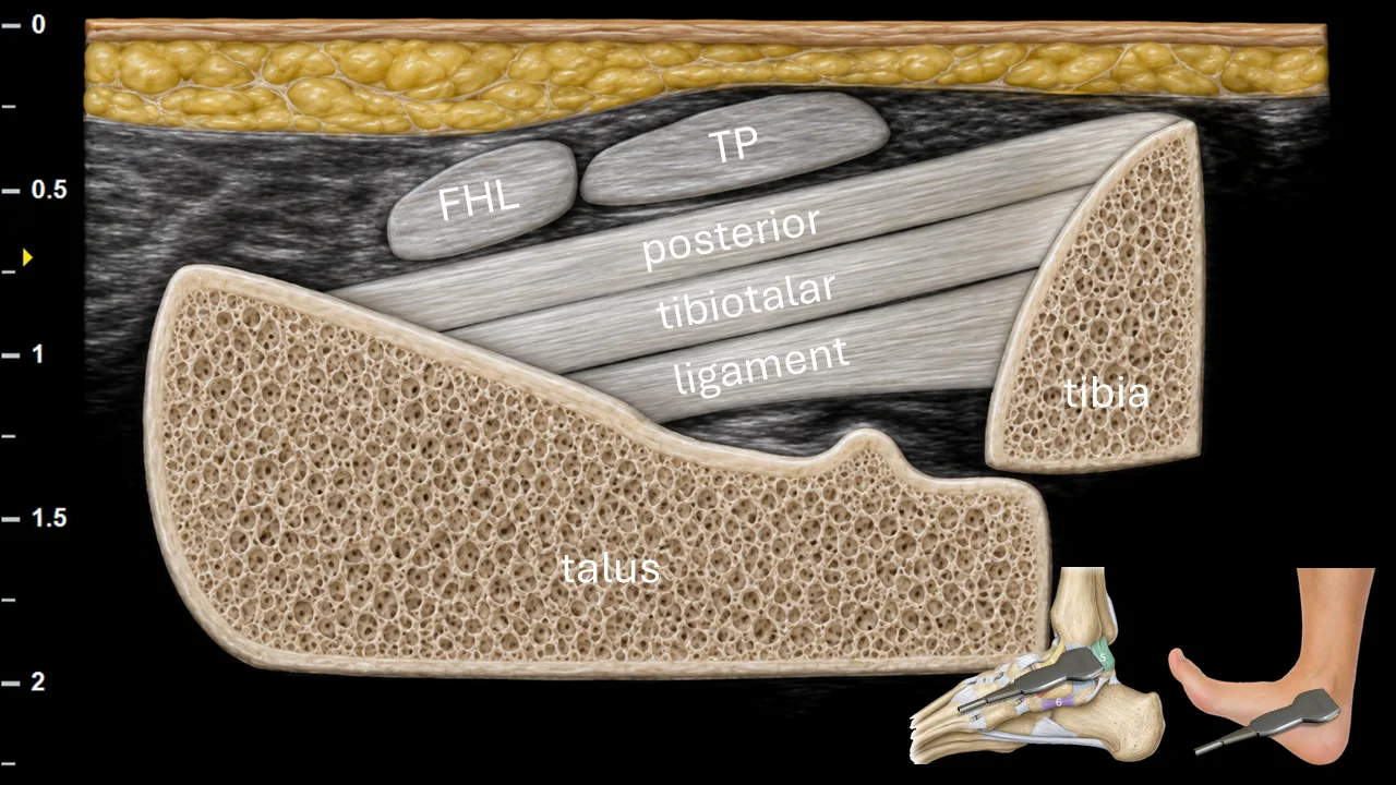
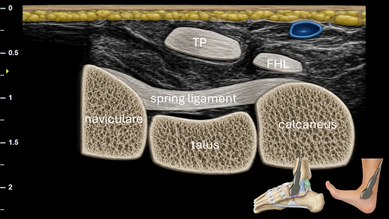
Ultrasound examination of the medial ankle ligaments is focused on the components of the deltoid ligament complex and the spring ligament. The anterior tibiotalar ligament, tibionavicular ligament, tibiospring ligament, tibiocalcaneal ligament, posterior tibiotalar ligament, and the superomedial part of the spring ligament should be assessed systematically in both short- and long-axis views.
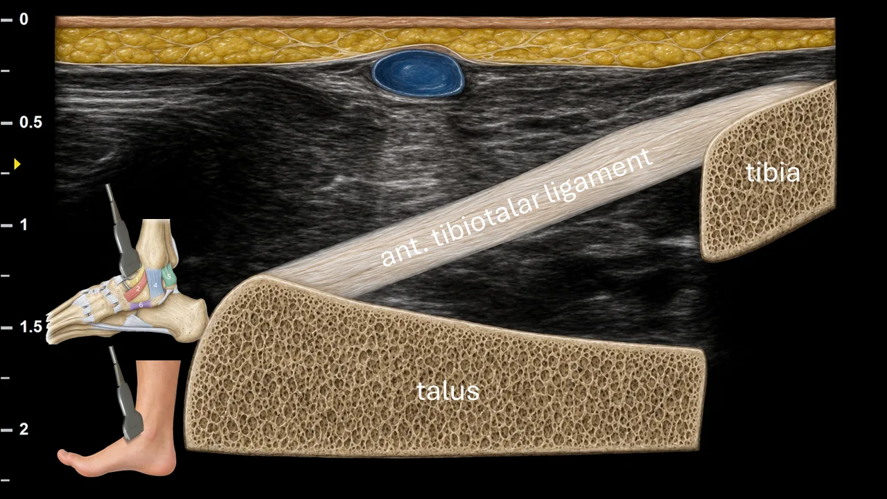
A key principle of the examination is that each ligament should be evaluated under appropriate tension. Therefore, correct positioning of the foot is essential. Depending on the ligament examined, the foot should be gently positioned to place the ligament on stretch, allowing better visualization of its fibrillar structure, thickness, continuity, and possible injury.
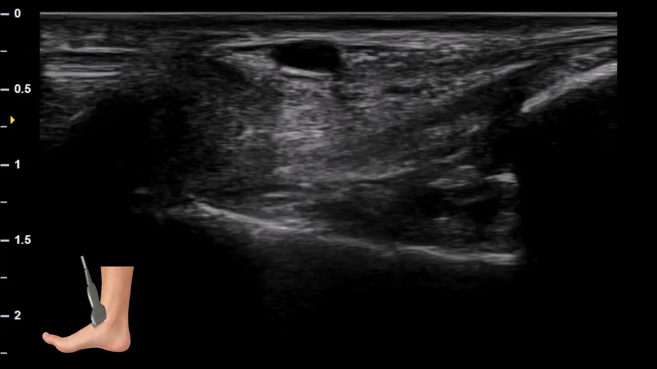
1. Ligamentum tibiotalare anterius: slight plantar flexion with mild eversion/external rotation of the foot.
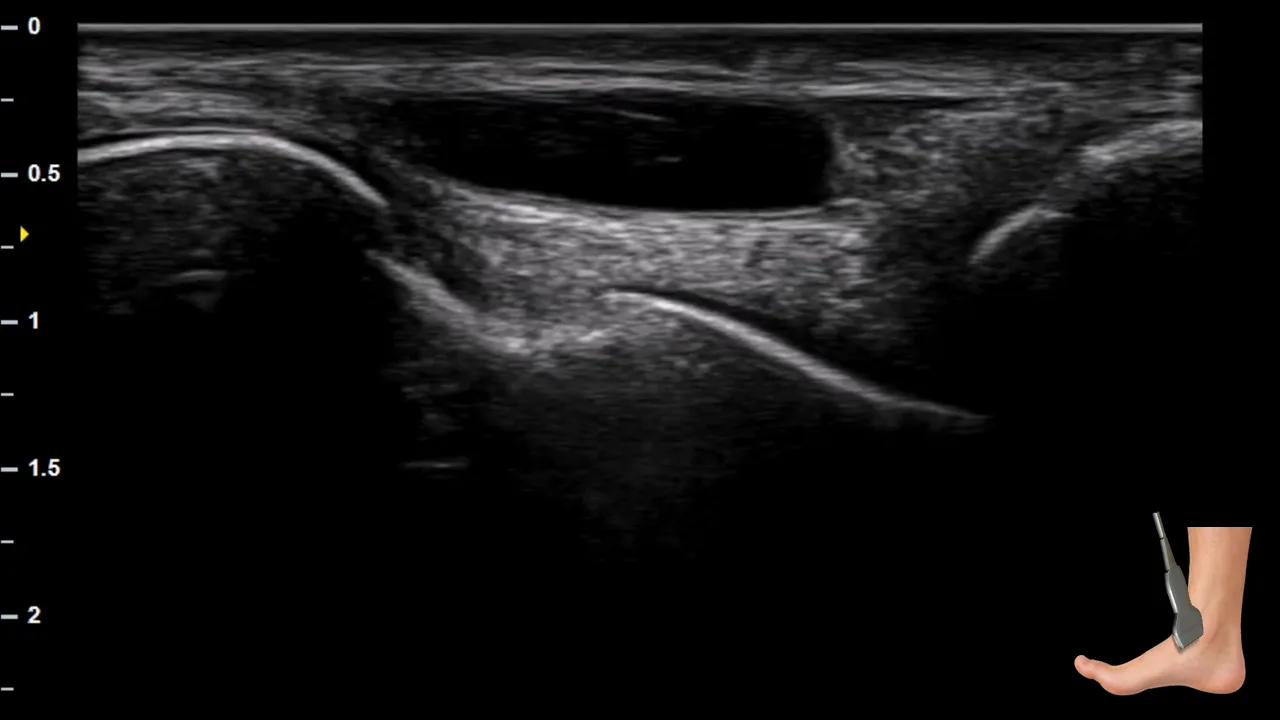
2. Ligamentum tibionaviculare: neutral position or slight plantar flexion; the medial arch should be gently opened and the ligament kept under light tension.
3. Ligamentum tibiospring: neutral to slight dorsiflexion with mild eversion of the foot.
4. Ligamentum tibiocalcaneale: dorsiflexion with mild eversion of the hindfoot.
5. Ligamentum tibiotalare posterius: dorsiflexion of the ankle; this position places the posterior part of the deltoid ligament under tension.
6. Spring ligament: slight dorsiflexion with mild eversion/pronation of the foot, so that the medial longitudinal arch and the calcaneonavicular complex are placed under tension.