Ultrasound examination
Examination protocol
Ventral view
- Transverse plane
- Sagittal plane
Medial view
- Frontal plane
Lateral view
- Frontal plane
Dorsal view
- Sagittal plane
Interaktive Funktion, verfügbar in der App
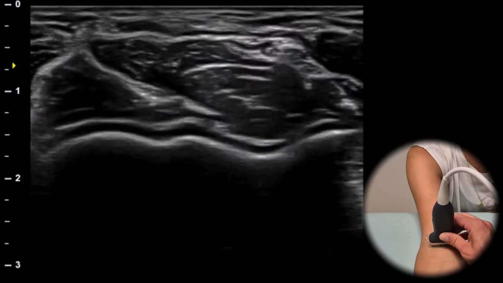
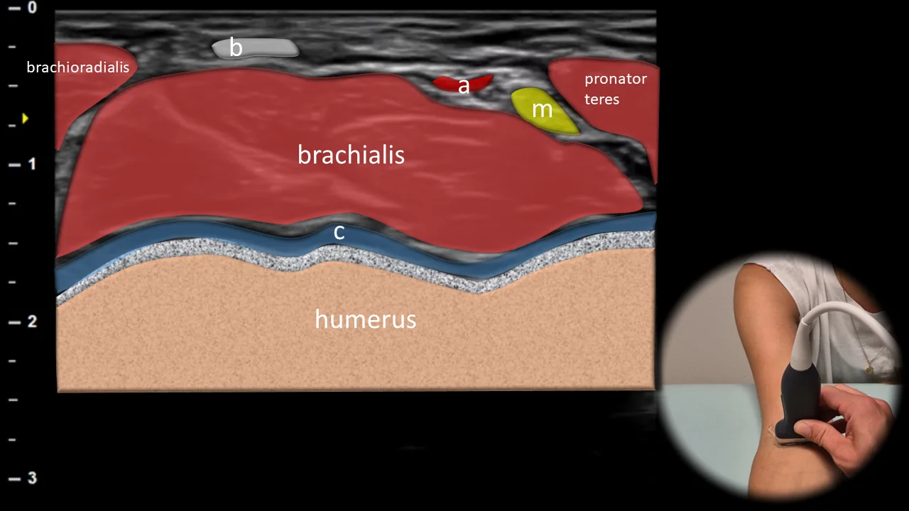
Figure 1. Ventral view, transverse plane. b: distal tendon of m. biceps brachii, a: a. brachialis, m: n. medianus, c: cartilage
Transverse ultrasound section of the anterior part of the elbow joint. The distal humerus is displayed as a hyperechoic cortical line covered by a hypoechoic cartilage layer. In the surrounding area, m. brachialis, m. pronator teres, m. brachioradialis and the distal tendon of m. biceps brachii (b) are visible. In this projection, neurovascular structures are also clearly visible, particularly a. brachialis (a) and n. medianus (m).
Clinical Note
This projection is suitable for orientation in the anterior soft tissues and for evaluating neurovascular structures.
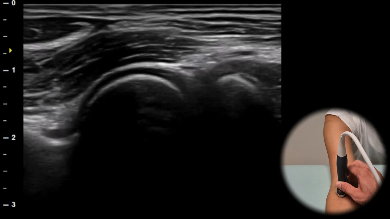
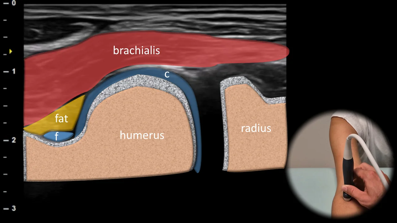
Figure 2. Ventral view, sagittal plane, radial side. c: cartilage, f: fluid in the elbow joint
Longitudinal ultrasound section of the anterior radial part of the elbow joint focusing on the radiohumeral articulation. The radial head, capitulum humeri and radial fossa are visible, which may contain a small amount of fat and fluid (f). A layer of hyaline cartilage (c) is shown on the surface of the humerus. Among the surrounding soft tissues, the brachialis muscle is most clearly visible.
Clinical Note
In this projection, we primarily look for intra-articular fluid in the area of the fossa radialis and anterior recess.
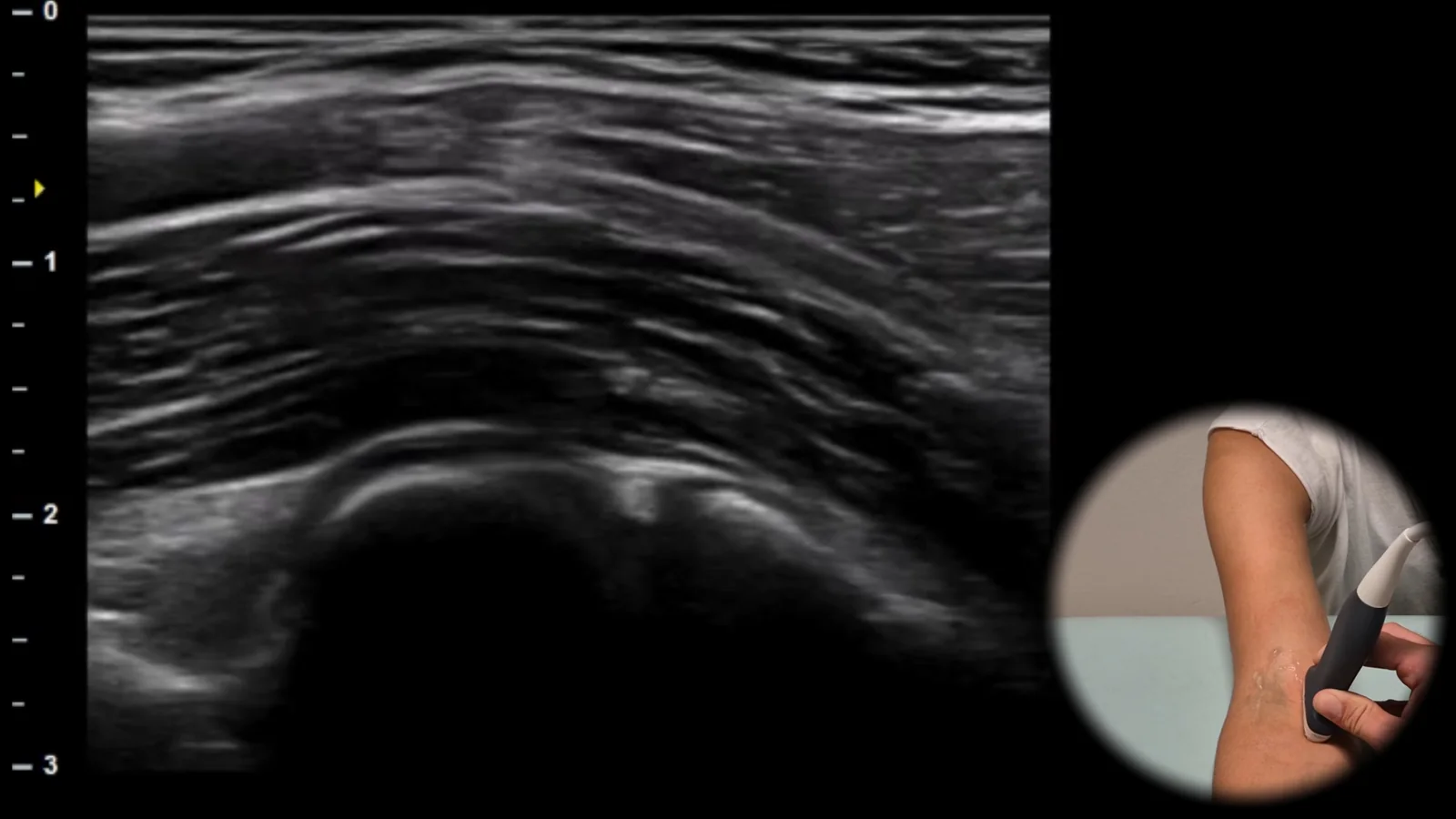
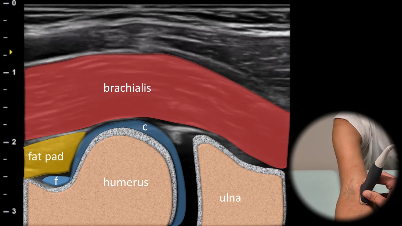
Figure 3. Ventral view, sagittal plane, ulnar side. c: cartilage, f: fluid in the elbow joint
Longitudinal ultrasound section of the anterior ulnar part of the elbow joint. Shows the humeroulnar articulation, particularly the trochlea humeri and processus coronoideus ulnae. We evaluate the joint space, fossa coronoidea and anterior synovial recess, where fluid (f) may accumulate. Among the surrounding soft tissues, primarily m. brachialis and m. pronator teres are visible in this projection.
Clinical Note
In this projection, we primarily look for joint effusion in the area of the anterior synovial recess.
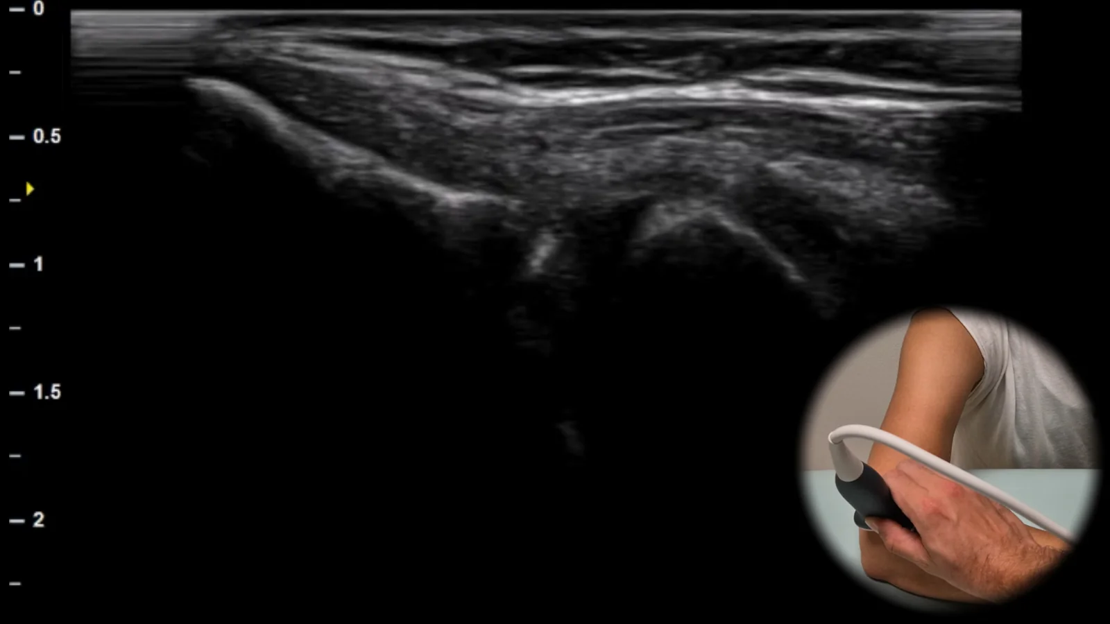
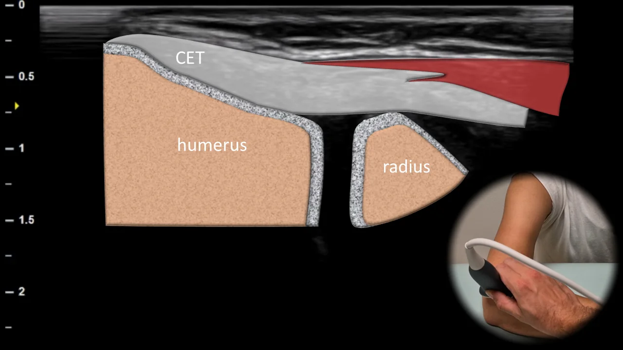
Figure 4. Medial view, frontal plane. CFT: common flexor tendon
Longitudinal ultrasound projection of the medial portion of the elbow joint. The probe is positioned over the medial epicondyle of the humerus along the long axis of the flexor-pronator muscle group. The common flexor tendon (CFT) is shown superficially, with the medial collateral ligament deeper. This projection allows evaluation of the bony surface and tendon insertions for tendinopathy, calcifications, enthesopathy, erosions, osteophytes, or ruptures.
Clinical Note
This projection is key for evaluating medial epicondylitis and the common flexor tendon.
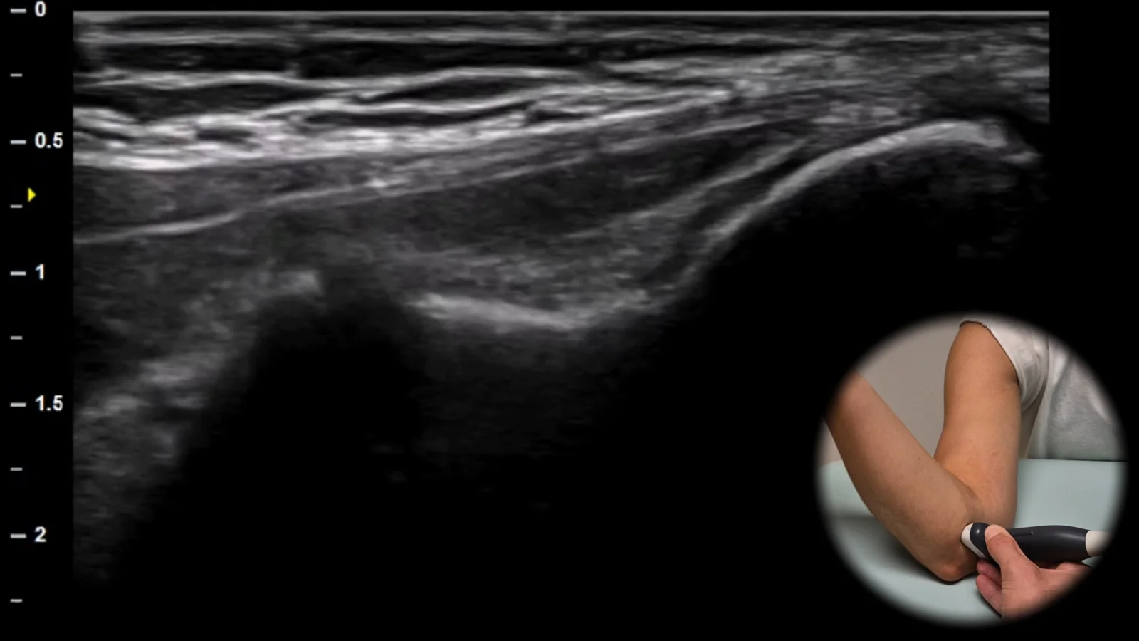
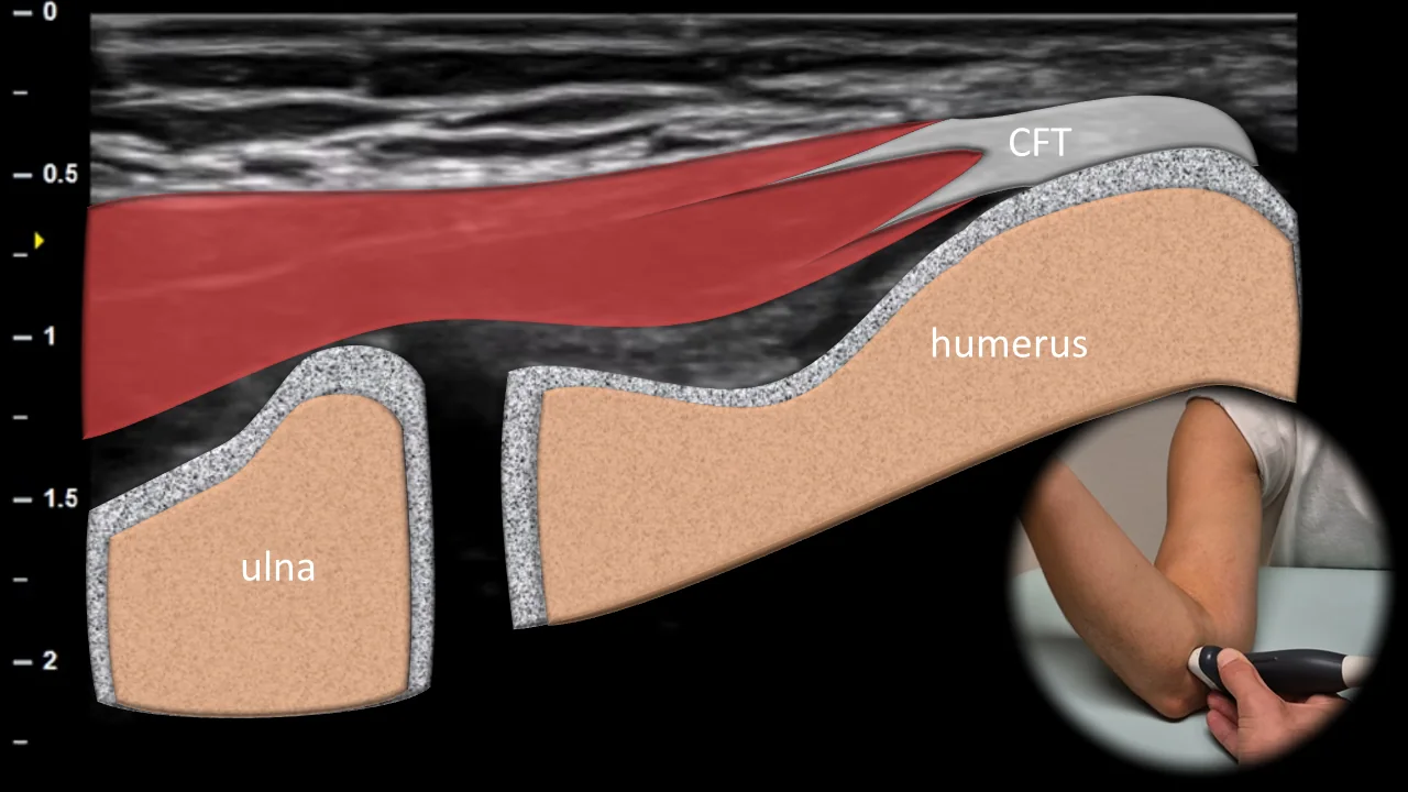
Figure 5. Lateral view, frontal plane. CET: common extensor tendon
Longitudinal ultrasound projection of the lateral aspect of the elbow joint. The probe is positioned along the long axis of the extensor muscle group originating from the lateral epicondyle of the humerus. Superficially, the common extensor tendon (CET) is depicted, with the lateral collateral ligament complex shown more deeply. This projection is used to assess the structure of the tendon, enthesis, and adjacent bone surface.
Clinical Note
In this projection, we focus primarily on lateral epicondylitis and the integrity of the common extensor tendon.
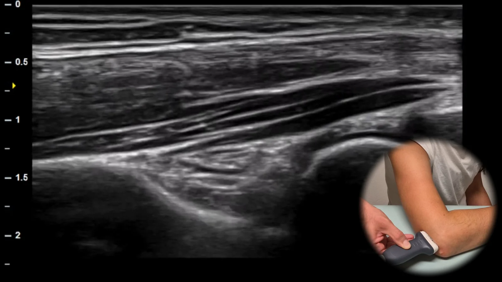
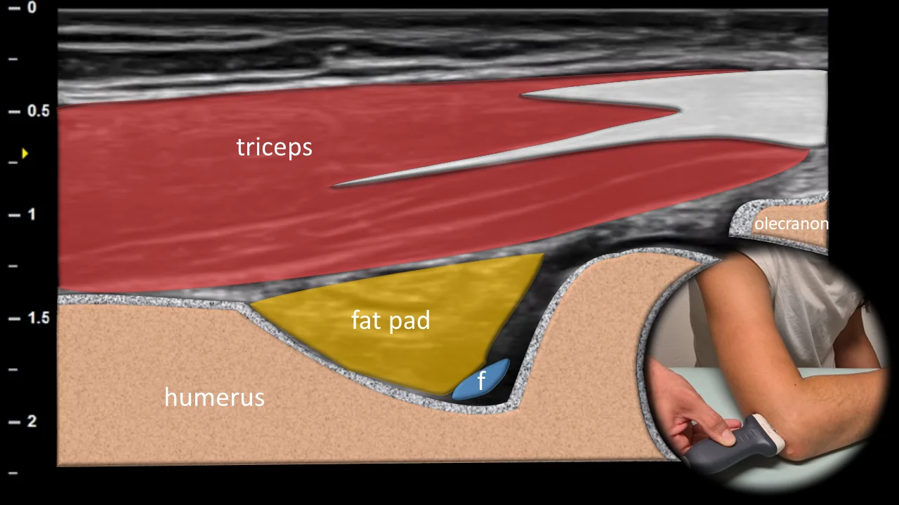
Figure 6. Dorsal view, sagittal plane. f: joint fluid
Longitudinal ultrasound projection of the posterior part of the elbow joint. The probe is positioned over the olecranon and the triceps brachii tendon. In the long axis, the triceps brachii muscle, its myotendinous junction, and the tendon inserting on the olecranon are visible. The posterior joint recess is located between the posterior fat pad and the humerus in the fossa olecrani area and may contain a small amount of fluid (f). This projection is also suitable for assessing the olecranon bursa.
Clinical Note
In this projection we mainly evaluate effusion in the posterior recess, olecranon bursitis and involvement of the triceps brachii tendon.
Schalten Sie die vollständige Health Library frei
Voller Zugriff auf Scan-Protokolle, Anatomie und klinische Referenzen. Jederzeit kündbar.
- Alle Protokolle und Anatomiereferenzen
- Originale Ultraschall-Illustrationen und Video-Demonstrationen
- Synchronisierung zwischen Mobilgerät und Web