 MSK Academy
4. Dorsale Ansicht
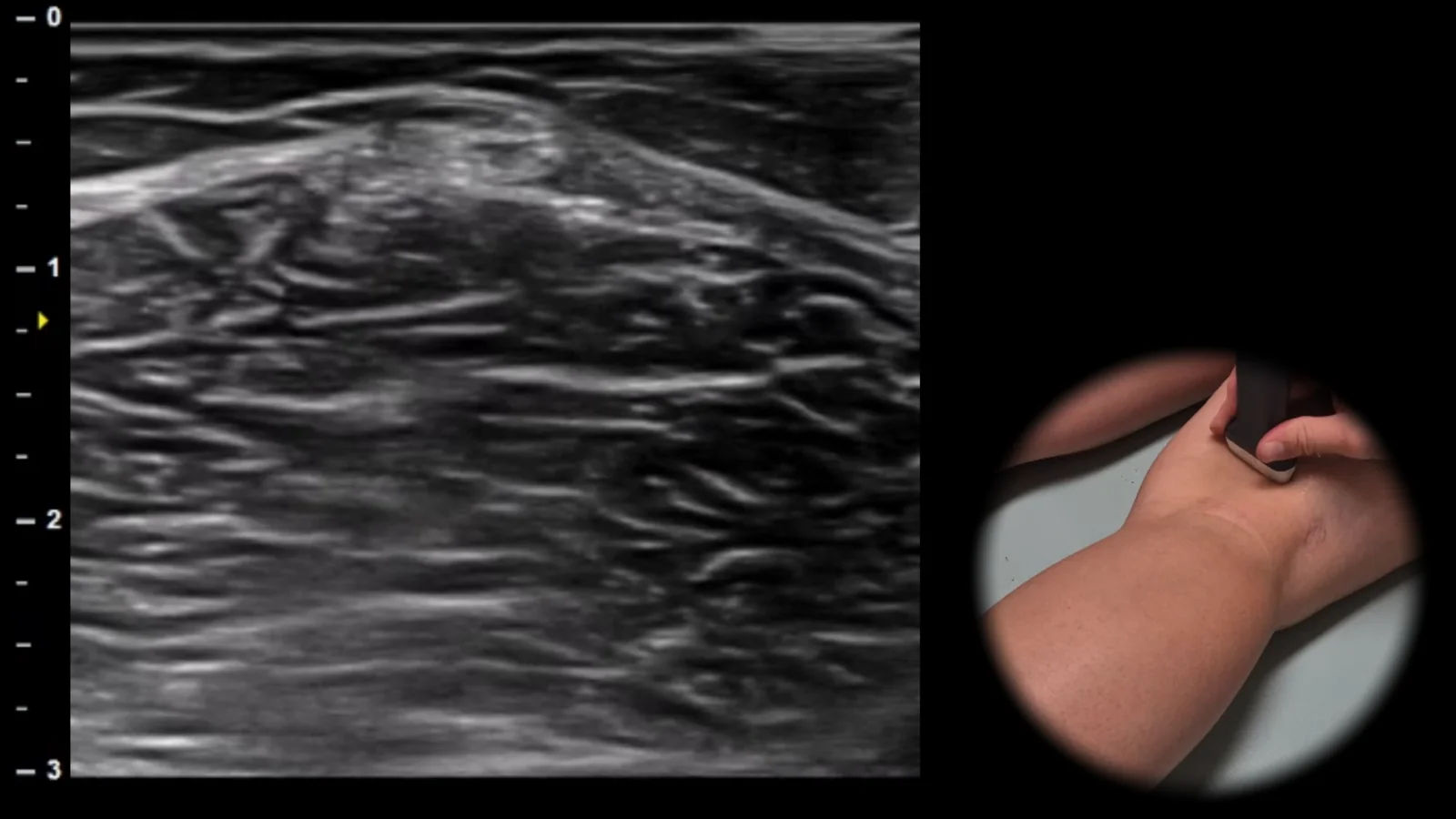
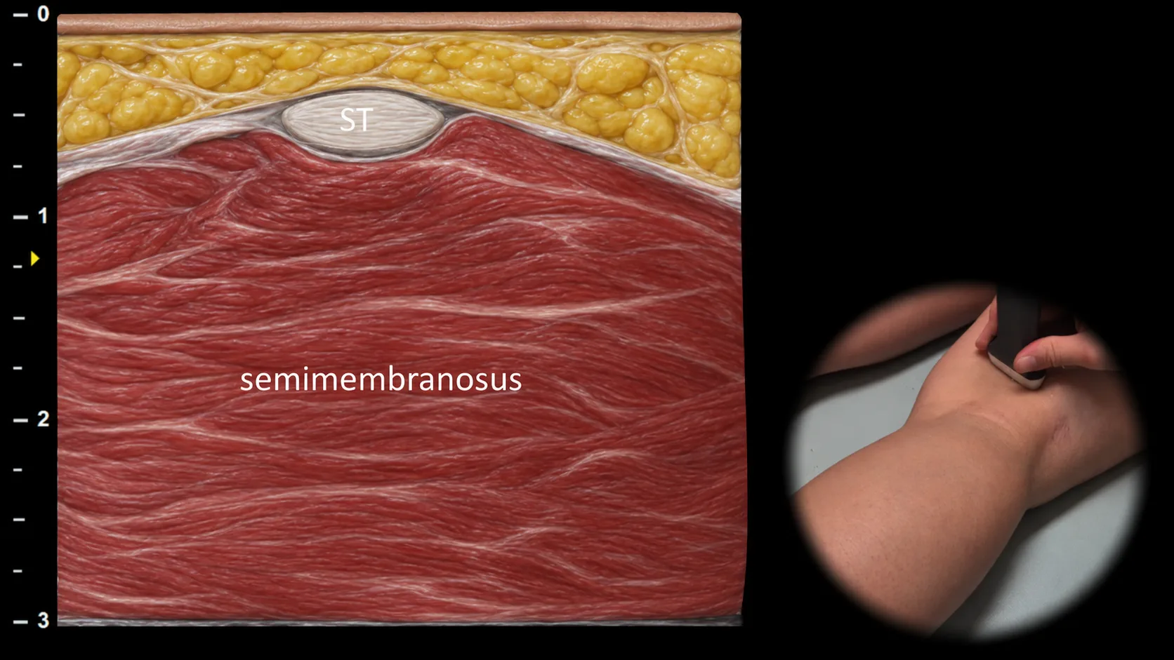
Abbildung 1. Dorsale Ansicht, Transversalebene. ST: Sehne des M. semitendinosus
Transversaler sonografischer Schnitt durch den posteromedialen Teil des Knies mit Darstellung der Sehne des M. semitendinosus (ST) als ovale Struktur, die oberflächlich auf dem M. semimembranosus liegt, der die breitere muskuläre Grundlage dieser Projektion bildet. Diese typische räumliche Anordnung ermöglicht eine eindeutige Identifizierung des medialen Anteils der ischiokruralen Muskulatur in der Poplitealregion. Diese Projektion ist als Orientierungspunkt für die Verlaufsdarstellung der medialen ischiokruralen Muskulatur und für die Beurteilung ihrer Sehnen- und Muskelläsionen wichtig. Klinischer Hinweis: Das charakteristische, als "cherry on cake" bezeichnete Erscheinungsbild erleichtert die schnelle sonografische Orientierung und Lokalisation der Sehne des M. semitendinosus.
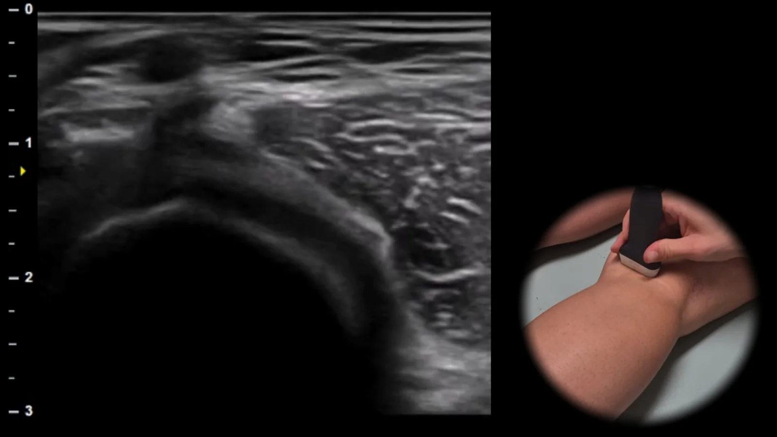
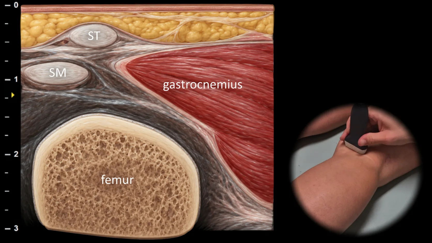
Abbildung 2. Dorsale Ansicht, Transversalebene. SM: Sehne des M. semimembranosus, ST: Sehne des M. semitendinosus
Transversaler sonografischer Schnitt durch den posteromedialen Anteil des Knies mit Darstellung des Bereichs zwischen der Sehne des M. semimembranosus (SM) und dem medialen Kopf des M. gastrocnemius. In dieser Projektion ist auch die Sehne des M. semitendinosus (ST) zusammen mit der in der Tiefe gelegenen knöchernen Kontur des Femurs als wichtigster anatomischer Orientierungspunkt sichtbar. Diese Projektion ist für die Beurteilung der Poplitealregion und insbesondere für den Nachweis einer Baker-Zyste essenziell, die sich typischerweise als Flüssigkeitsansammlung zwischen der Sehne des M. semimembranosus und dem medialen Kopf des M. gastrocnemius darstellt.
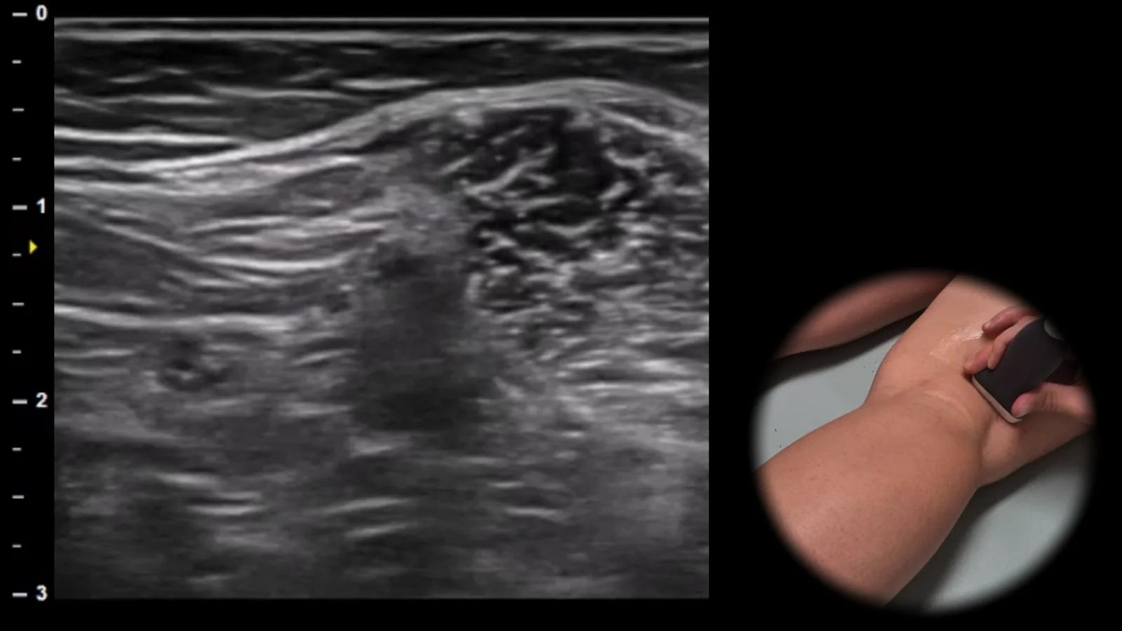
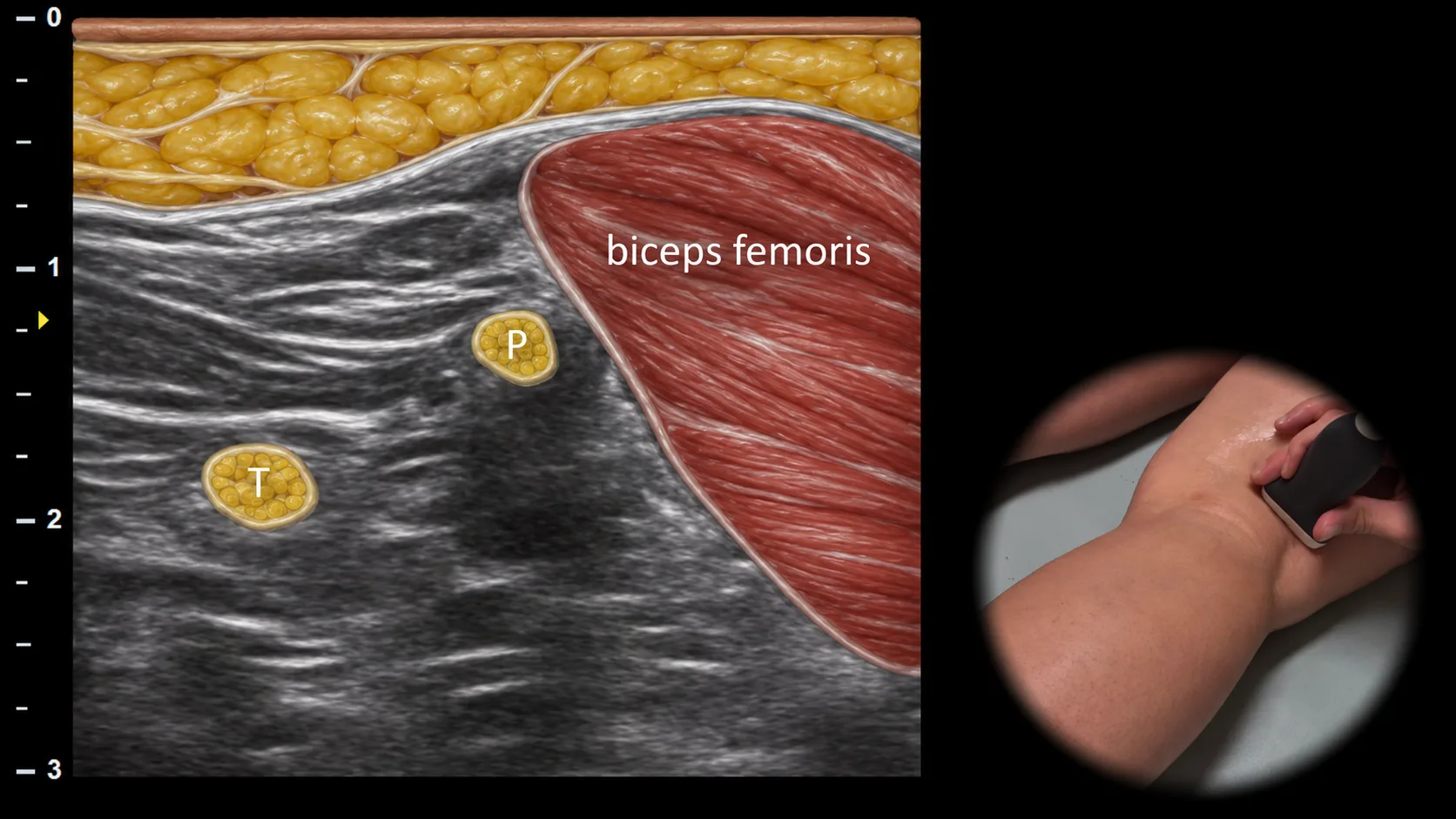
Abbildung 3. Dorsale Ansicht, Transversalebene. T: N. tibialis, P: N. fibularis communis
Transversaler Ultraschallschnitt durch den posterolateralen Teil des Knies im Bereich der Fossa poplitea. In dieser Projektion ist der M. biceps femoris als wichtiger muskulärer Orientierungspunkt sichtbar, in dessen Nähe der N. fibularis communis (P) als ovale Struktur mit typischem faszikulärem Erscheinungsbild dargestellt werden kann, die nach lateral abzweigt, während der N. tibialis (T) weiter medial verläuft. Diese Projektion ist für die Beurteilung der Bifurkation des N. ischiadicus und den Nachweis einer Einklemmung, Kompression oder traumatischen Verletzung peripherer Nerven im posterolateralen Kniebereich von entscheidender Bedeutung.
Zugang zur gesamten Bibliothek
Voller Zugriff auf Scan-Protokolle, Anatomie und klinische Referenzen. Jederzeit kündbar.
- Alle Protokolle und Anatomiereferenzen
- Originale Ultraschall-Illustrationen und Video-Demonstrationen
- Synchronisierung zwischen Mobilgerät und Web