Anatomy
The median nerve arises from the union of fibers from the lateral and medial fascicles of the plexus brachialis, most commonly from roots C6–T1. It is the main nerve of the anterior muscle group of the forearm and a significant nerve of the hand. Cross-sectional anatomy is key to understanding the sonographic appearance, as ultrasound displays structures in individual slices. Below we will review five key levels of cross-sectional imaging that we will follow along the nerve.
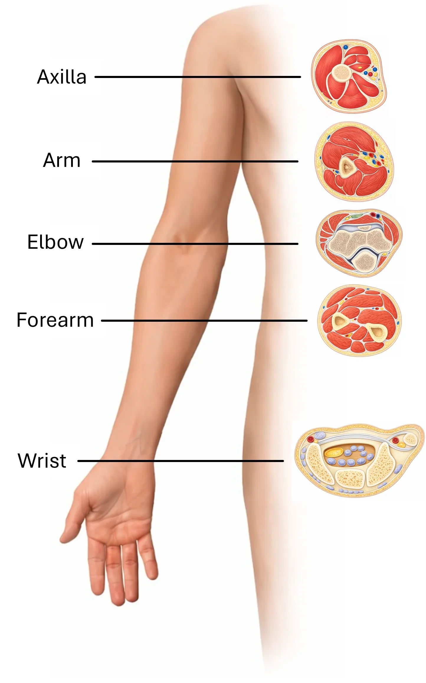
Axilla
In the axilla, the n. medianus is formed by the union of the radix lateralis from the fasciculus lateralis and the radix medialis from the fasciculus medialis of the plexus brachialis. On ultrasound examination, it is usually well recognizable as the most anteriorly located nerve relative to the axillary artery.
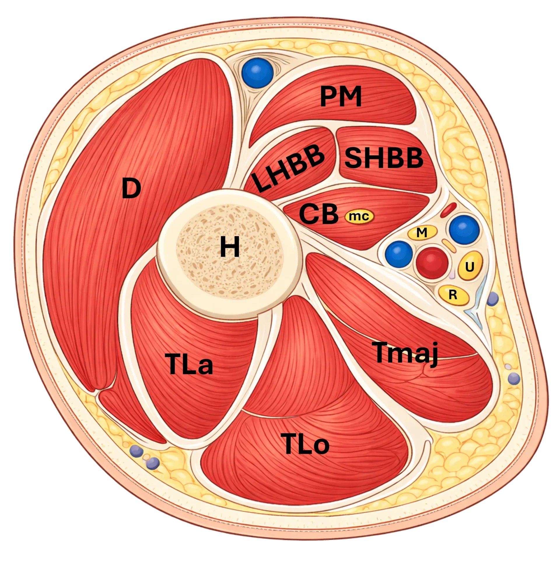
Figure 1: Cross-section of the axilla. D - m. deltoideus, PM - m. pectoralis major, LHBB - caput longum m. biceps brachii, SHBB - caput breve m. biceps brachii, CB - m. coracobrachialis, mc - n. musculocutaneus, M - n. medianus, U - n. ulnaris, R - n. radialis, Tmaj - m. teres major, TLa - caput laterale m. triceps brachii, TLo - caput longum m. triceps brachii, H - humerus
Arm
In the arm, it runs in the neurovascular bundle together with the a. brachialis. Proximally it is more often lateral to the artery, distally it crosses to its medial side. It lies between the m. biceps brachii and m. triceps brachii. For ultrasound, it is crucial: the n. medianus is a typical satellite of the a. brachialis in the brachium.
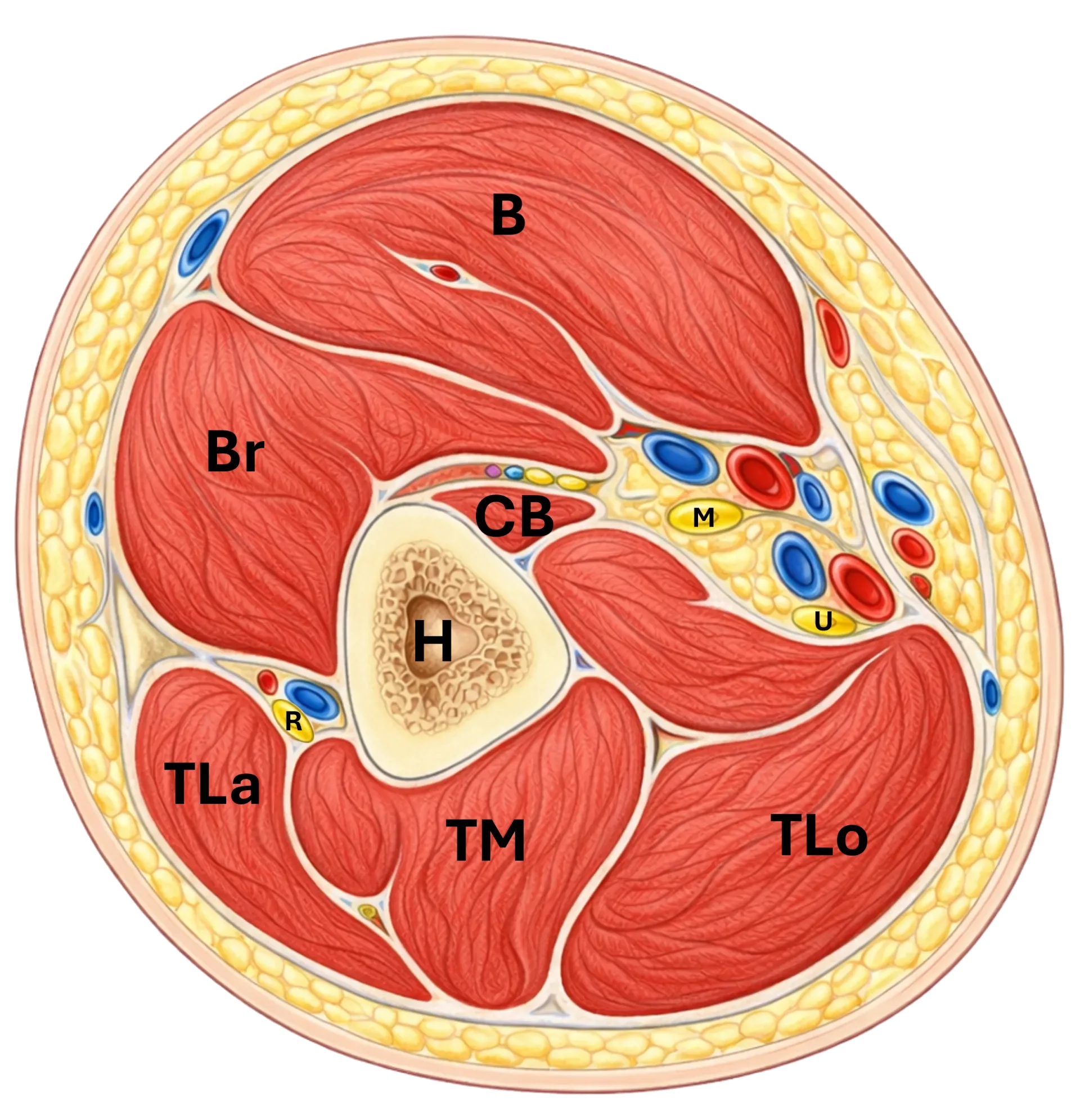
Figure 2: Cross-section of the arm. B – m. biceps brachii, Br – m. brachialis, CB – m. coracobrachialis, H – humerus, TLa – caput laterale m. triceps brachii, TM – caput mediale m. triceps brachii, TLo – caput longum m. triceps brachii, R – n. radialis, M – n. medianus, U – n. ulnaris
Elbow
In the cubital fossa, the n. medianus lies medial to the a. brachialis and beneath the lacertus fibrosus. In this region, the orientational scheme BAM applies well: biceps tendon – brachial artery – median nerve. Distally, it continues between the humeral and ulnar heads of the m. pronator teres, which is one of the common sites of possible compression.
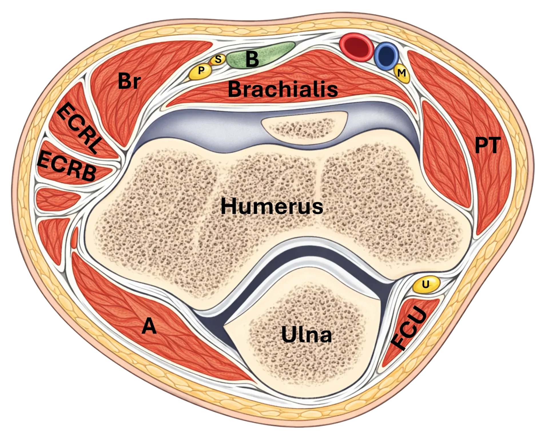
Figure 3: Cross-section of the elbow. Br – m. brachioradialis, ECRL – m. extensor carpi radialis longus, ECRB – m. extensor carpi radialis brevis, B – tendon of m. biceps brachii, S – r. superficialis n. radialis, P – r. profundus n. radialis (n. interosseus posterior), M – n. medianus, PT – m. pronator teres, U – n. ulnaris, FCU – m. flexor carpi ulnaris, A – m. anconeus
Forearm
In the forearm, it first runs under the arch of the m. flexor digitorum superficialis and then between the superficial and deep flexor layers. In the middle part of the forearm, it is mainly related to the FDS, FDP, and FPL. Distally, it becomes more superficial and lies close to the tendon of the m. flexor carpi radialis.
Important branches:
n. interosseus anterior – arises in the proximal forearm and runs deeper along the interosseous membrane
ramus cutaneus palmaris – arises proximal to the carpal tunnel and travels superficially to the palm
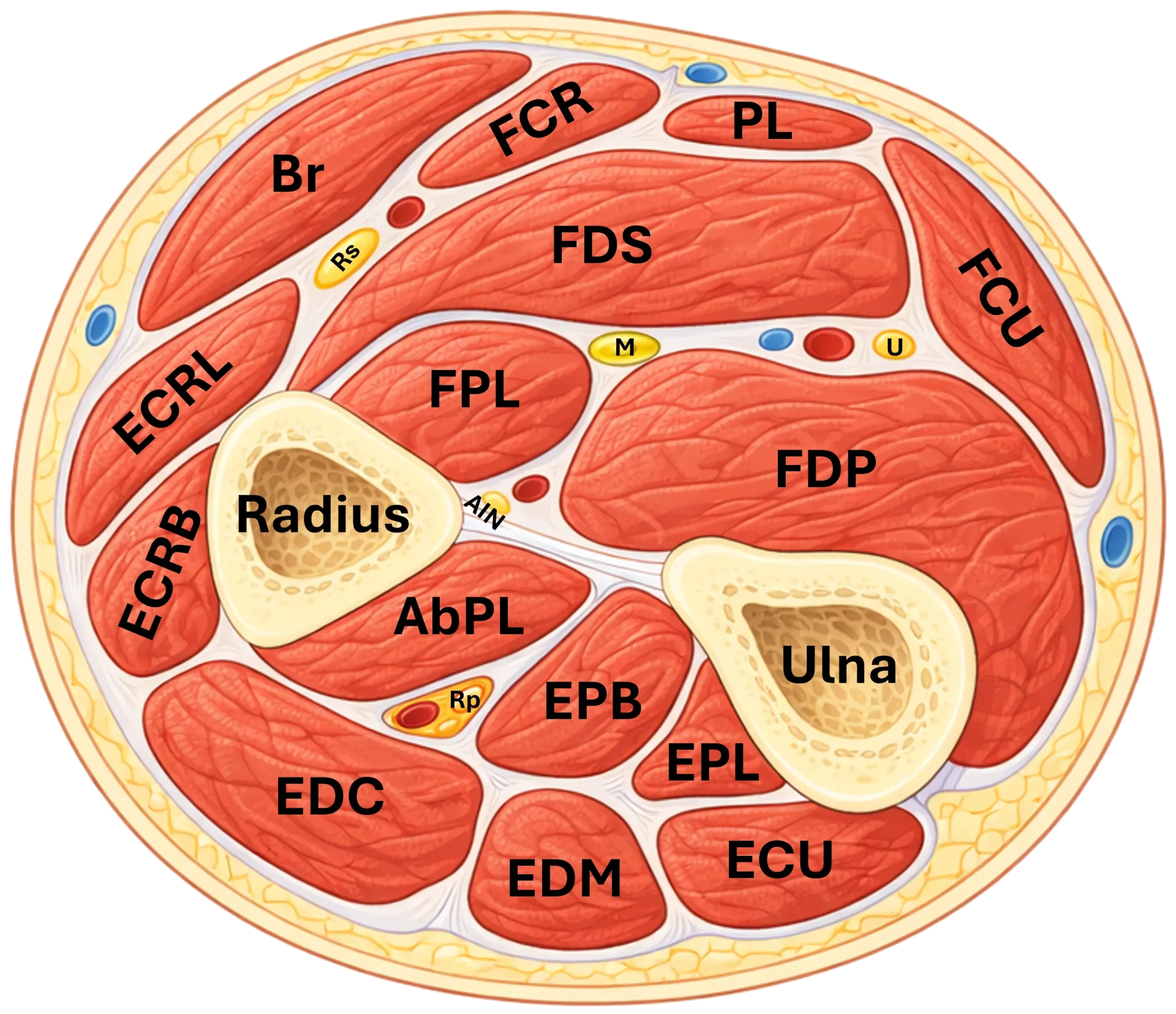
Figure 4: Cross-section of the forearm. Br – m. brachioradialis, ECRL – m. extensor carpi radialis longus, ECRB – m. extensor carpi radialis brevis, EDC – m. extensor digitorum communis, EDM – m. extensor digiti minimi, ECU – m. extensor carpi ulnaris, AbPL – m. abductor pollicis longus, EPB – m. extensor pollicis brevis, EPL – m. extensor pollicis longus, FCR – m. flexor carpi radialis, PL – m. palmaris longus, FDS – m. flexor digitorum superficialis, FPL – m. flexor pollicis longus, FDP – m. flexor digitorum profundus, FCU – m. flexor carpi ulnaris, M – n. medianus, U (nerve) – n. ulnaris, Rs – r. superficialis n. radialis, Rp – r. profundus n. radialis (n. interosseus posterior), AIN – n. interosseus anterior, R – radius, U (bone) – ulna
Wrist
In the wrist region, the n. medianus enters the carpal tunnel beneath the flexor retinaculum. This is its most important entrapment location.
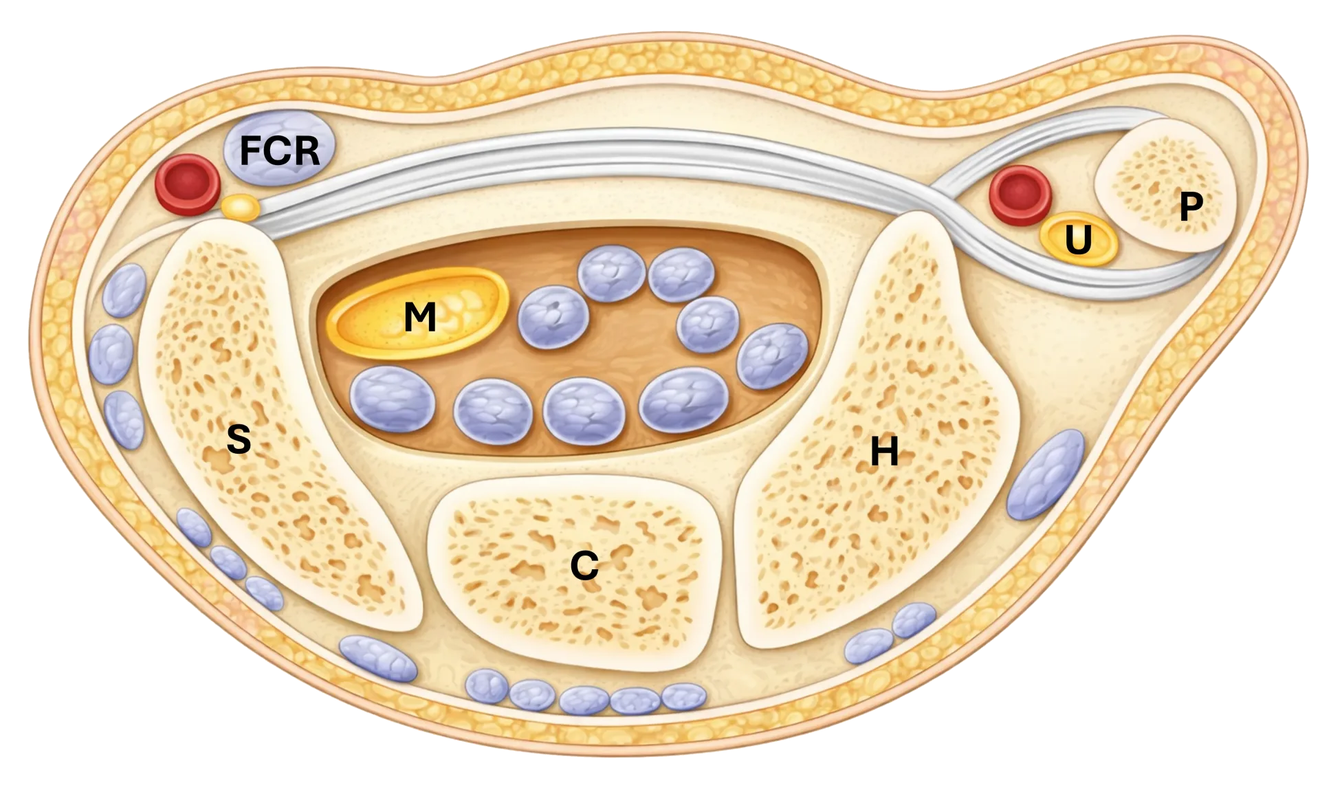
Figure 5: Cross-section of the wrist. FCR – m. flexor carpi radialis, M – n. medianus, U – n. ulnaris, P – os pisiforme, H – os hamatum, C – os capitatum, S – os scaphoideum
Practical anatomical landmarks for US
in axilla: anterior to a. axillaris
in arm: adjacent to a. brachialis
in cubital fossa: BAM, beneath lacertus fibrosus
distal to elbow: between heads of m. pronator teres
in forearm: under the arch of FDS
in wrist: in carpal tunnel beneath retinaculum