Ultrasound examination
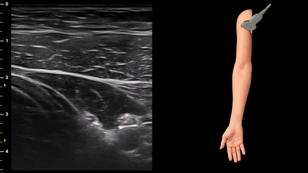
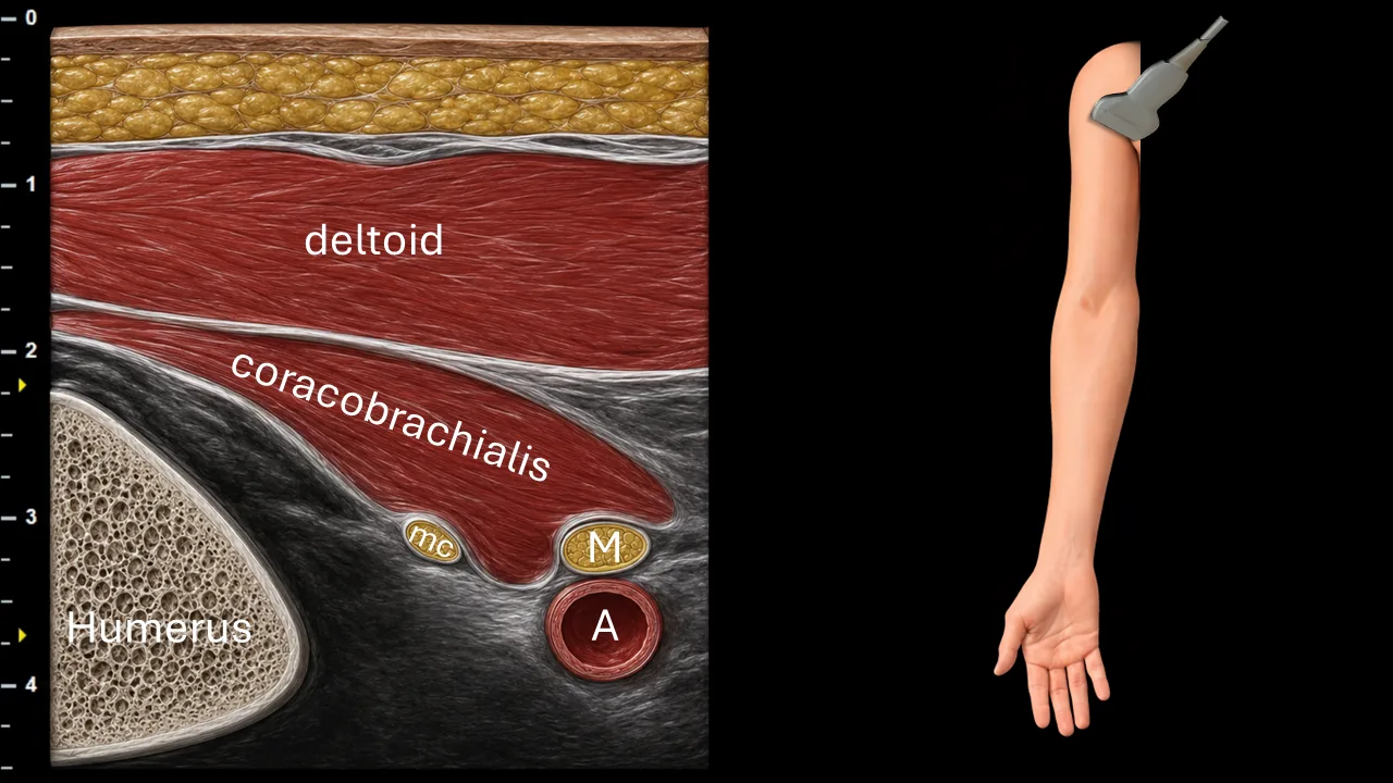
Figure 1. Axilla / proximal arm, transverse plane. A: a. axillaris, M: n. medianus, mc: n. musculocutaneus
Transverse ultrasound section of the axillary region focused on visualization of the n. musculocutaneus. The image shows the a. axillaris (A) as a prominent vascular structure, with the n. medianus (M) located ventrally and superficially in relation to the artery. The n. musculocutaneus (mc) is visible as a small oval nerve structure positioned laterally to the neurovascular bundle, in close relationship to the m. coracobrachialis. In this projection, the nerve is seen near the deep surface of the coracobrachialis muscle, which is an important landmark for its identification. The image also demonstrates the surrounding muscular structures, particularly the m. coracobrachialis and m. deltoideus, as well as the contour of the humerus.
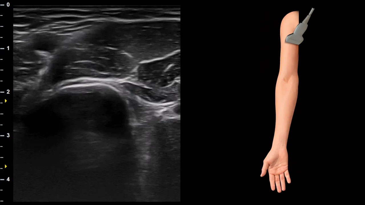
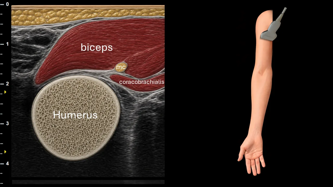
Figure 2. Proximal arm, transverse plane. mc: n. musculocutaneus
Transverse ultrasound section of the proximal arm focused on visualization of the n. musculocutaneus (mc). The nerve is seen as a small oval hyperechoic fascicular structure positioned between the m. biceps brachii and m. coracobrachialis, in close contact with the deep aspect of the biceps and superficial to the coracobrachialis muscle. In this projection, the humerus forms the main bony landmark deep to the muscular structures. The image demonstrates the typical course of the n. musculocutaneus after leaving the axillary region, where it runs within the anterior compartment of the arm in relation to the coracobrachialis and biceps brachii muscles.
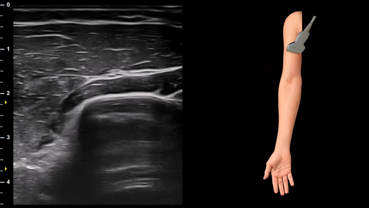
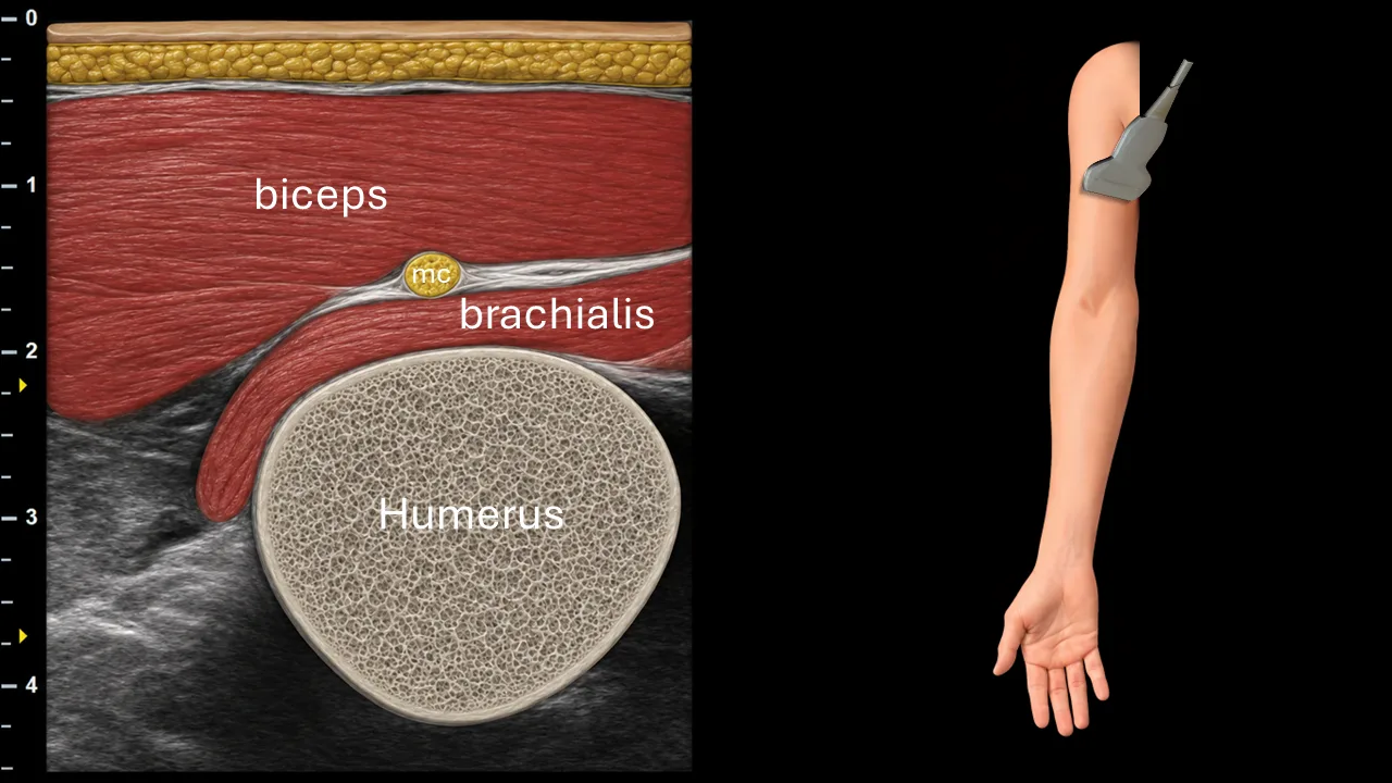
Figure 3. Arm, transverse plane. mc: n. musculocutaneus
Transverse ultrasound section of the arm focused on visualization of the n. musculocutaneus (mc). The nerve is seen as a small oval fascicular structure located between the m. biceps brachii and the m. brachialis, within the anterior compartment of the arm. In this projection, the n. musculocutaneus has already passed distally from the coracobrachialis region and runs in the intermuscular plane between biceps and brachialis. The humerus forms the main deep bony landmark, with the brachialis muscle lying directly superficial to it. The image demonstrates the typical distal course of the n. musculocutaneus before it continues toward the lateral aspect of the forearm as the lateral antebrachial cutaneous nerve.
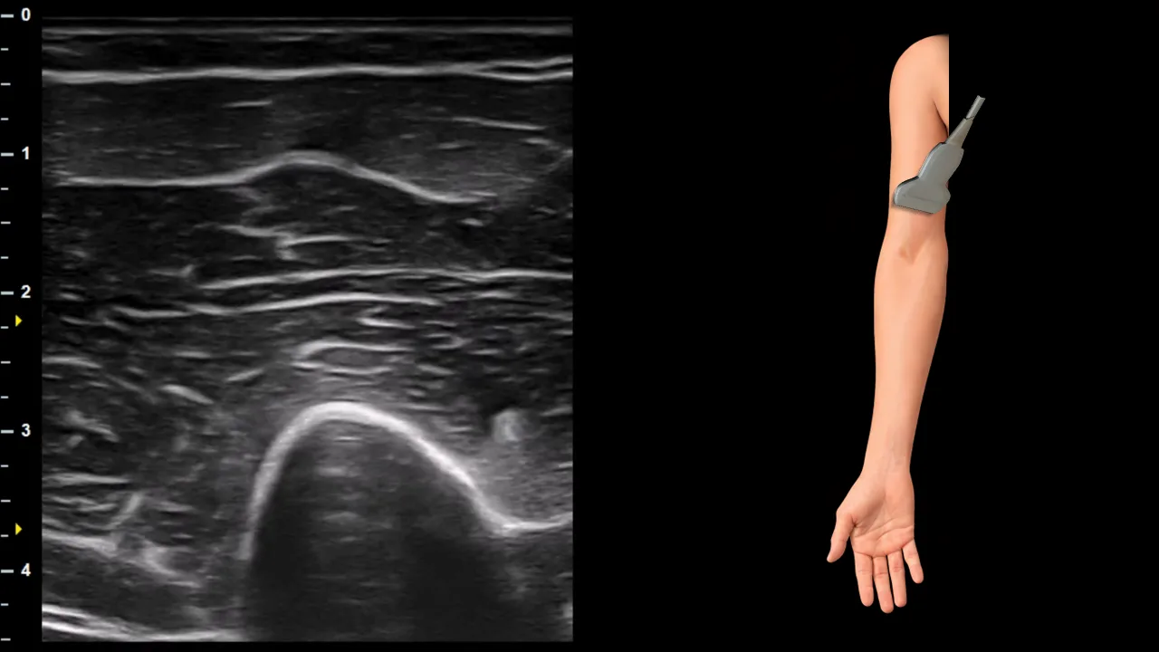
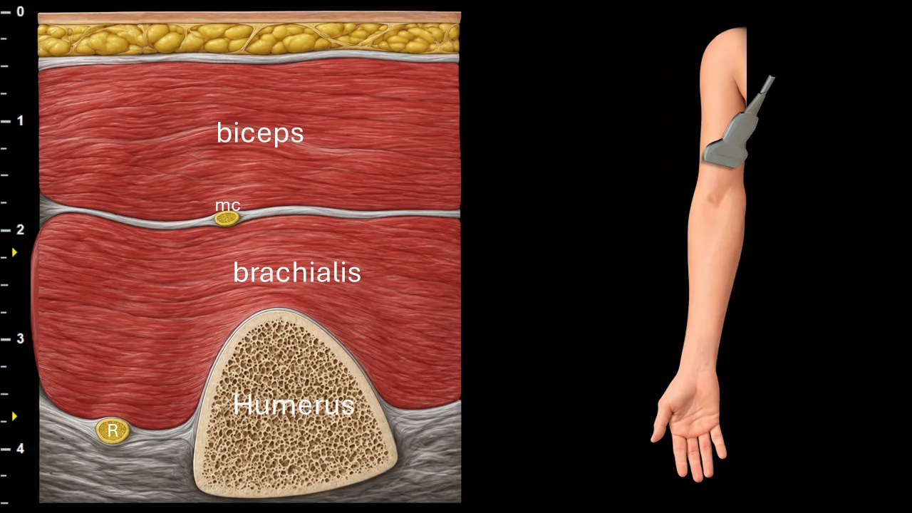
Figure 4. Mid arm, transverse plane. mc: n. musculocutaneus, R: n. radialis
Transverse ultrasound section of the arm focused on visualization of the n. musculocutaneus (mc). The nerve is seen as a small oval fascicular structure located in the intermuscular plane between the m. biceps brachii and the m. brachialis. In this projection, the n. musculocutaneus runs distally within the anterior compartment of the arm, superficial to the brachialis muscle and deep to the biceps brachii. The humerus forms the main deep bony landmark, with the brachialis muscle lying directly superficial to its anterior contour. The image also shows the n. radialis (R) positioned more deeply and laterally in relation to the humerus, separated from the musculocutaneous nerve by the brachialis muscle.
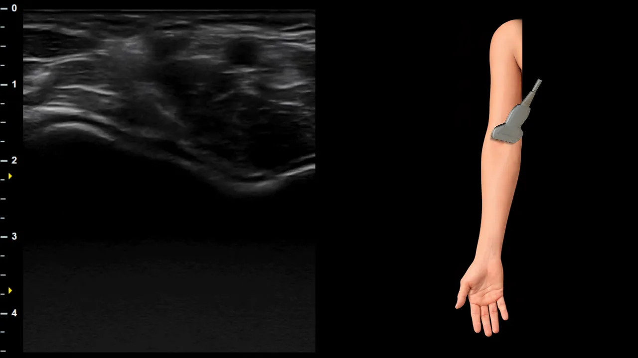
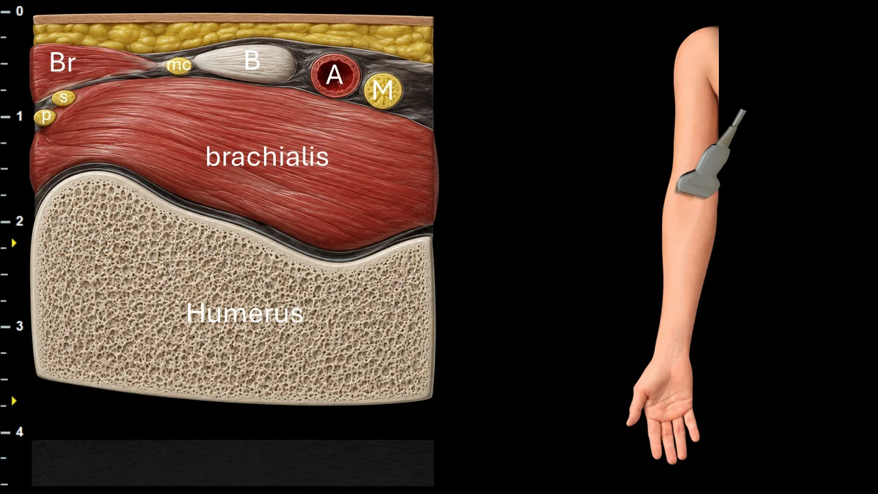
Figure 5. Distal arm / cubital region, transverse plane. mc: n. musculocutaneus / n. cutaneus antebrachii lateralis, A: a. brachialis, M: n. medianus, B: biceps tendon, Br: m. brachioradialis
Transverse ultrasound section of the distal arm focused on visualization of the n. musculocutaneus (mc) as it approaches the cubital region. In this projection, the nerve is seen superficially and laterally, close to the biceps tendon (B) and near the medial border of the m. brachioradialis (Br). Distally, the n. musculocutaneus continues as the n. cutaneus antebrachii lateralis, emerging toward the superficial plane of the lateral forearm.
The image also shows the a. brachialis (A) and the n. medianus (M) located more medially within the neurovascular bundle. The m. brachialis lies directly superficial to the anterior contour of the humerus, which forms the main deep bony landmark. This level demonstrates the transition of the musculocutaneous nerve from its course between the biceps brachii and brachialis muscles toward its terminal sensory branch in the lateral forearm.
Schalten Sie die vollständige Health Library frei
Voller Zugriff auf Scan-Protokolle, Anatomie und klinische Referenzen. Jederzeit kündbar.
- Alle Protokolle und Anatomiereferenzen
- Originale Ultraschall-Illustrationen und Video-Demonstrationen
- Synchronisierung zwischen Mobilgerät und Web