September 2015
EURO-MUSCULUS/USPRM. Basic Scanning Protocols for Ankle and foot
Autoren: L Özçakar, M Kara, K V Chang, A Bayram Çarli, C Y Hung, F Tok, C H Wu, N Akkaya, M Y Hsiao, L Tekin, T G Wang, A M Ulaşlı, W S Chen, M De Muynck
Zeitschrift: European Journal of Physical and Rehabilitation Medicine
Veröffentlicht: September 2015
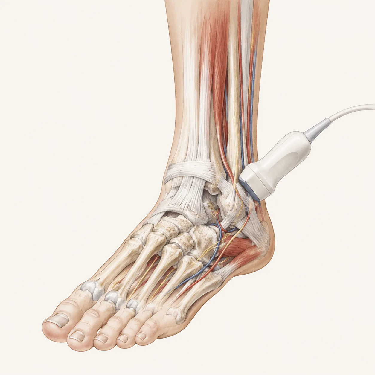
This article presents a practical ultrasound protocol for examining the ankle and foot. Its main strength is the clear step-by-step approach to patient and probe positioning, making it useful for learning a standardized ankle and foot scan.
Interesting highlights:
The protocol shows how to assess the anterior ankle joint, tarsal joints, and extensor tendons, including dynamic movements to help identify individual tendons.
It covers key medial and lateral structures, including the tarsal tunnel, tibial nerve, flexor tendons, deltoid ligament, fibularis tendons, and lateral ankle ligaments.
It also includes posterior and plantar scanning, especially the Achilles tendon, retrocalcaneal bursae, plantar fascia, and forefoot evaluation for Morton’s neuroma using Mulder’s maneuver.
Most useful for:
MSK ultrasound beginners, rehabilitation physicians, sports medicine clinicians, radiologists, orthopedic physicians, podiatrists, and anyone learning a structured approach to ankle and foot ultrasound.