September 2015
EURO-MUSCULUS/USPRM. Basic scanning protocols for knee
Autoren: L Özçakar, M Kara, K V Chang, F Tok, C Y Hung, N Akkaya, C H Wu, A B Çarli, M Y Hsiao, L Tekin, T G Wang, A M Ulaşlı, W S Chen, M De Muynck
Zeitschrift: European Journal of Physical and Rehabilitation Medicine
Veröffentlicht: September 2015
This article presents a practical ultrasound protocol for examining the knee. Its main strength is the clear step-by-step approach to patient and probe positioning, making it useful for learning a standardized knee scan.
Interesting highlights:
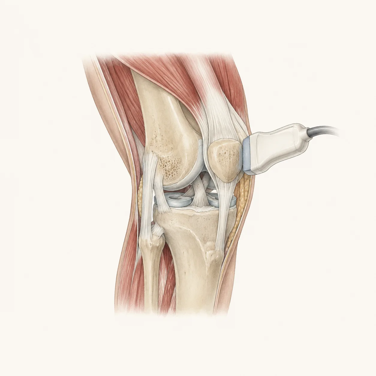
The protocol starts with the suprapatellar region, where joint fluid is commonly assessed, and also shows how to evaluate the quadriceps tendon and femoral cartilage.
It covers the anterior knee structures, including the patellar tendon, Hoffa’s fat pad, infrapatellar bursae, and even the anterior cruciate ligament in maximum knee flexion.
It also includes medial, lateral, and posterior knee scanning, including the menisci, collateral ligaments, pes anserine region, iliotibial band, biceps femoris tendon, Baker’s cyst region, and posterior cruciate ligament.
Most useful for:
MSK ultrasound beginners, rehabilitation physicians, sports medicine clinicians, radiologists, orthopedic physicians, and anyone learning a structured approach to knee ultrasound.