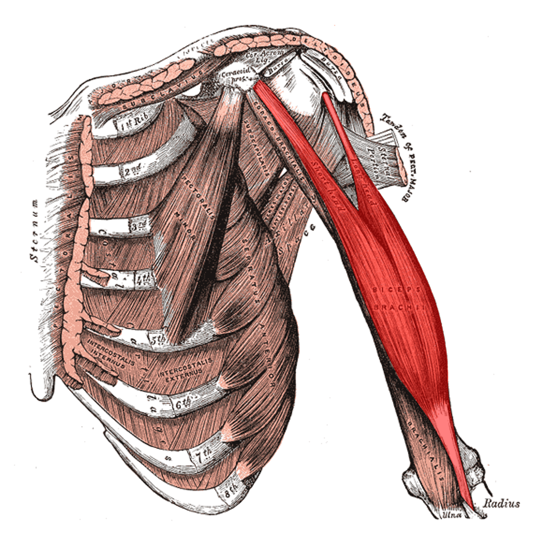
1. Name: biceps brachii 2. Origin: A. LHBB: supraglenoid tubercle of the scapula B. SHBB: coracoid process of the scapula 3. Insertion: A. Radial tuberosity B. Fascia of the forearm via the bicipital aponeurosis 4. Function: A. Elbow flexion B. Forearm supination C. Shoulder flexion 5. Inervation: A. Peripheral: musculocutaneous nerve B. Trunks: C5 and C6
Anatomy
Scanning protocol
1. Proximal part A. LHBB – transverse – longitudinal B. SHBB – transverse – longitudinal 2. Muscle belly – transverse – longitudinal 3. Distal part A. Distal tendon – transverse – longitudinal – pronator window – cobra view – dorsal view – lateral roll view B. Lacertus fibrosus
Proximal part and muscle belly
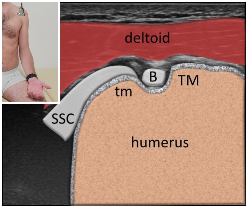
Subscapularis tendon, LHBB tendon short axis
Greater (TM) and lesser ™ tubercle is visualized moving the probe more cranially. In between (in bicipital sulcus) there is the long head of biceps brachii tendon (B). In this part, collection of the joint fluid can be seen around the biceps tendon. Subscapularis tendon (SSC) attaching the lesser tubercle can be also seen in this projection.
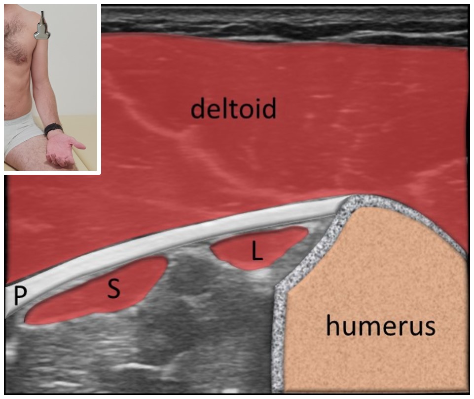
Pectoralis major tendon
Further cranially the pectoralis major tendon (P) between the deltoid and the short (S) and long (L) head of biceps brachii.
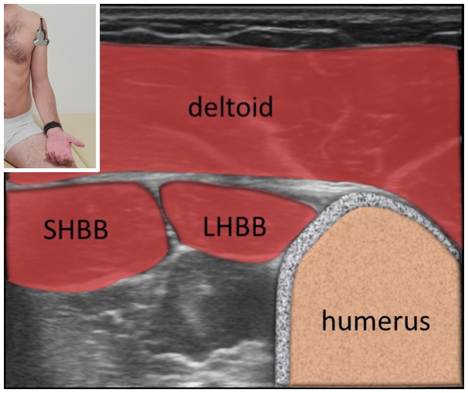
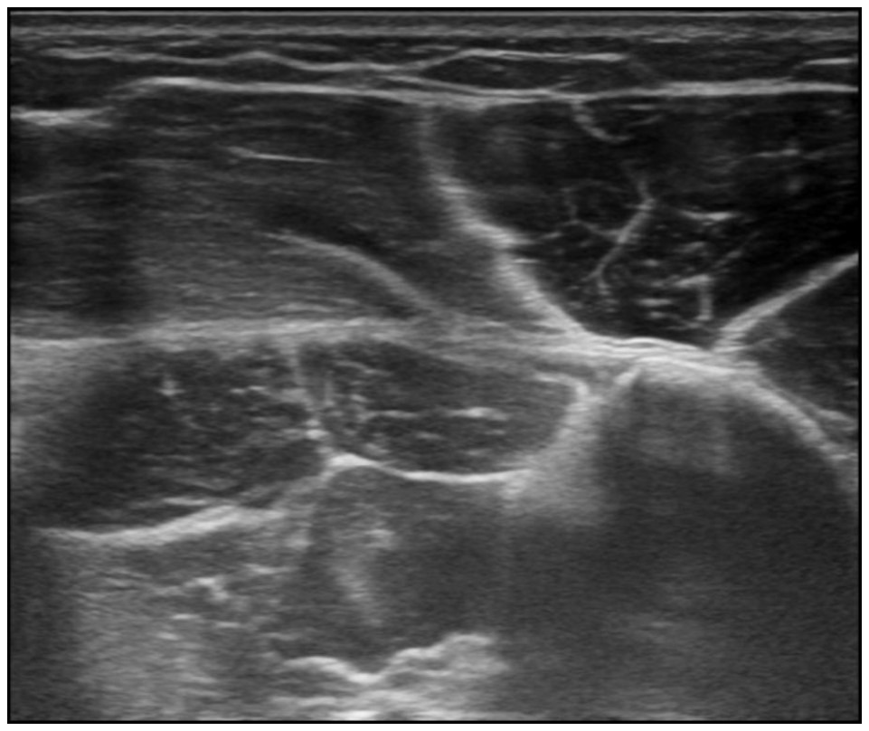
Biceps brachii long and short head
Moving the probe cranially the short (SHBB) and the long (LHBB) head of biceps brachii can be identified.
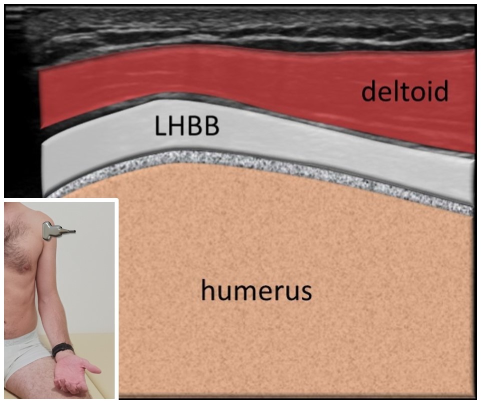
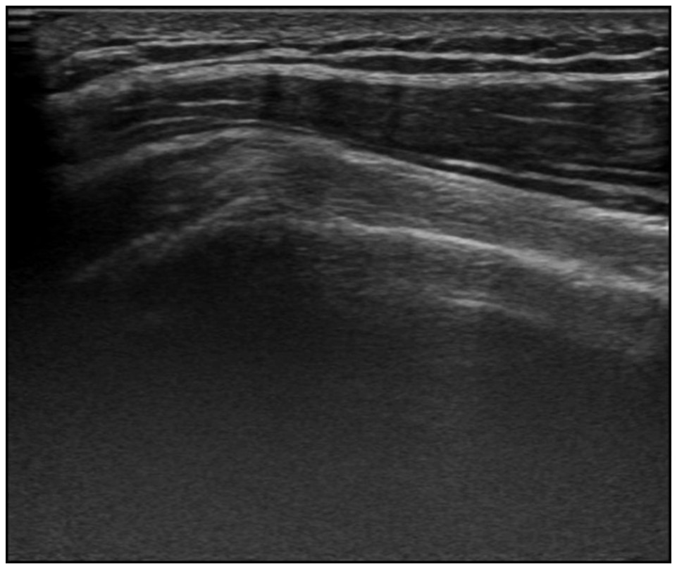
LHBB tendon long axis
Rotating the probe 90° long head of biceps bracahii tendon (LHBB) can be visualized in long axis. Integrity or effusion of the tendon can be evaluated. Healthy tendon has fibralar appearence similar to spaghetti.
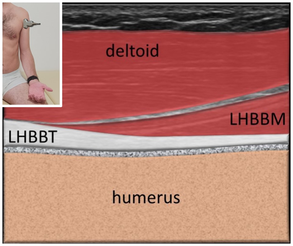
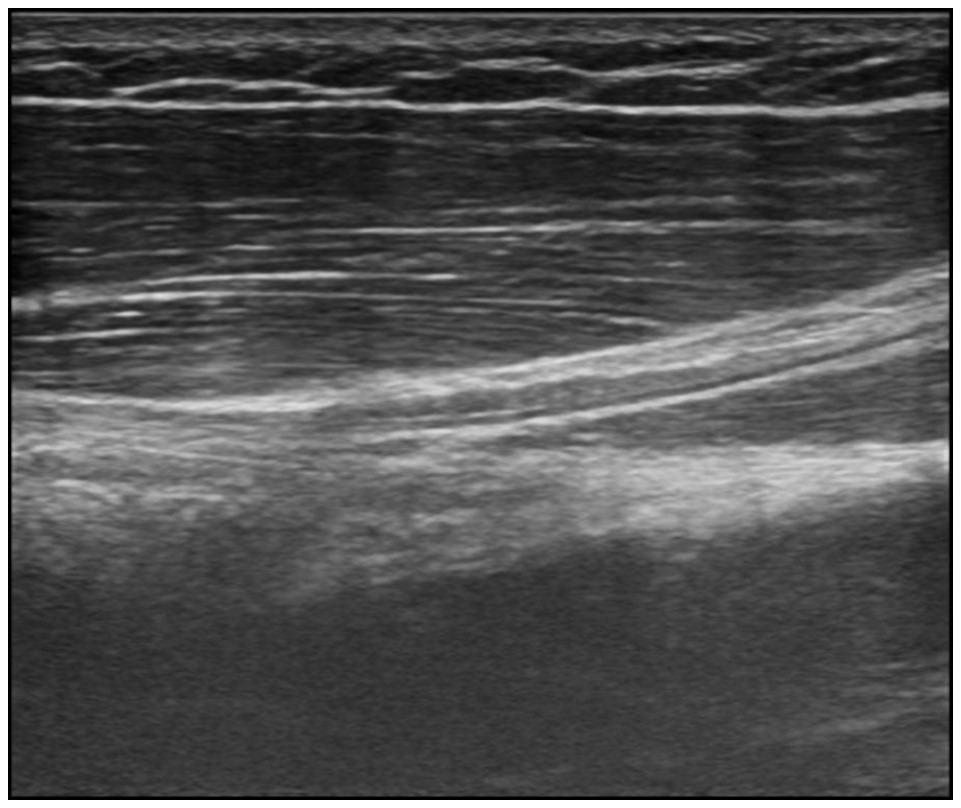
Biceps brachii myotendineous junction
Moving the probe caudally the myotendineous junction (connection of the tendon – LHBBT and the muscle – LHBBM) of the biceps brachii can be visualized.
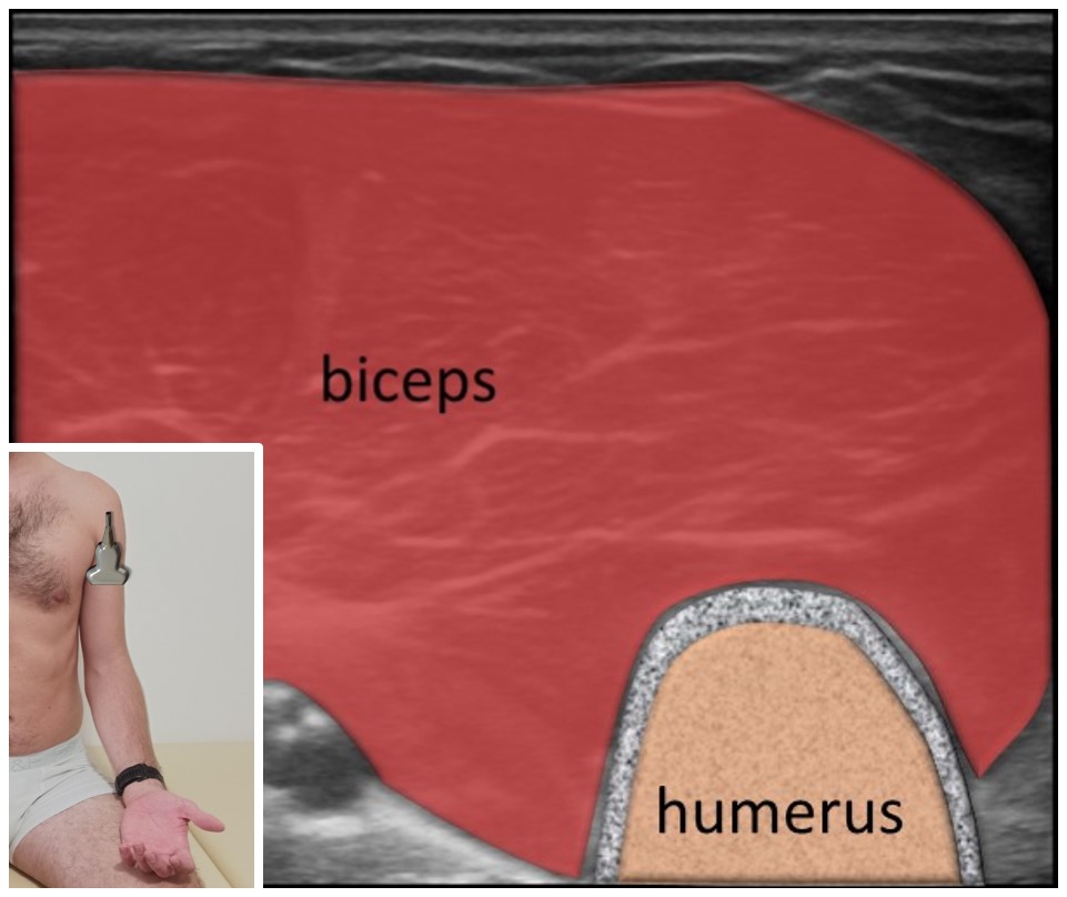
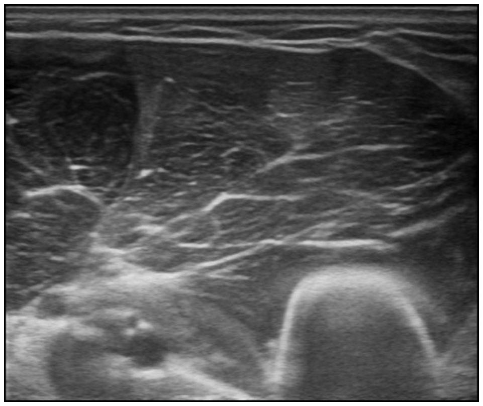
Biceps brachii belly
The probe is placed in horizontal plane at the arm and biceps brachii muscle belly is visualized.