 MSK Academy
Your MSK ultrasound reference, always within reach
Review examination protocols and anatomy before or during scanning. Follow probe positions and identify anatomical structures in the ultrasound image.
Also available on iOS and Android


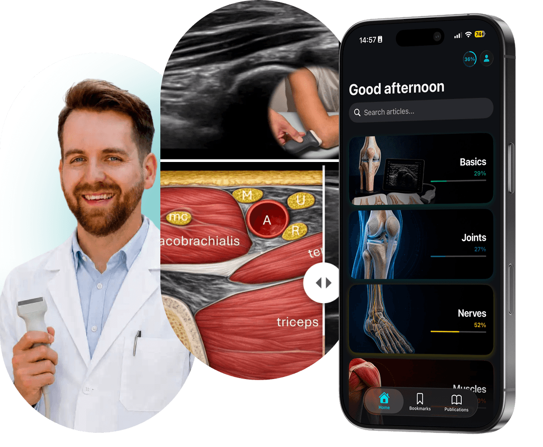
Jakub Jacisko, MD, PhD
Author
100+
Original illustrations
Regular updates
New content added monthly
100+
Educational illustrations
15+
Videos
Made by a clinician, for clinicians
Try the shoulder examination protocol
Choose a view, then drag the slider to compare the ultrasound image with its labeled anatomy.
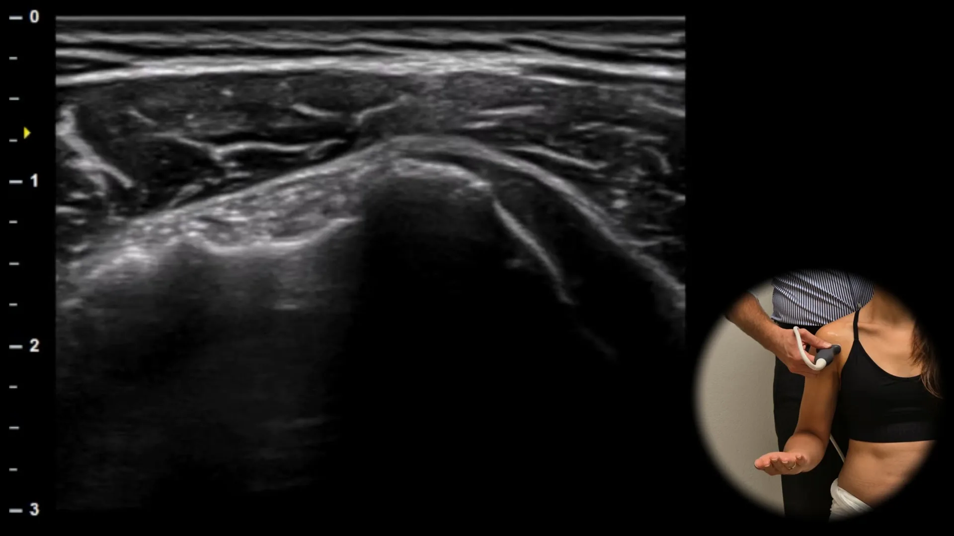
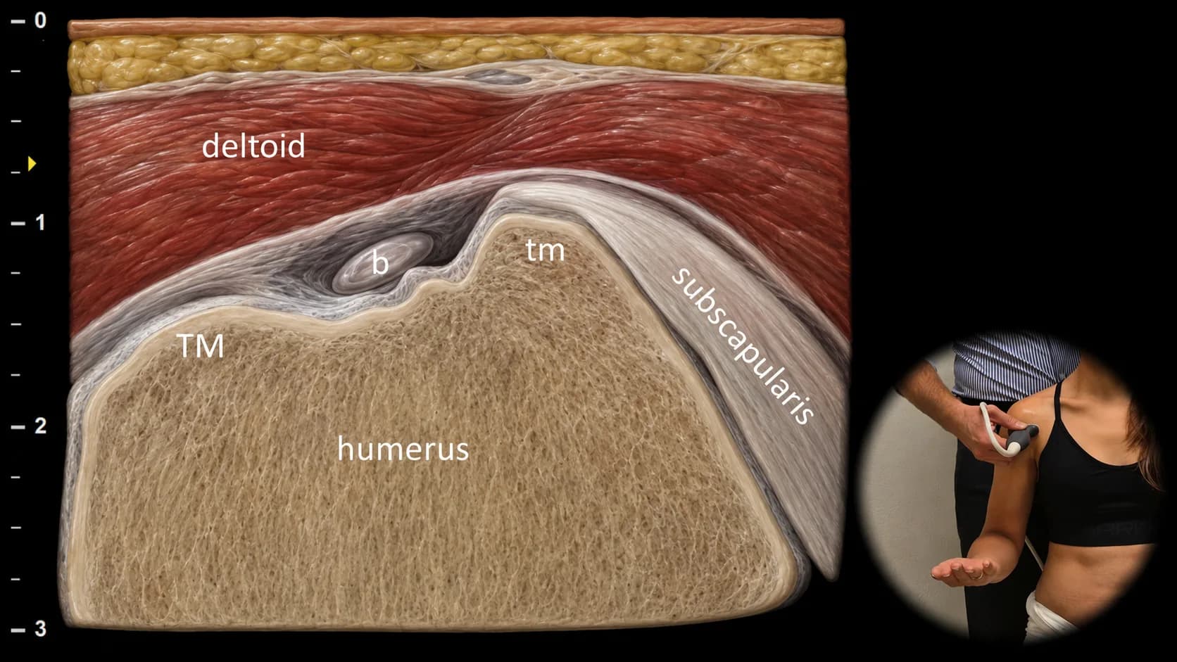
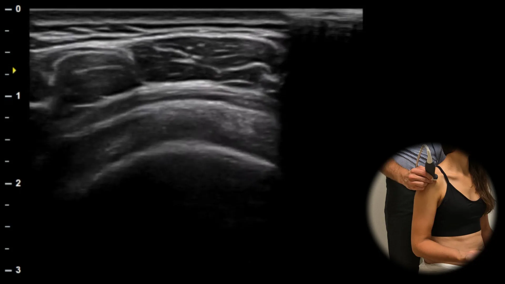
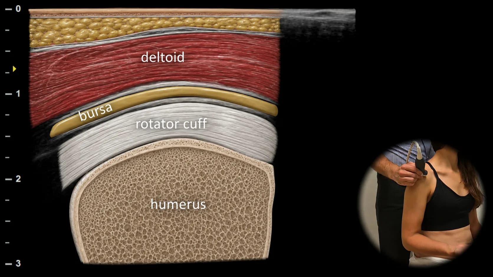
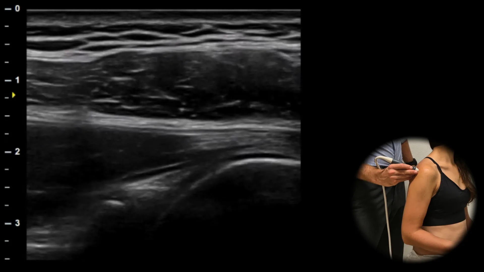
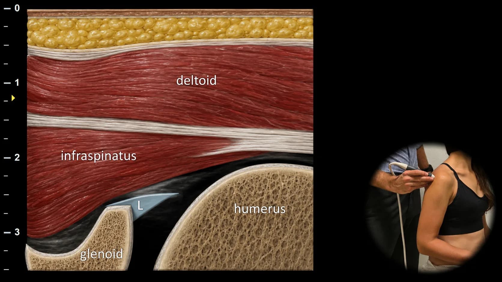
Drag the slider to reveal the labeled anatomy
TM and tm are the main anterior bony landmarks. The LHBB runs between them in the bicipital groove, a common location for detectable fluid because the tendon sheath communicates with the glenohumeral joint. In this view, also assess the subscapularis tendon, superficial deltoid layer, and fluid amount (physiologic versus synovitis).
b: long head of biceps tendon, TM: greater tubercle, tm: lesser tubercle.
More examination protocols
The app guides you step by step through 9 more examination protocols, from probe position to labeled anatomy:
- Median nerve
- Radial nerve
- Suprascapular nerve
- Sciatic nerve
- Common peroneal nerve
- Ulnar nerve
- Axillary nerve
- Musculocutaneous nerve
- Tibial nerve
Video example
See the examination in practice
Watch Dr. Jakub Jacisko demonstrate an ultrasound examination of the shoulder.
Musculoskeletal ultrasound education for clinical practice
Examination protocols
Follow examination protocols for individual joints and regions, with probe positions, anatomical landmarks, and illustrated ultrasound examples.
Anatomical illustrations
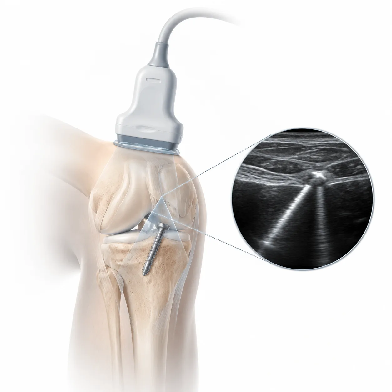
Compare ultrasound and anatomical images side by side during the examination. Check which structure you are looking at and how it should appear on screen.
On your phone, tablet, or computer
On your phone between patients, on a tablet in the examination room, or on your computer at home. Available for iOS, Android, and web.

Jakub Jacisko, MD, PhD
Specialist in Physical and Rehabilitation Medicine. He works in Prague at the Department of Rehabilitation and Sports Medicine, Motol University Hospital. He also works at Pavel Kolar's Center for movement medicine. His clinical and academic focus is musculoskeletal sonography and dynamic ultrasound examination methods.
- 10+ peer-reviewed publications in MSK sonography
- Ultrasound teaching for medical students and physicians
- Currently preparing for board certification in Pain Medicine
Built for clinicians who use ultrasound
Physical and Rehabilitation Medicine
Review examination protocols and anatomy before the examination or directly during scanning. Covers musculoskeletal diagnostics and ultrasound-guided procedures.
Orthopedics
Review tendons, muscles, joints, and periarticular structures on ultrasound during outpatient assessment and clinical decision-making.
Neurology
Follow a systematic approach to scanning peripheral nerves and the surrounding anatomical structures.
Radiology
Learn and review MSK ultrasound examination protocols for individual joints and regions.
Sports Medicine
Review examination protocols for the shoulder, knee, and ankle in the clinic or when working with athletes.
Physiotherapy
Build ultrasound skills for musculoskeletal assessment. Identify anatomical structures and connect the ultrasound image with the clinical findings.
Recent research in musculoskeletal ultrasound
Start with the shoulder protocol
Free access to foundational content. Available on iOS, Android, and web.