Anatomy
The hip joint is one of the most stable joints in the human body. It is adapted to bear the weight of the trunk during standing, walking, and running. This high stability is, however, achieved at the cost of reduced mobility, which is why the hip is less prone to dislocation but is often affected by overuse, tendinopathies, and degenerative changes. For ultrasound examination, orientation in bony landmarks, joint capsule, tendon insertions, bursae, and surrounding neurovascular structures is essential.
Bone Landmarks
Bone landmarks are fundamental reference points during scanning. They help guide the probe correctly and quickly distinguish normal anatomy from pathology.
- Medial epicondyle - orientation for the common flexor attachment and n. ulnaris.
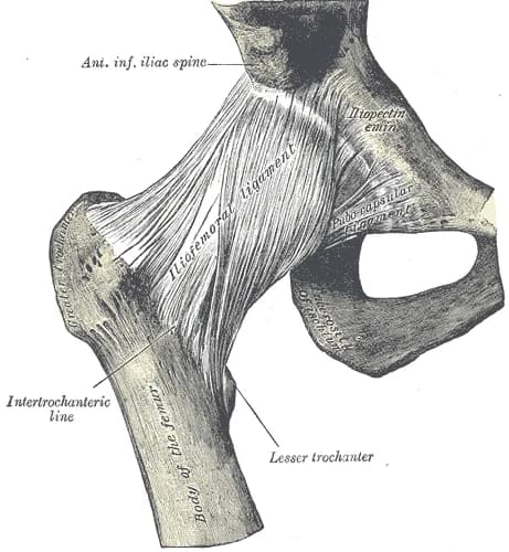
- Spina iliaca anterior inferior (SIAI) – located below the SIAS, the origin site of the straight head of m. rectus femoris.
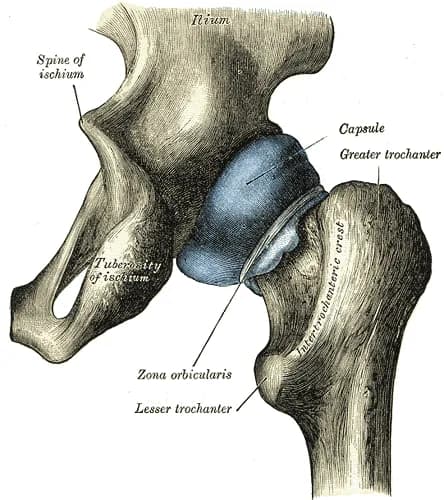
- Femoral head – spherical proximal end of the femur, part of the hip joint, assessable during examination of joint effusion and anterior recess.
- Femoral neck – connects to the head, an important landmark when imaging the anterior part of the hip joint.
- Trochanter major – large, easily palpable lateral process of the femur, insertion site of gluteal tendons, key point when examining the lateral side of the hip.
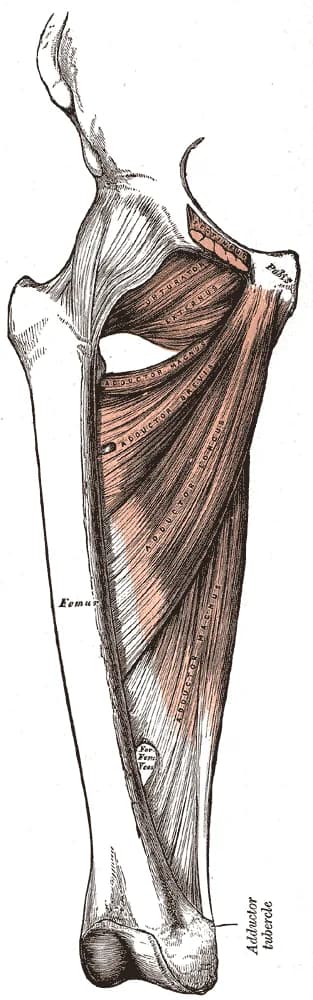
- Trochanter minor – smaller bony prominence on the mediocaudal side of the proximal femur, insertion site of the m. iliopsoas.
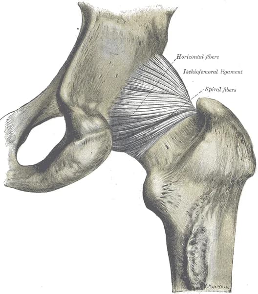
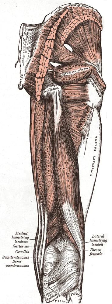
- Tuber ischiadicum – bony prominence on the posterior lower part of the pelvis, origin of the hamstrings, important when examining the posterior aspect of the hip.
Muscles
Muscles – anterior group
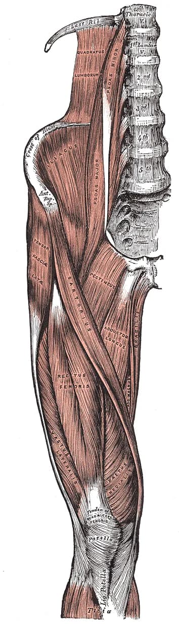
- Iliopsoas – primary hip flexor.
- Rectus femoris – hip flexion and knee extension.
- Sartorius – flexion, abduction and external rotation of the hip, along with flexion of the knee.
Muscles – lateral group
- Gluteus medius – hip abduction, internal rotation, pelvic stabilization during walking.
- Gluteus minimus – hip abduction and internal rotation.
- Tensor fasciae latae – assists with flexion, abduction, and internal rotation of the hip.
- Gluteus maximus – powerful hip extensor and external rotator, with upper fibers also contributing to abduction.
Muscles – posterior group
- Hamstrings (semitendinosus, semimembranosus, biceps femoris caput longum) – hip extension and knee flexion.
- Piriformis – external rotation of the hip in extension, assists with abduction during flexion.
Schalten Sie die vollständige Health Library frei
Voller Zugriff auf Scan-Protokolle, Anatomie und klinische Referenzen. Jederzeit kündbar.
- Alle Protokolle und Anatomiereferenzen
- Originale Ultraschall-Illustrationen und Video-Demonstrationen
- Synchronisierung zwischen Mobilgerät und Web