Ultrasound examination
Examination Protocol
Proximal part
- Transverse plane
- Sagittal plane
Middle section
- Transverse plane
- Sagittal plane
Distal portion
- Transverse plane
- Sagittal plane
- Pronator quadratus window
- Dorsal view - Lateral roll view
- Cobra view
- Lacertus fibrosus
Interaktive Funktion, verfügbar in der App
1. Proximal part
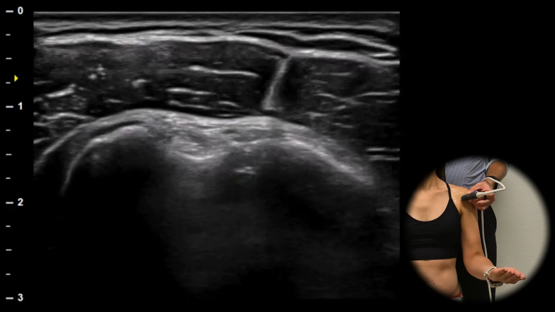
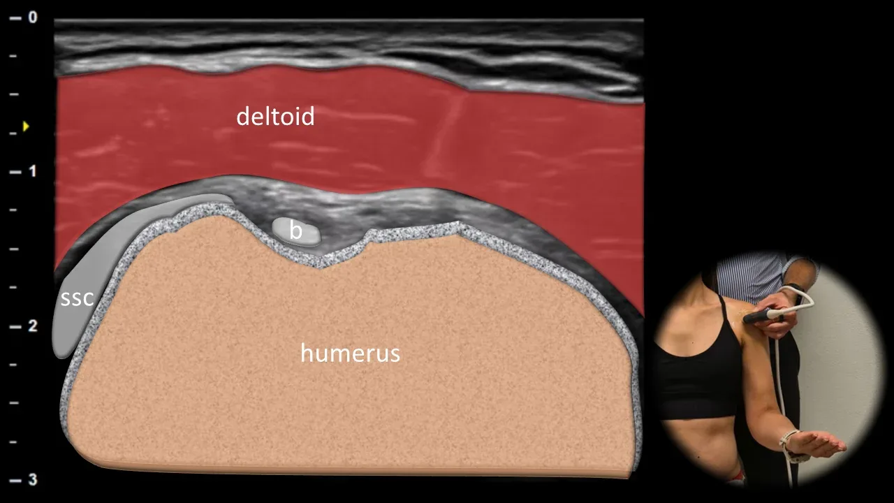
Figure 1. Ventral view, transverse plane. b: tendon of the long head of biceps brachii, ssc: tendon of subscapularis.
In the intertubercular sulcus of the humerus lies the tendon of the long head of biceps brachii (b), which appears as a well-demarcated oval hyperechoic fibrillar structure. Around the tendon, a thin hypoechoic rim of the synovial sheath is visible. The greater tubercle is shown laterally, the lesser tubercle medially. At the medial edge of the sulcus, part of the tendon of subscapularis (ssc) is visible, inserting on the lesser tubercle of the humerus. Superficially lies the deltoid muscle.
Clinical Note
Fluid around the long head of the biceps tendon may be a manifestation of involvement of the sheath itself, but may also originate from the glenohumeral joint due to communication of the synovial sheath with the joint cavity.
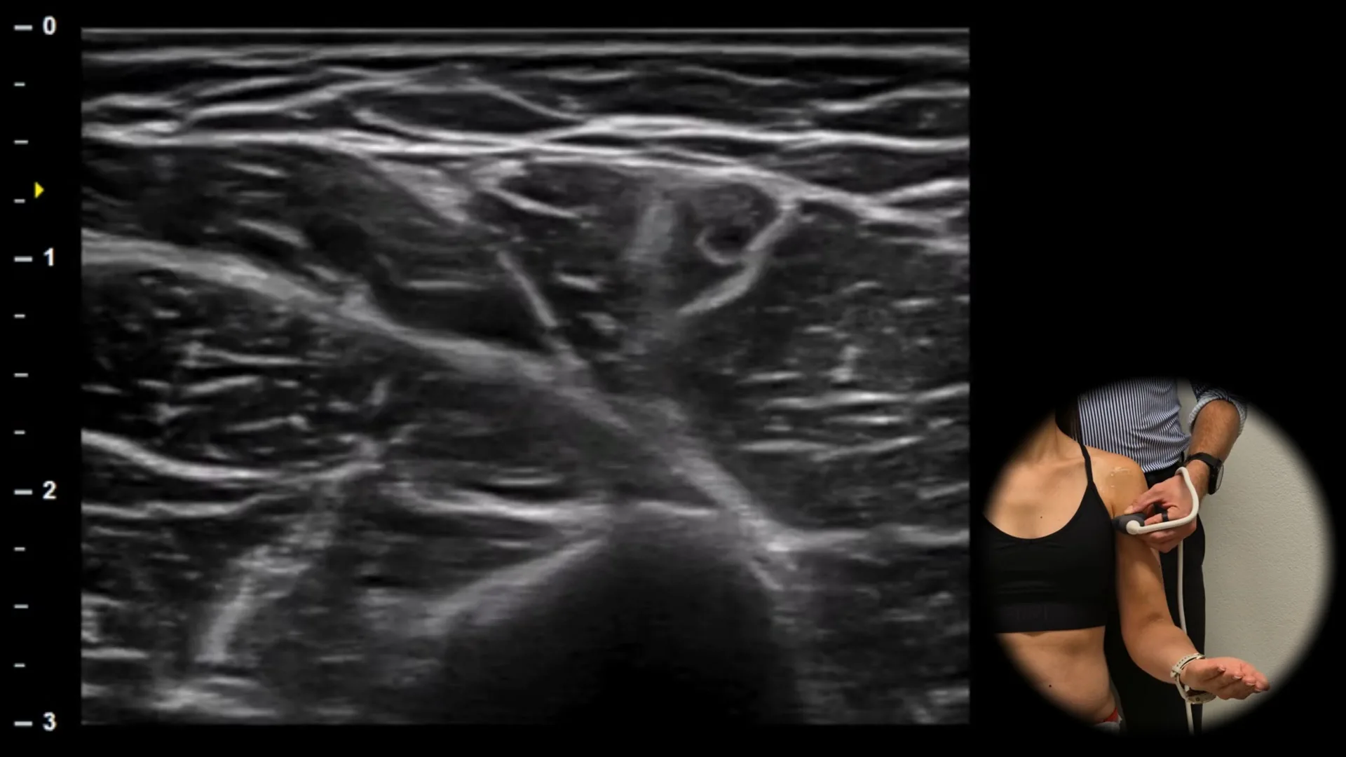
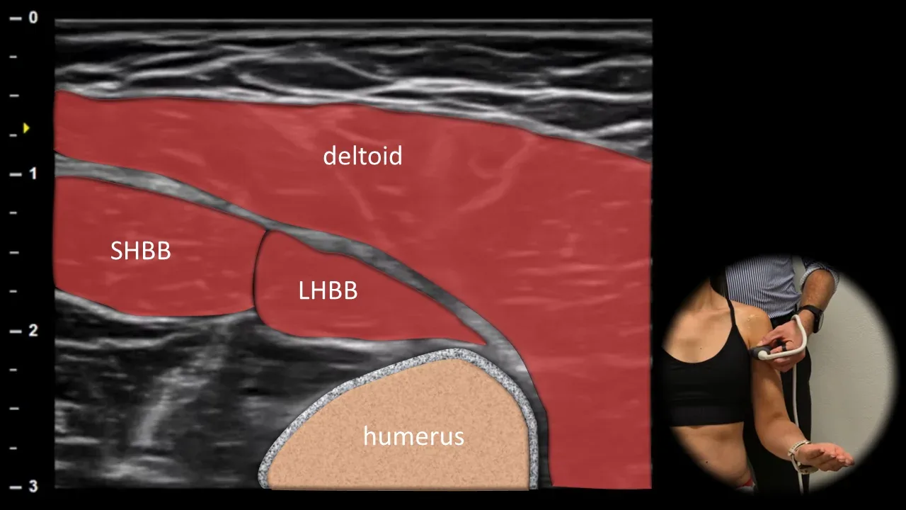
Figure 2. Ventral view, transverse plane. LHBB: muscle belly of the long head of biceps brachii, SHBB: muscle belly of the short head of biceps brachii
A more distal transverse ultrasound section of the anterior arm shows the muscle bellies of both heads of biceps brachii. The long head (LHBB) is located laterally, the short head (SHBB) medially. The deltoid muscle is positioned superficially, and the proximal humerus is visible deep. This projection is suitable for evaluating the integrity of muscle tissue, mutual symmetry of both biceps heads, and dynamic muscle contraction.
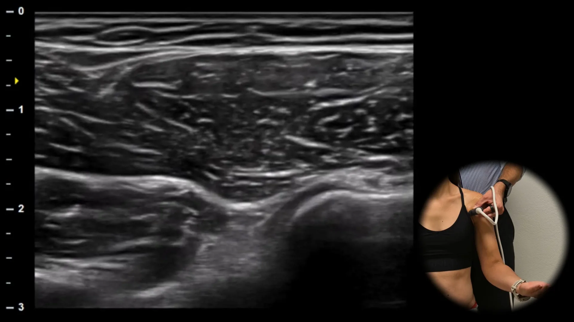
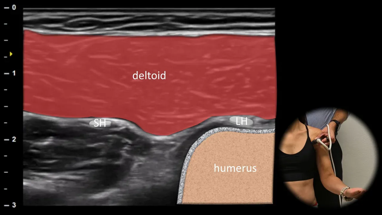
Figure 3. Ventral view, transverse plane. LH: long head of biceps brachii, SH: short head of biceps brachii
A more medial and proximal transverse ultrasound section of the anterior shoulder shows the short head of biceps brachii (SH) as a round hypoechoic structure located beneath the deltoid muscle and medial to the long head (LH). The short head here extends toward its origin in the region of the coracoid process. The deltoid muscle is located superficially, while the proximal humerus is visible deep. This projection is important for complete evaluation of the proximal anatomy of the biceps and for distinguishing isolated involvement of the short and long heads.
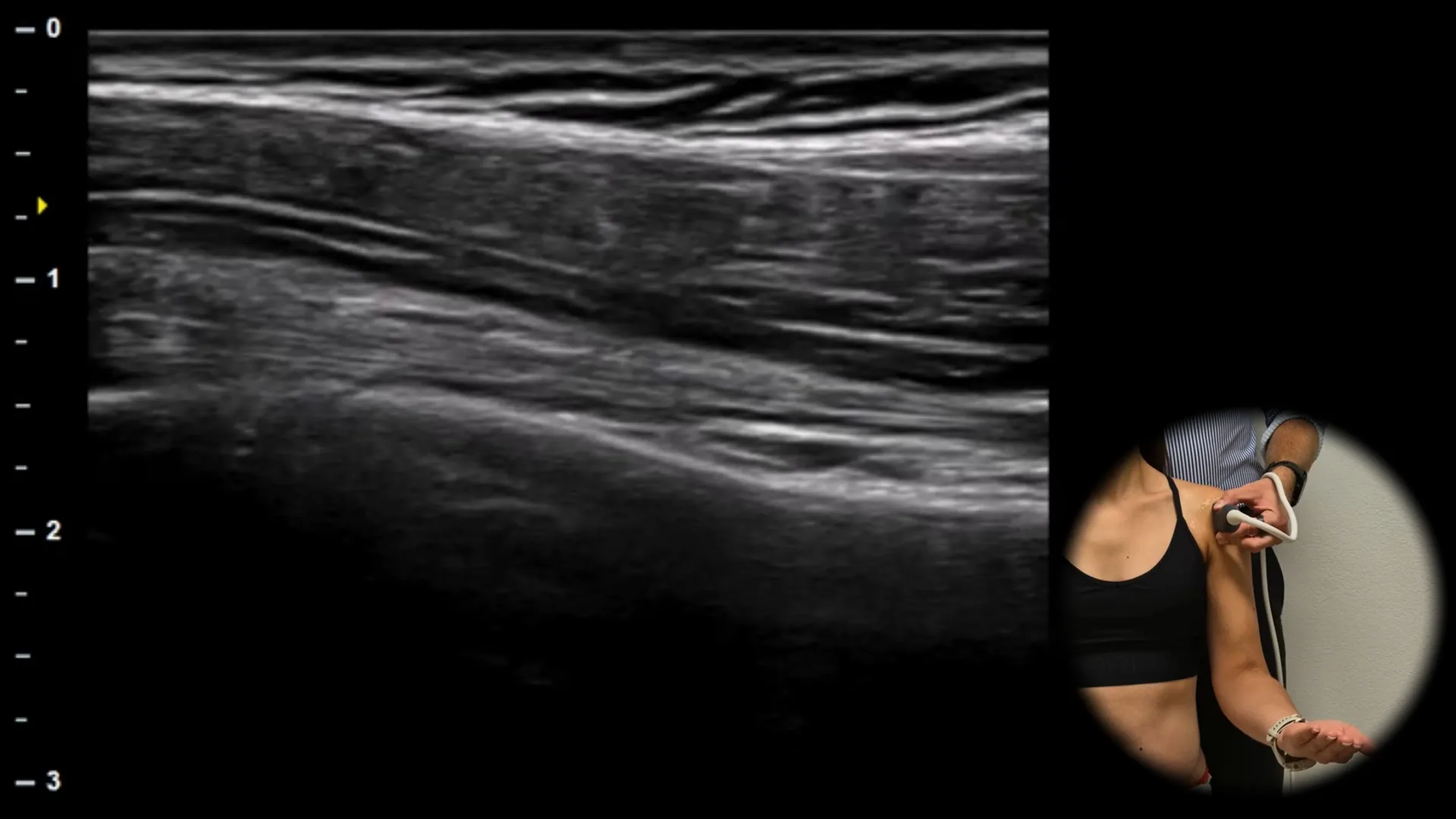
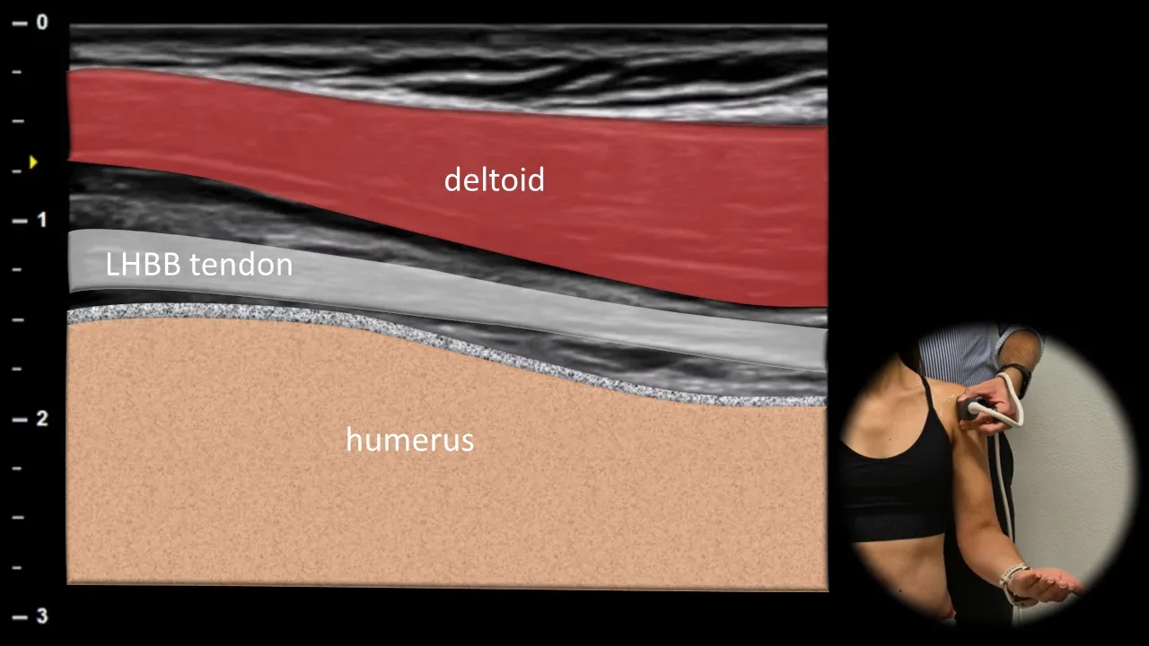
Figure 4. Ventral view, longitudinal plane. LHBB tendon: long head of biceps brachii tendon
The longitudinal ultrasound section of the anterior shoulder region shows the long head of biceps brachii tendon as a hyperechoic fibrillar structure running in the intertubercular sulcus of the humerus. The bony walls of the sulcus are formed by the cortical margins of the tuberculum majus and tuberculum minus, which provide reliable anatomical orientation. The deltoid muscle is located superficially. In this projection, we primarily evaluate the continuity, contour and thickness of the tendon, as well as the potential presence of fluid in the sheath.
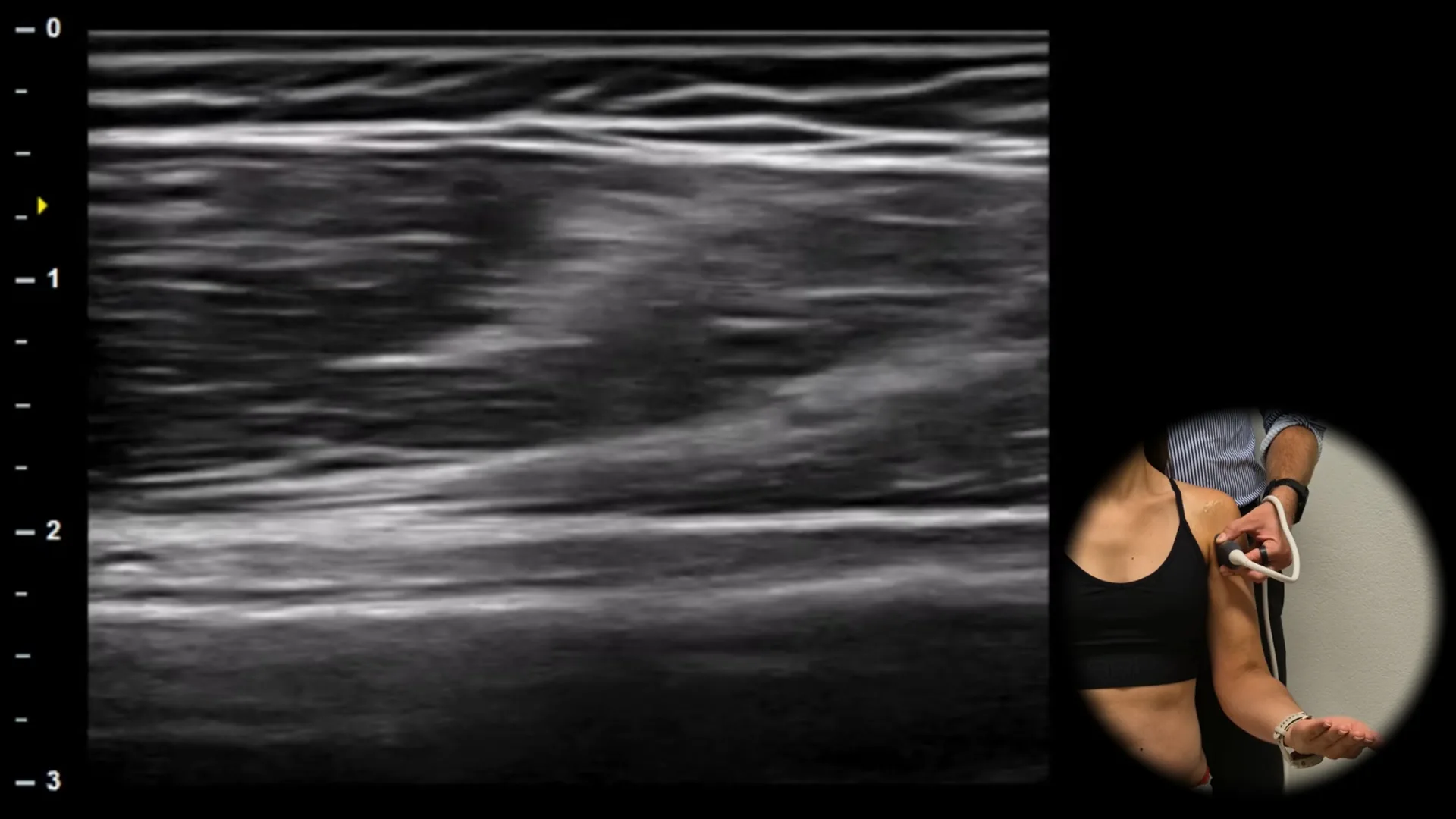
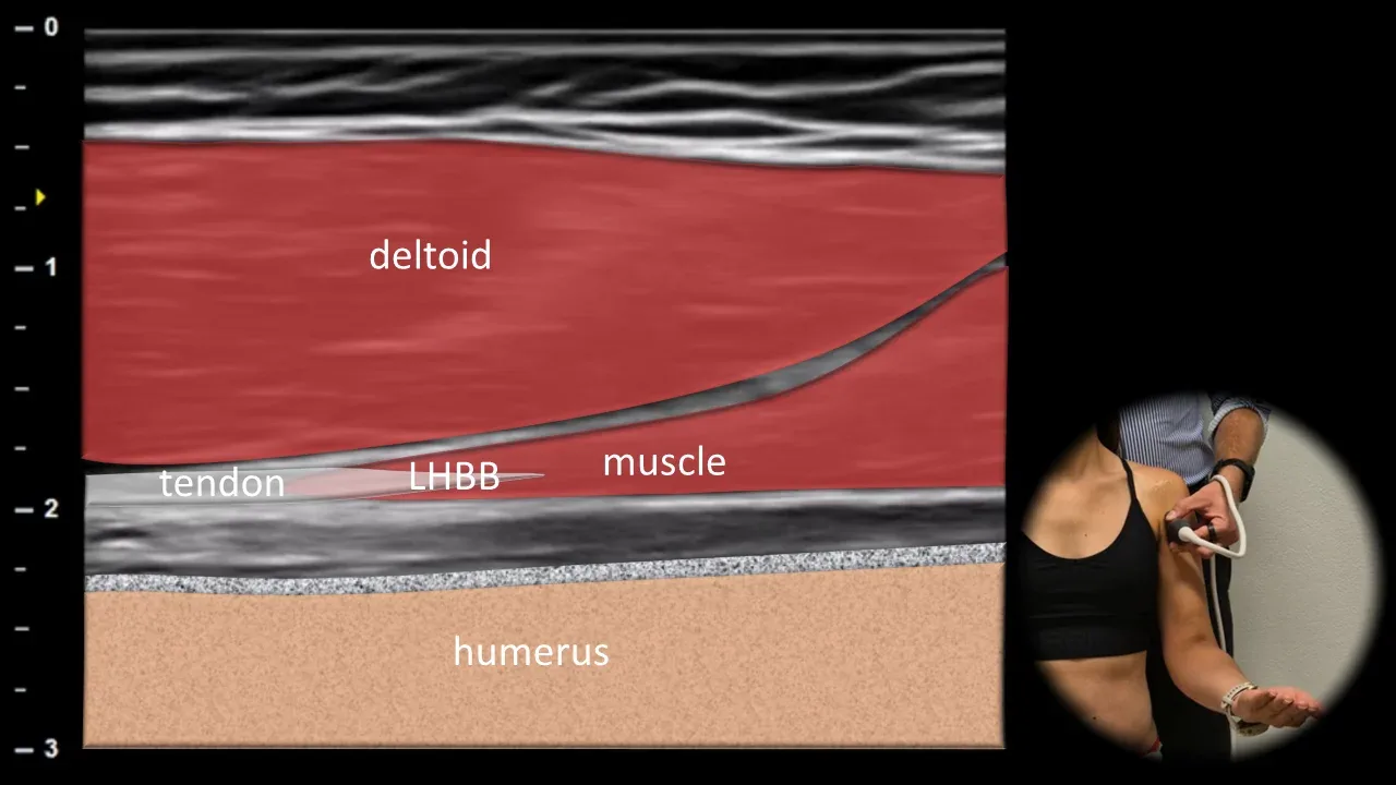
Figure 5. Ventral view, longitudinal plane. LHBB: long head of biceps brachii muscle
Longitudinal ultrasound section of the anterior part of the shoulder in the area of the myotendinous junction of the long head of biceps brachii muscle. A smooth transition from tendon to muscle belly is evident, representing a change from the hyperechoic fibrillar structure of the tendon to hypoechoic muscle tissue with fine striated architecture. The deltoid muscle is located superficially, and the humerus is visible deep.
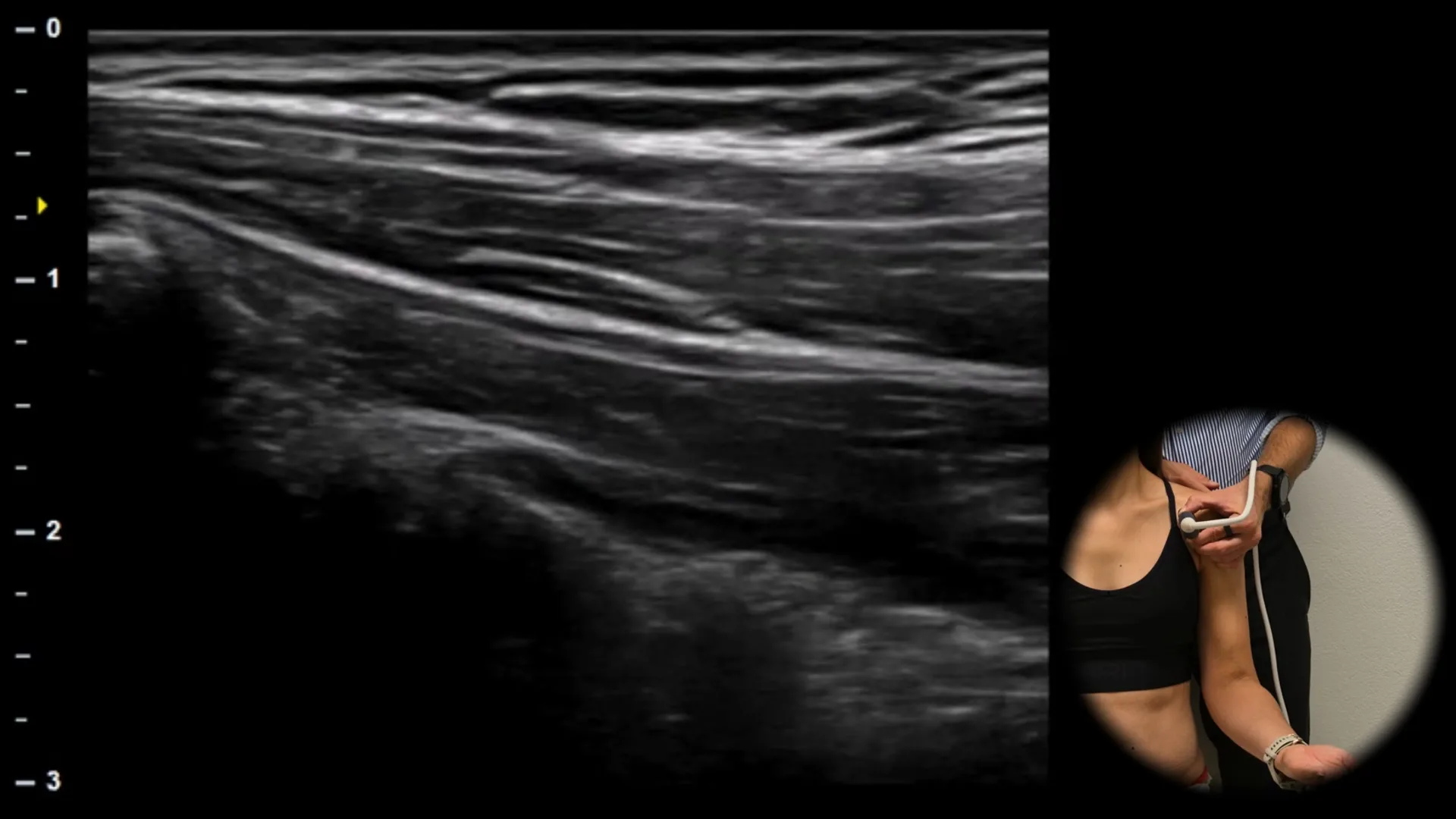
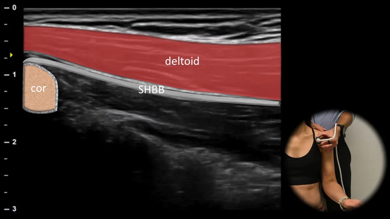
Figure 6. Ventral view, longitudinal plane. SHBB: short head of m. biceps brachii, cor: processus coracoideus
The longitudinal ultrasound section of the anterior part of the shoulder displays the short head of m. biceps brachii (SHBB) running proximally to its origin on the processus coracoideus of the scapula (cor). The muscle appears as a hypoechoic structure with fine striated architecture, adjacent to the cortical surface of the coracoid. The m. deltoideus is located superficially. This projection is useful for differentiating the short and long heads of the biceps and for evaluating the area in cases of proximal biceps pain or suspected traumatic avulsion injury.
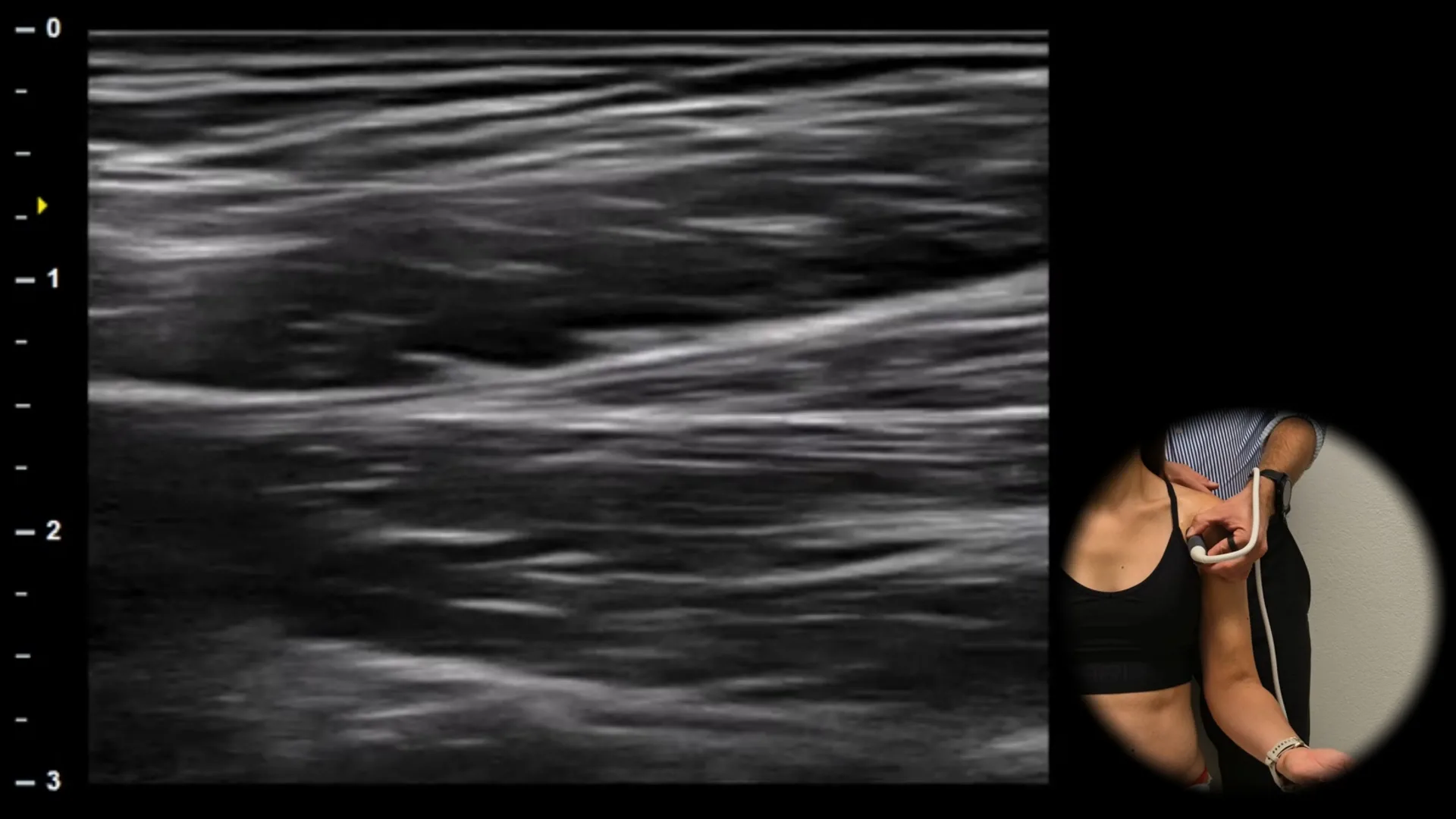
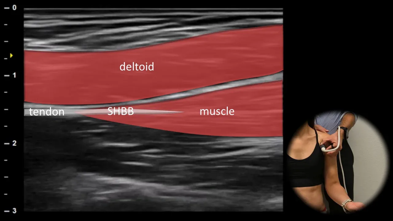
Figure 7. Ventral view, longitudinal plane. SHBB: short head of biceps brachii muscle
Longitudinal ultrasound section of the anterior part of the arm in the region of the myotendinous junction of the short head of biceps brachii muscle (SHBB). A smooth transition of tendon fibers into the proximal muscle belly is visible, which has homogeneous echogenicity and fine striated architecture. The deltoid muscle is located superficially. In this projection, we primarily evaluate the continuity and structure of the myotendinous junction.
2. Middle Part

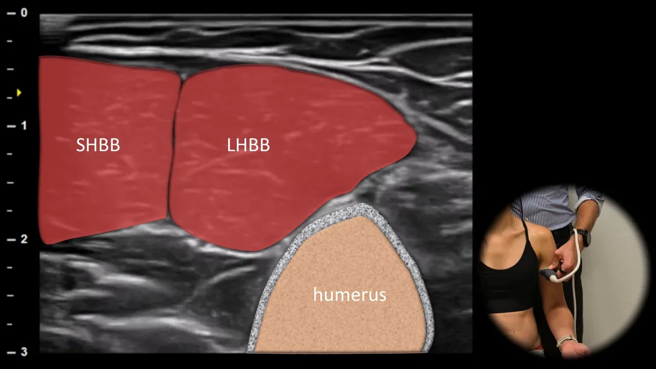
Figure 8. Ventral view, transverse plane. LHBB: muscle belly of the long head of biceps brachii, SHBB: muscle belly of the short head of biceps brachii
The transverse ultrasound section through the middle portion of biceps brachii shows muscle bellies of the long and short heads of the biceps. The muscle tissue has the typical "starry sky" appearance, where hypoechoic muscle fibers are interspersed with fine hyperechoic septa of connective tissue and perimysium, creating the finely granular pattern of healthy muscle. The long head (LHBB) is positioned laterally, the short head (SHBB) medially. The humerus is visible in the depth. This view is suitable for evaluating symmetry, volume and echotexture of muscle tissue and for detecting edema, atrophy or focal lesions.
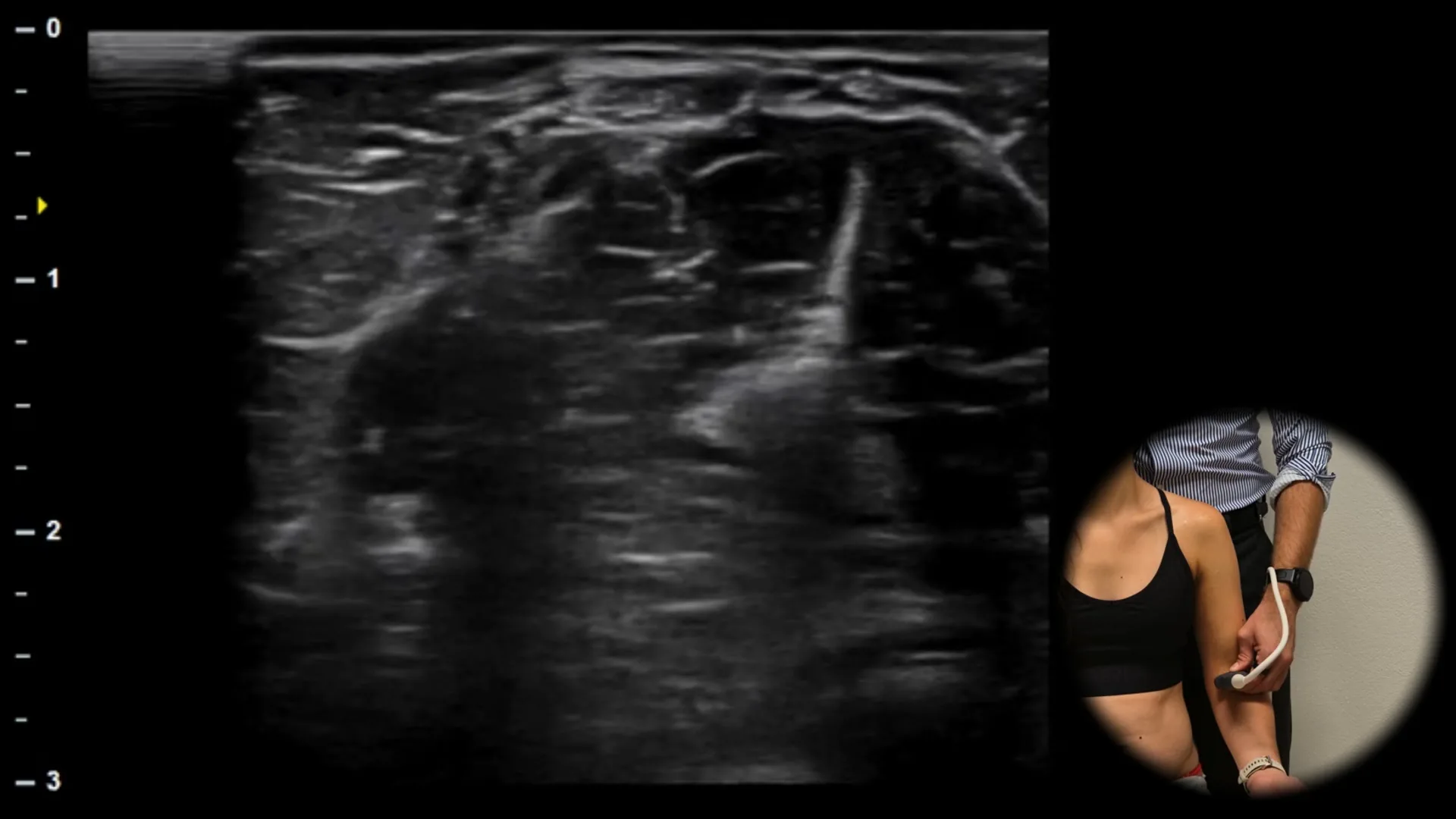
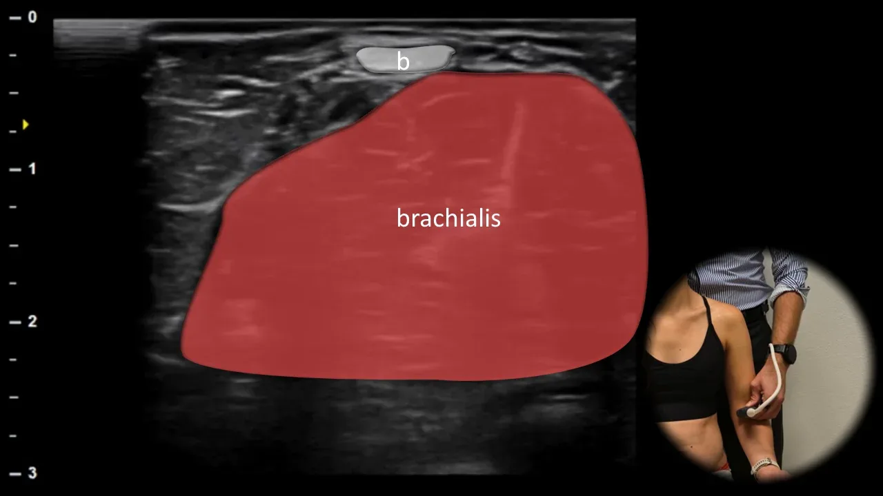
Figure 9. Ventral view, transverse plane. b: biceps brachii tendon, brachialis: m. brachialis
A more distal transverse ultrasound section through the anterior arm displays the superficially located biceps brachii tendon (b) as a hyperechoic fibrillar structure. Beneath it, the m. brachialis is visible, appearing as a broader, less echogenic muscle belly in close relation to the humerus. Clear differentiation of the biceps tendon from the deeper m. brachialis enables precise orientation in this area and is important for evaluating tendon injuries, myotendinous lesions, and fluid collections between muscle layers.
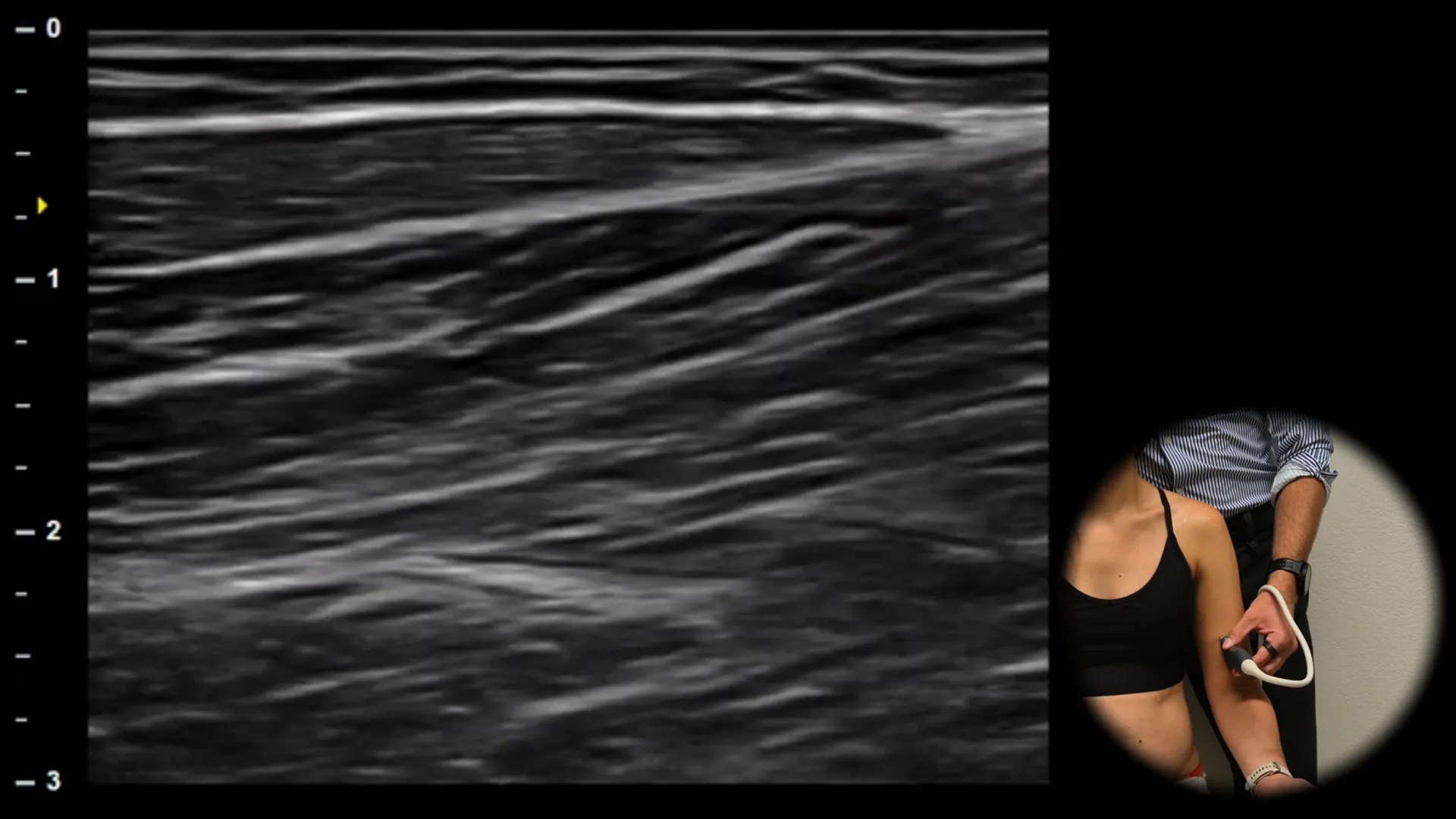
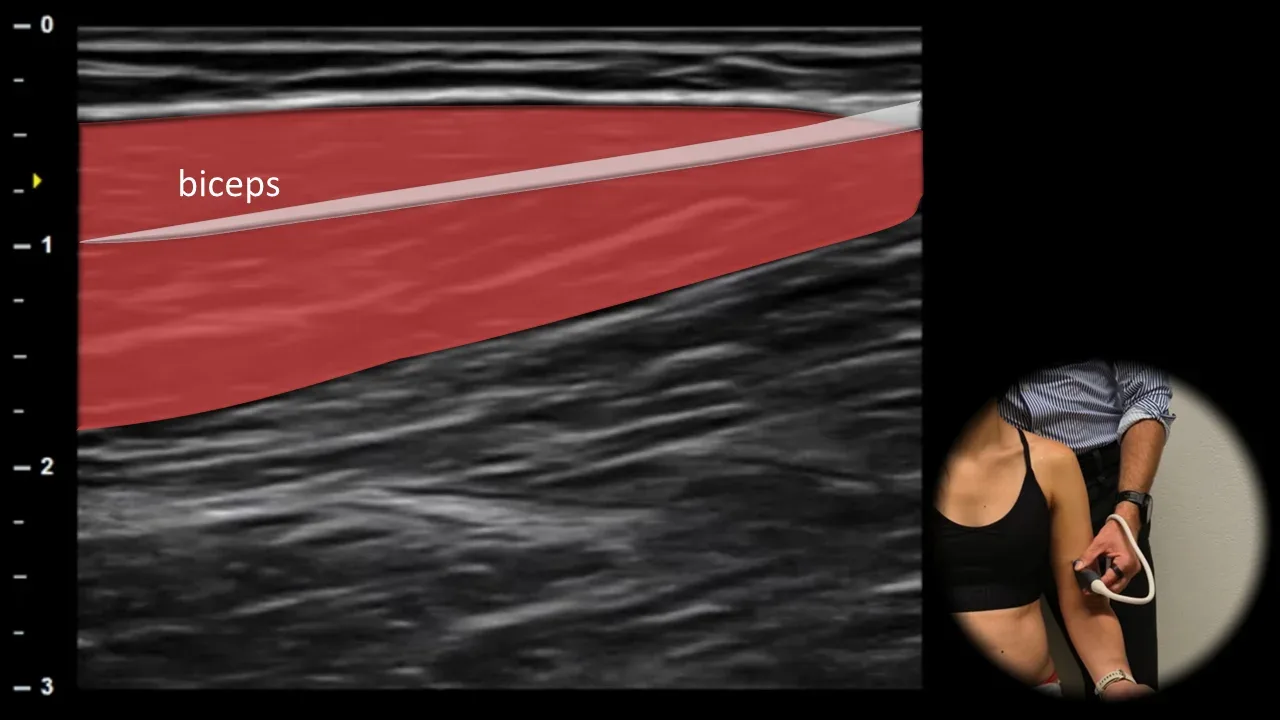
Figure 10. Ventral view, longitudinal plane. biceps: m. biceps brachii
Longitudinal ultrasound section of the distal portion of m. biceps brachii shows the distal myotendinous junction, where muscle fibers gradually narrow and transition into the distal tendon. The tendon appears as a well-defined hyperechoic fibrillar structure extending distally to its insertion. Proximally, the transition of muscle tissue into this dense, regularly organized fibrillar architecture is evident. Deeper lies the m. brachialis, which has lower echogenicity compared to the tendon. This projection is important for evaluating partial and complete lesions of the myotendinous junction, monitoring healing, and planning further treatment or rehabilitation.
3. Distal part
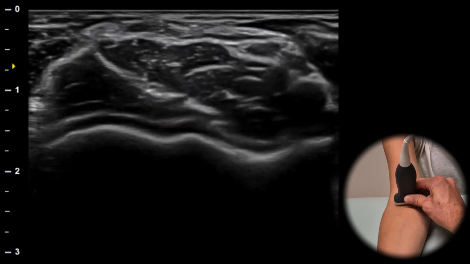
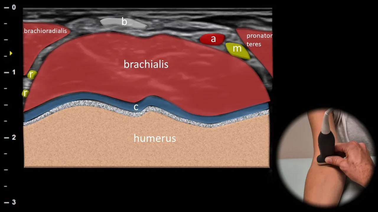
Figure 11. Ventral view, transverse plane. b: distal tendon of m. biceps brachii, m: n. medianus, r: n. radialis, a: a. brachialis, c: humeral cartilage
Transverse ultrasound section through the anterior portion of the distal arm shows the distal tendon of m. biceps brachii (b) in cross-section as a superficially located hyperechoic oval structure. Immediately beneath it, the m. brachialis is visible, which has a more homogeneous and less echogenic appearance. Laterally the m. brachioradialis is located, medially the m. pronator teres. In this area, the n. medianus (m) located medially to the tendon and the n. radialis (r) running laterally along the m. brachioradialis are also well identifiable; the a. brachialis (a) is also visible in close proximity. This projection is important for anatomical orientation, nerve mapping and for safe planning of interventional procedures in the cubital fossa region.
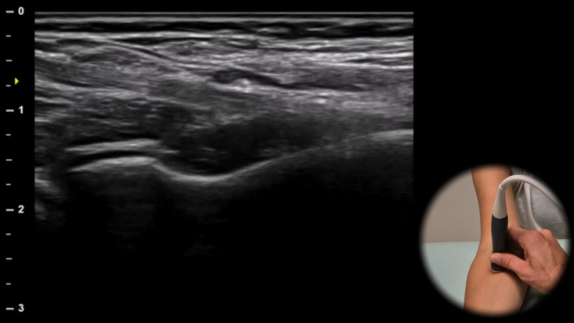
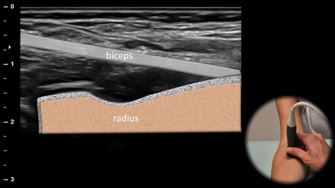
Figure 12. Ventral view, longitudinal oblique plane. biceps: distal tendon of m. biceps brachii
The longitudinal oblique ultrasound section follows the natural course of the distal tendon of m. biceps brachii to its insertion on the tuberositas radii. The tendon appears as a hyperechoic fibrillar structure with parallel linear architecture that narrows distally. The probe orientation corresponds to the oblique course of the tendon. Deep to the tendon, the m. brachialis is visible, with the cortex of the radius forming the bottom of the image. This projection is essential for assessing the continuity of the distal biceps tendon and for demonstrating partial or complete ruptures.
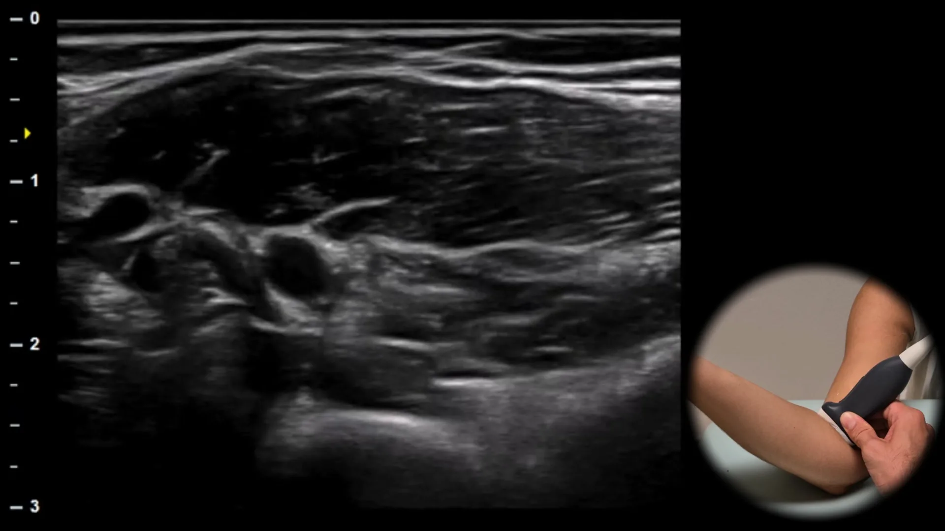
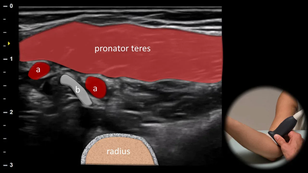
Figure 13. Ventral view, transverse plane – pronator window. b: distal tendon of biceps brachii muscle, a: brachialis artery
Transverse ultrasound section in the pronator window area shows the distal tendon of biceps brachii muscle (b) in cross-section during its course to insertion on the tuberositas radii. The tendon appears as an oval hyperechoic fibrillar structure. Deep beneath the tendon, the radius is visible as a hyperechoic cortical line with dorsal acoustic shadowing. Superficially, the pronator teres muscle is located, which creates a suitable acoustic window for imaging this area. This projection is particularly useful for evaluating distal biceps tendon pathology, especially partial ruptures and enthesopathy.
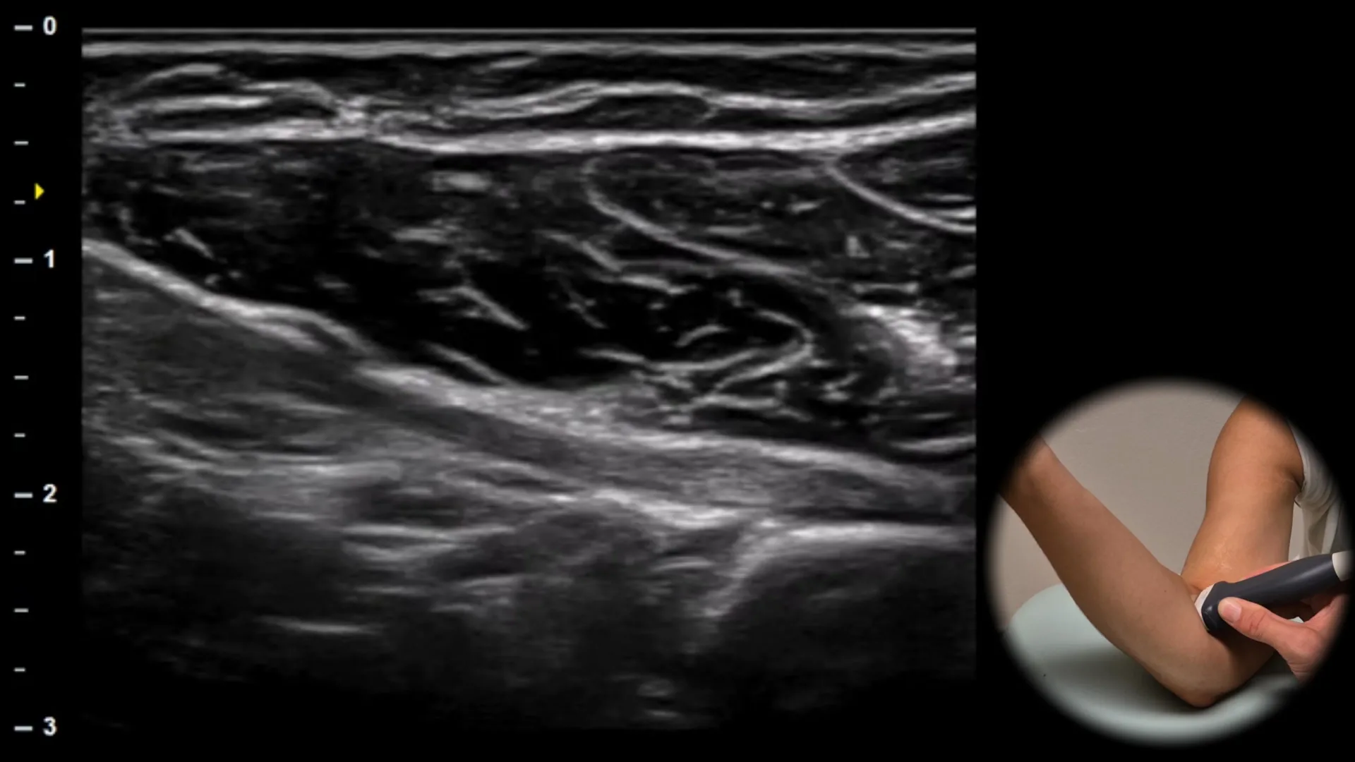
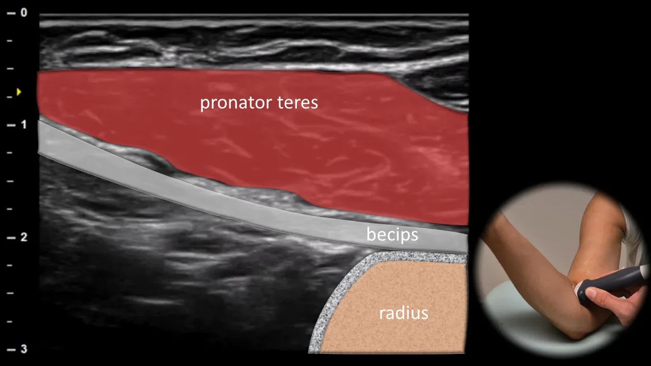
Figure 14. Ventral view, longitudinal plane. biceps: distal tendon of m. biceps brachii
The longitudinal ultrasound section displays the distal tendon of m. biceps brachii in its oblique course from the distal myotendinous junction to its insertion on the tuberositas radii. The tendon appears as a linear hyperechoic fibrillary structure running beneath the m. pronator teres. The tuberositas radii is visible as a bright continuous cortical line where the tendon inserts. This projection allows for assessment of tendon continuity, dynamic examination and evaluation of tendinopathy or partial and complete ruptures.
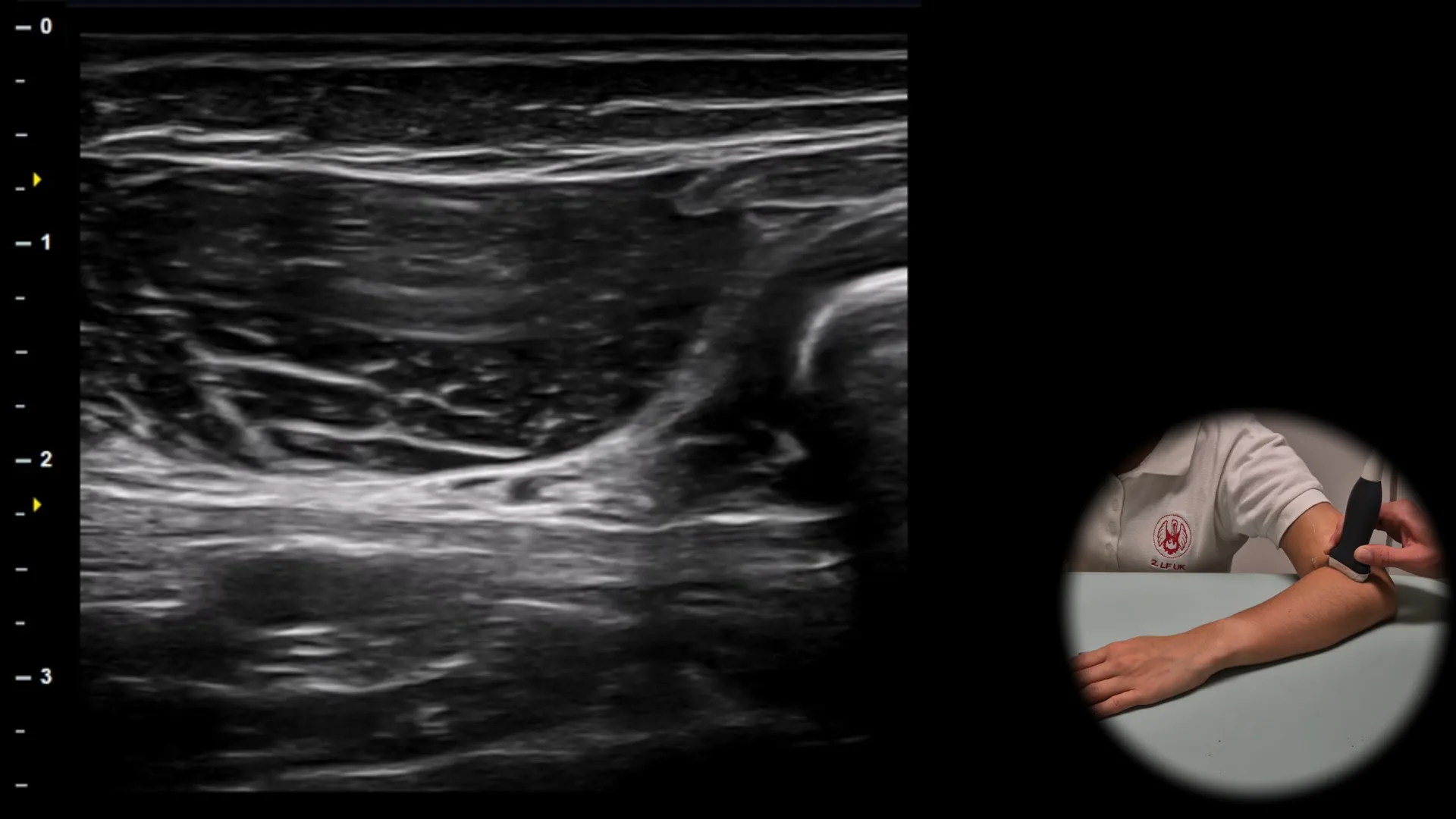
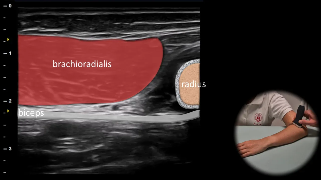
Figure 15. Lateral roll position, longitudinal plane. biceps: distal tendon of m. biceps brachii
Longitudinal ultrasound section in lateral roll position shows the distal tendon of m. biceps brachii as a hyperechoic fibrillar structure running obliquely from the myotendinous junction to its insertion on the tuberositas radii. In this projection, the tendon is located beneath m. brachioradialis. This view is suitable for dynamic examination, assessment of distal tendon continuity and evaluation of the myotendinous junction area.
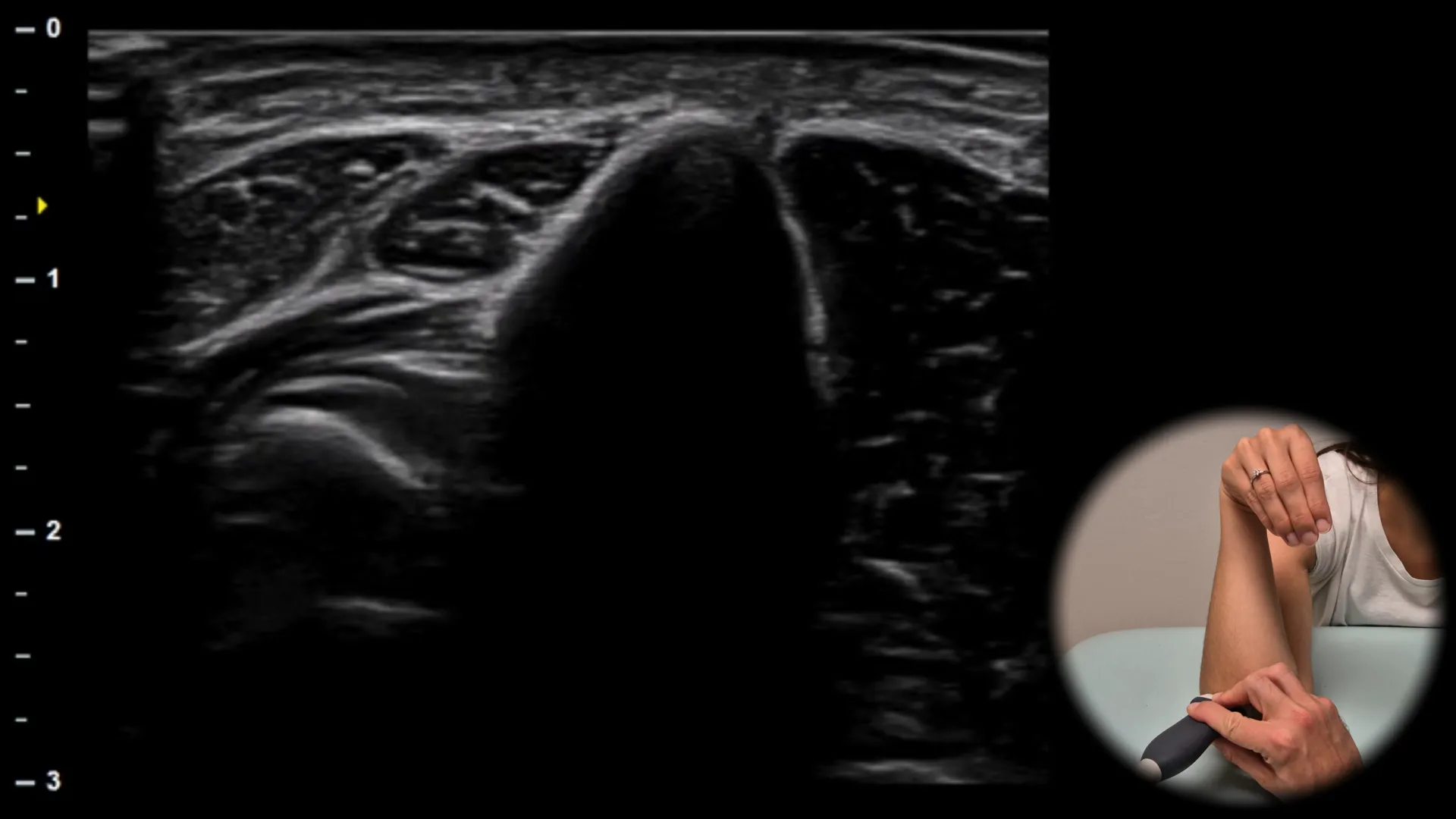
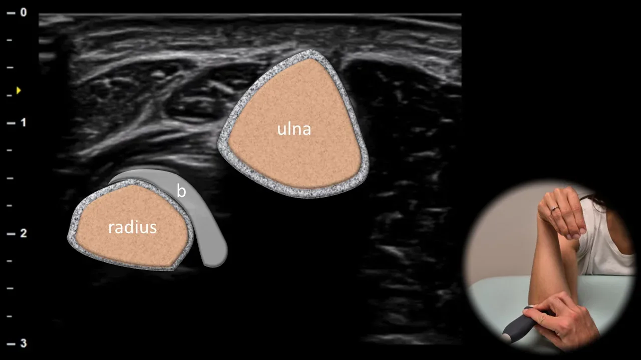
Figure 16. Cobra position, transverse plane. b: distal tendon of biceps brachii muscle
Transverse ultrasound section in cobra position (elbow flexion and forearm pronation with hand placed palm down at the shoulder) shows the distal tendon of biceps brachii muscle (b) during its curved course to insertion on the tuberositas radii. The tendon is visible between the radius and ulna, which allows clearer visualization of its distal portion and relationship to the insertion site. This projection is particularly useful for assessing continuity of the distal biceps tendon and in diagnosing partial or complete ruptures.
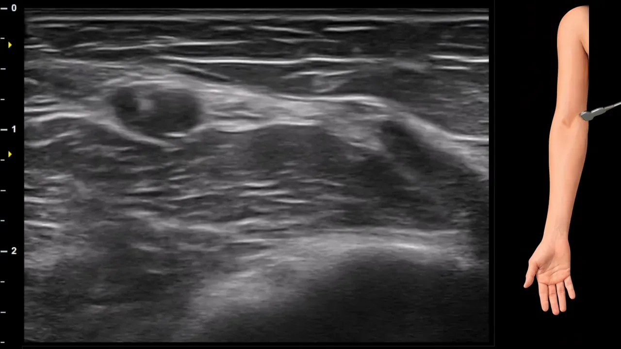
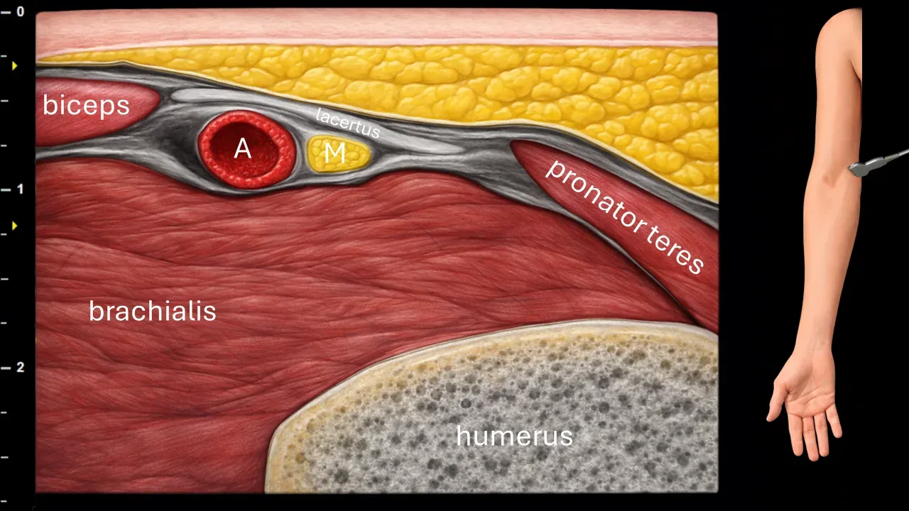
Figure 17. Cubitum, transverse plane. A: a. brachialis, M: n. medianus.
Transverse ultrasound section in the cubital fossa region. The image shows the a. brachialis (A) and n. medianus (M), which is positioned medially to the a. brachialis in this projection. The n. medianus further runs beneath the lacertus fibrosus and is located in the space between the m. brachialis and m. pronator teres. The image also captures the m. biceps brachii, m. brachialis, m. pronator teres and the contour of the humerus.
Schalten Sie die vollständige Health Library frei
Voller Zugriff auf Scan-Protokolle, Anatomie und klinische Referenzen. Jederzeit kündbar.
- Alle Protokolle und Anatomiereferenzen
- Originale Ultraschall-Illustrationen und Video-Demonstrationen
- Synchronisierung zwischen Mobilgerät und Web