Entrapment sites
Most common sites of peripheral nerve compression
- Struthers ligament - fibrous band near a supracondylar process, proximal median nerve compression.
- Lacertus fibrosus - biceps aponeurosis in the cubital fossa, can compress the nerve.
- Pronator teres - passage between two heads, classic site of pronator syndrome.
- FDS arcade - fibrous arch of flexor digitorum superficialis.
- Gantzer muscle - accessory head of flexor pollicis longus.
- AINS (Kiloh-Nevin) - anterior interosseous neuropathy.
- Carpal tunnel - course under the flexor retinaculum, most common distal compression.
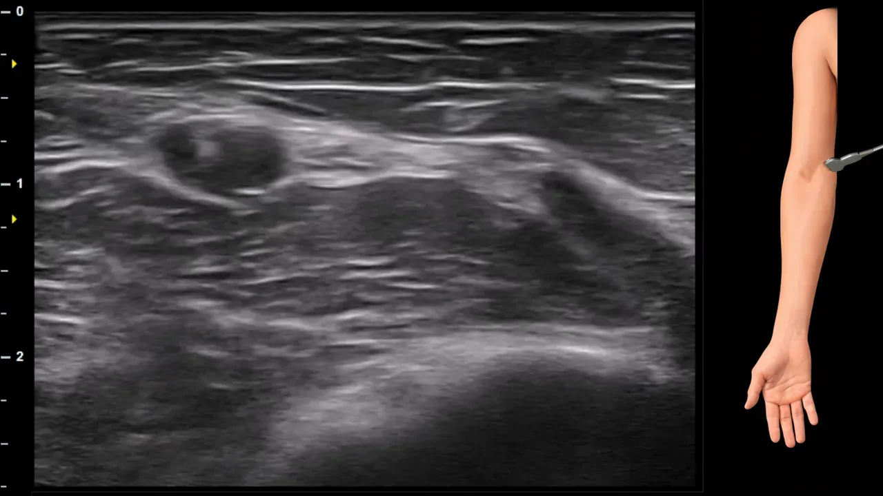
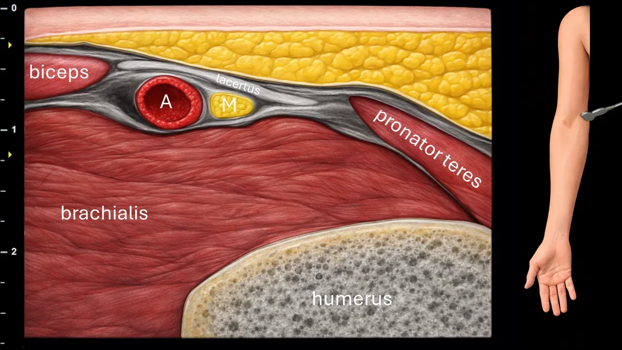
Figure 1. Cubitum, transverse plane, passage of n. medianus under lacertus fibrosus. A: a. brachialis, M: n. medianus.
Transverse ultrasound section in the cubital fossa region. The image shows the a. brachialis (A) and n. medianus (M), which is positioned medially to the a. brachialis in this projection. The n. medianus further runs under the lacertus fibrosus and is located in the space between the m. brachialis and m. pronator teres. The image also captures the m. biceps brachii, m. brachialis, m. pronator teres and the contour of the humerus.
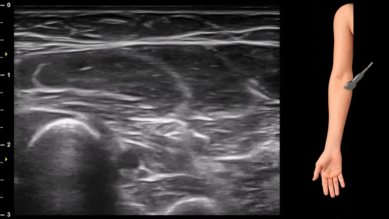
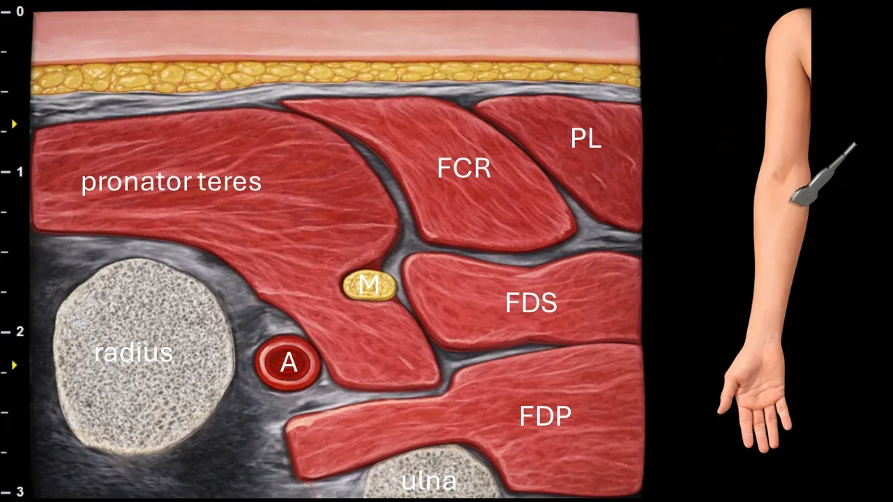
Figure 2. Elbow, transverse plane, course of median nerve between two heads of pronator teres muscle. A: ulnar artery, M: median nerve, FCR: flexor carpi radialis muscle, PL: palmaris longus muscle, FDS: flexor digitorum superficialis muscle, FDP: flexor digitorum profundus muscle.
Transverse ultrasound section in the proximal forearm region. The median nerve (M) in this projection runs between the two heads of the pronator teres muscle, the superficial (humeral) and deep (ulnar) heads. The deep head of the pronator teres muscle separates the median nerve from the ulnar artery (A) and is present in approximately 80% of individuals. The image also shows the flexor carpi radialis muscle (FCR), palmaris longus muscle (PL), flexor digitorum superficialis muscle (FDS), flexor digitorum profundus muscle (FDP), as well as the radius and ulna.
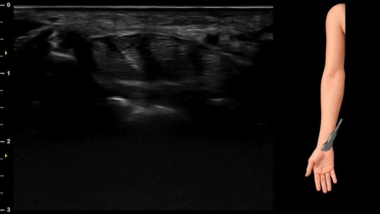
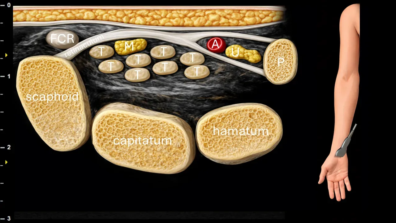
Figure 3. Carpus, transverse plane, passage of n. medianus through carpal tunnel. M: n. medianus, T: flexor tendons, A: a. ulnaris, U: n. ulnaris, P: os pisiforme, FCR: tendon of m. flexor carpi radialis.
Transverse ultrasound section in the carpal region. N. medianus (M) runs beneath the flexor retinaculum in the carpal tunnel, where it is positioned superficially above the flexor tendons (T). The image also shows the tendon of m. flexor carpi radialis (FCR), a. ulnaris (A) and n. ulnaris (U) located ulnarly outside the carpal tunnel, as well as os pisiforme (P) and carpal bones, particularly os scaphoideum, os capitatum and os hamatum.