Ultrasound examination
Examination Protocol
Nervus medianus throughout the entire upper extremity
- Axilla
- Brachium
- Elbow
- Forearm
- Carpus
Anterior interosseus nerve
- Distance of AIN from the main trunk of n. medianus
Palmar cutaneus nerve
- Distance of PCN from the main trunk of n. medianus
- PCN passage through fascia
Terminal branches and ramus recurrens
- Terminal branching of the nervus medianus
Interaktive Funktion, verfügbar in der App
1. Nervus medianus along the entire upper extremity
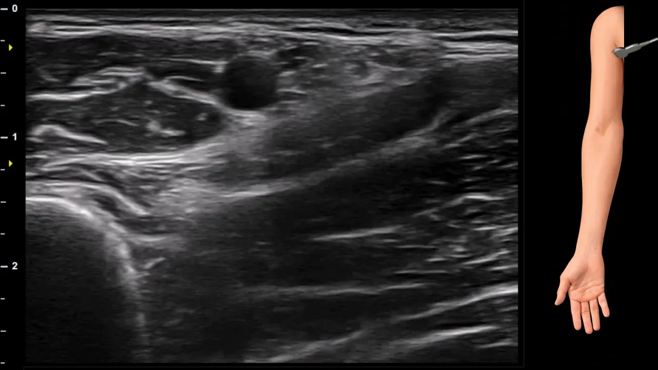
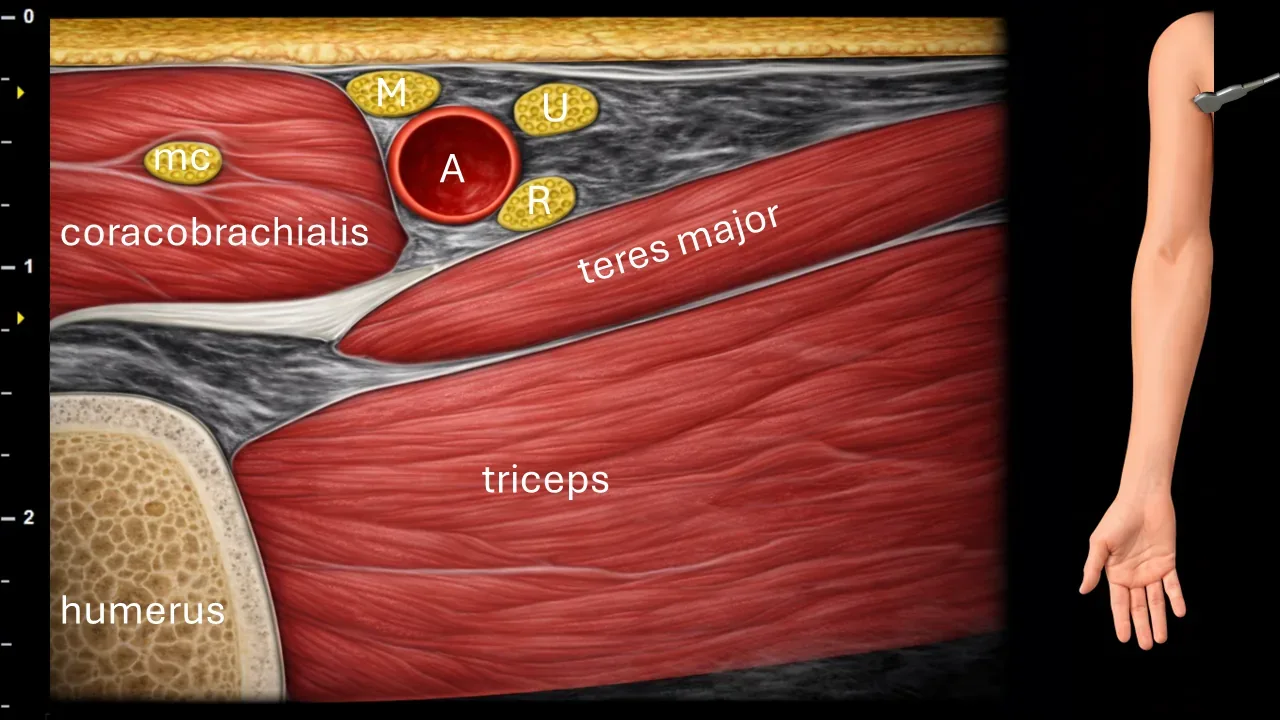
Figure 1. Axilla, transverse plane. A: a. axillaris, M: n. medianus, R: n. radialis, U: n. ulnaris, mc: n. musculocutaneus
Transverse ultrasound section of the axilla. The image shows the a. axillaris (A), around which the nerve structures of the brachial plexus are located. The n. medianus (M) is positioned in this projection slightly ventrally, medially and superficially to the a. axillaris. Also visible in the vicinity are the n. radialis (R), n. ulnaris (U) and n. musculocutaneus (mc). The image also captures muscle structures, particularly the m. coracobrachialis, m. teres major and m. triceps brachii, as well as the contour of the humerus.
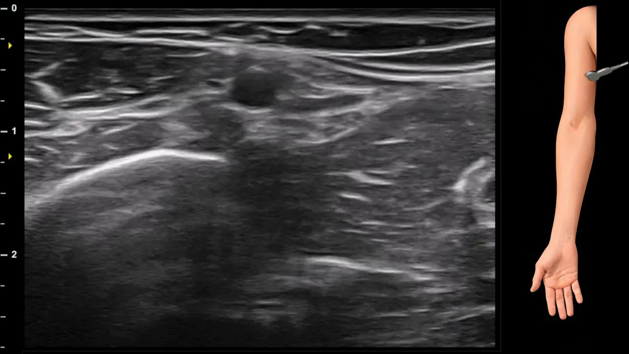
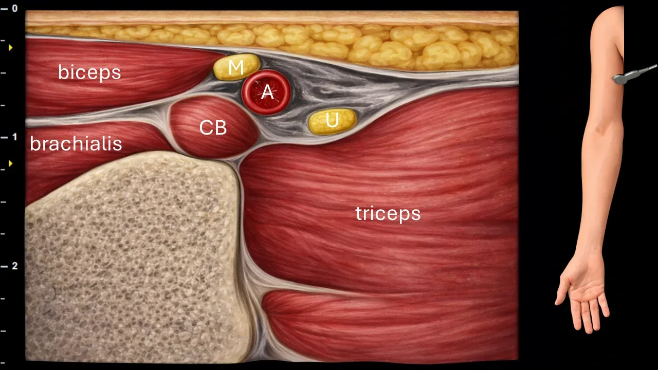
Figure 2. Brachium, transverse plane. A: a. brachialis, M: n. medianus, U: n. ulnaris, CB: m. coracobrachialis.
Transverse ultrasound section of the arm. The image shows the a. brachialis (A), which runs in close proximity to neural structures. The n. medianus (M) is positioned adjacent to the a. brachialis in this projection and runs together with it in the arm as part of the neurovascular bundle. The image also shows the n. ulnaris (U) and muscular structures, particularly the m. coracobrachialis (CB), m. biceps brachii, m. brachialis and m. triceps brachii.
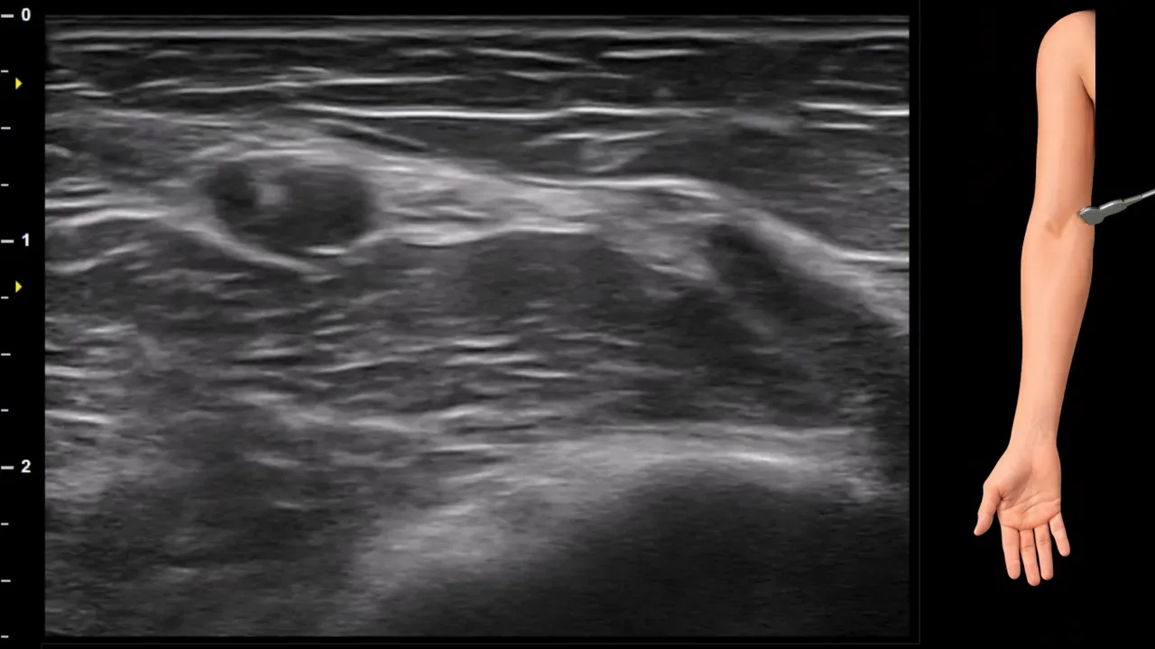
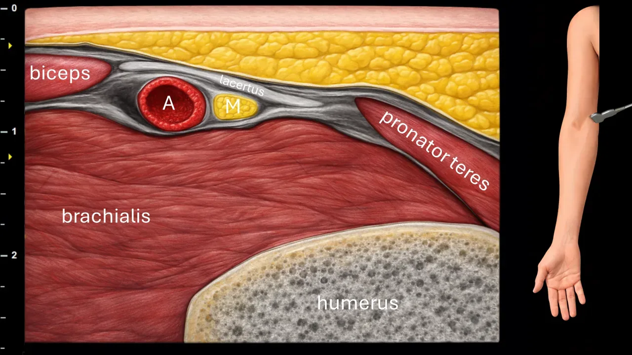
Figure 3. Cubitum, transverse plane. A: a. brachialis, M: n. medianus.
Transverse ultrasound section in the cubital fossa region. The image shows the a. brachialis (A) and n. medianus (M), which in this projection is located medially to the a. brachialis. The n. medianus further runs beneath the lacertus fibrosus and is located in the space between the m. brachialis and m. pronator teres. The image also captures the m. biceps brachii, m. brachialis, m. pronator teres and the contour of the humerus.
Clinical Note
The area under the lacertus fibrosus represents one of the possible sites of median nerve compression.
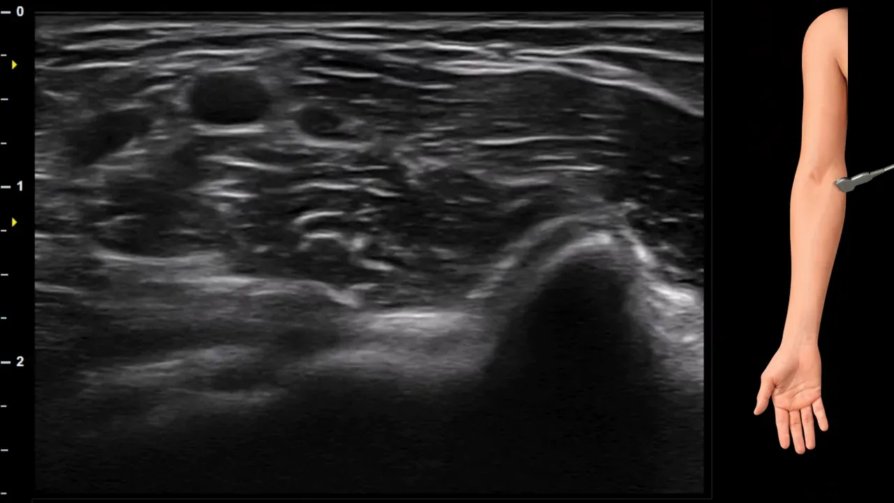
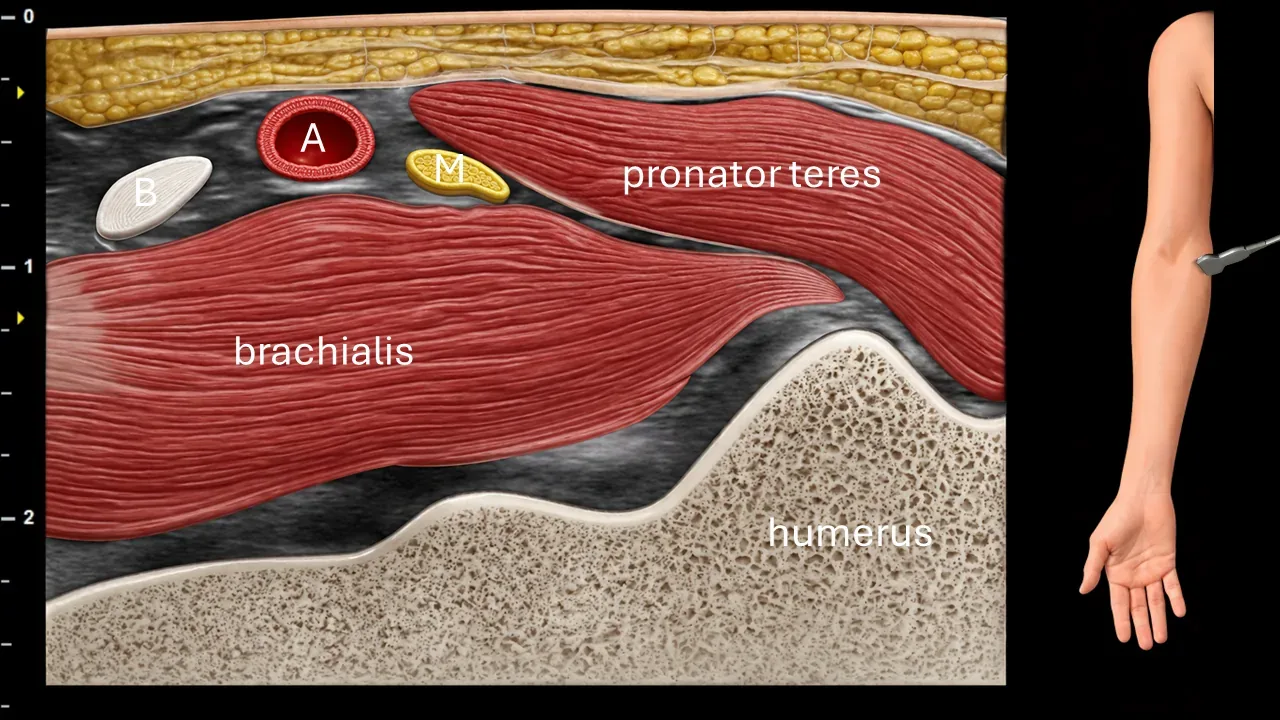
Figure 4. Elbow, transverse plane. A: brachial artery, M: median nerve, B: distal tendon of biceps brachii muscle.
Transverse ultrasound section in the cubital fossa region. The image shows the brachial artery (A), distal tendon of biceps brachii muscle (B) and median nerve (M). The median nerve in this region passes between the brachialis muscle and pronator teres muscle. The image also captures the brachialis muscle, pronator teres muscle and the contour of the humerus.
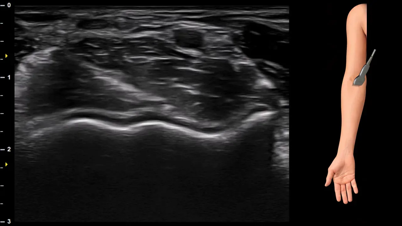
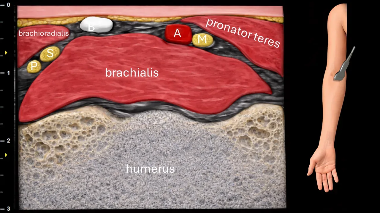
Figure 5. Elbow, transverse plane. A: a. brachialis, M: n. medianus, B: distal tendon of m. biceps brachii, S: ramus superficialis n. radialis, P: ramus profundus n. radialis.
Transverse ultrasound section in the cubital fossa region during basic elbow imaging. The image shows the a. brachialis (A), distal tendon of m. biceps brachii (B) and n. medianus (M), which is located between m. brachialis and m. pronator teres. Laterally, branches of the n. radialis are also visible, specifically the ramus superficialis n. radialis (S) and ramus profundus n. radialis (P). The image also captures m. brachialis, m. brachioradialis, m. pronator teres and the contour of the humerus.
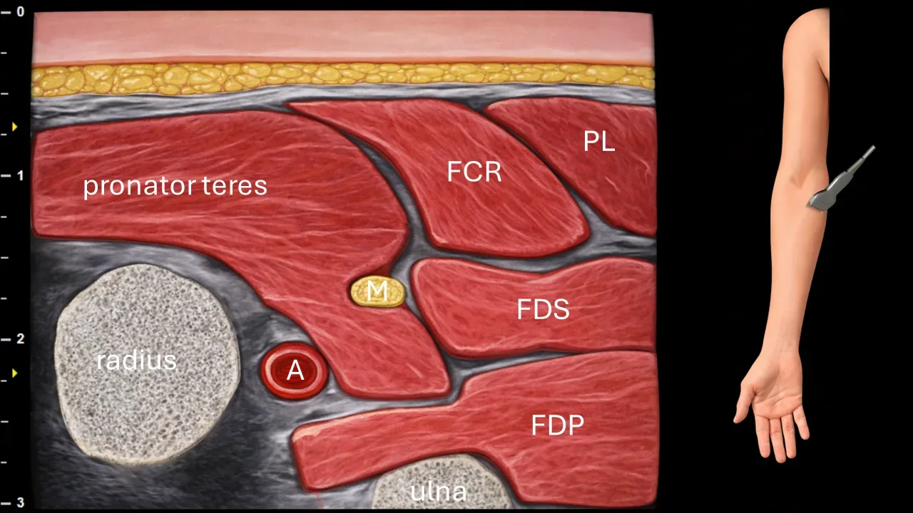
Figure 6. Cubitum, transverse plane. A: a. ulnaris, M: n. medianus, FCR: m. flexor carpi radialis, PL: m. palmaris longus, FDS: m. flexor digitorum superficialis, FDP: m. flexor digitorum profundus.
Transverse ultrasound section in the area of the proximal forearm. N. medianus (M) in this projection runs between two heads of m. pronator teres, the superficial (humeral) and deep (ulnar) heads. The deep head of m. pronator teres separates n. medianus from a. ulnaris (A) and is present in approximately 80% of individuals. The image also shows m. flexor carpi radialis (FCR), m. palmaris longus (PL), m. flexor digitorum superficialis (FDS), m. flexor digitorum profundus (FDP), as well as the radius and ulna.
Clinical Note
In this area, compression of the median nerve may occur between the heads of the pronator teres muscle, which can be the cause of pronator syndrome.
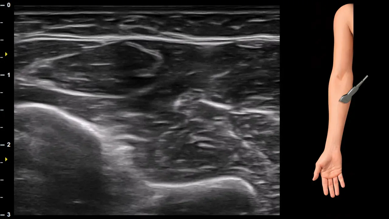
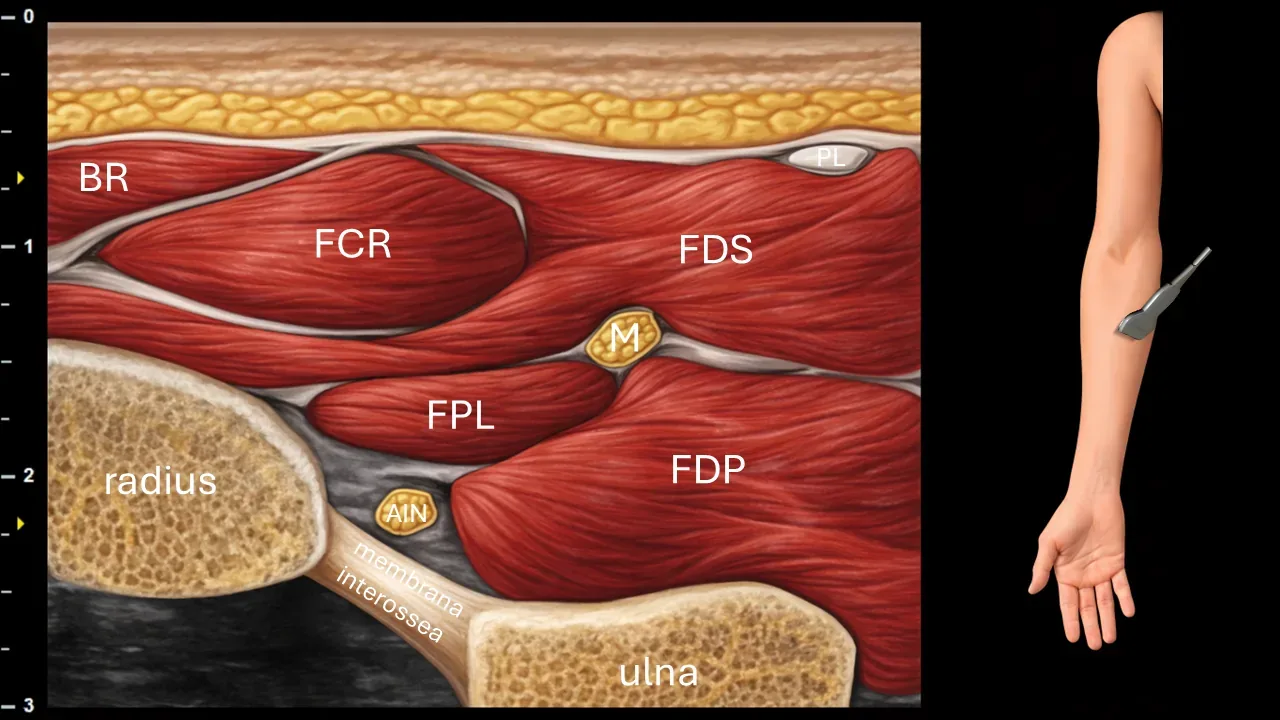
Figure 7. Forearm, transverse plane. M: median nerve, AIN: anterior interosseous nerve, BR: brachioradialis muscle, FCR: flexor carpi radialis muscle, PL: palmaris longus muscle, FDS: flexor digitorum superficialis muscle, FDP: flexor digitorum profundus muscle, FPL: flexor pollicis longus muscle.
Transverse ultrasound section in the forearm region. The median nerve (M) in this projection runs between the flexor digitorum superficialis muscle (FDS), flexor digitorum profundus muscle (FDP) and flexor pollicis longus muscle (FPL). The anterior interosseous nerve (AIN) branches from the median nerve here, descending between the flexor muscles toward the interosseous membrane. The image also shows the brachioradialis muscle (BR), flexor carpi radialis muscle (FCR), palmaris longus muscle (PL), radius, ulna and interosseous membrane.
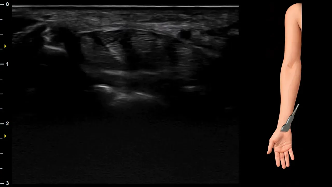
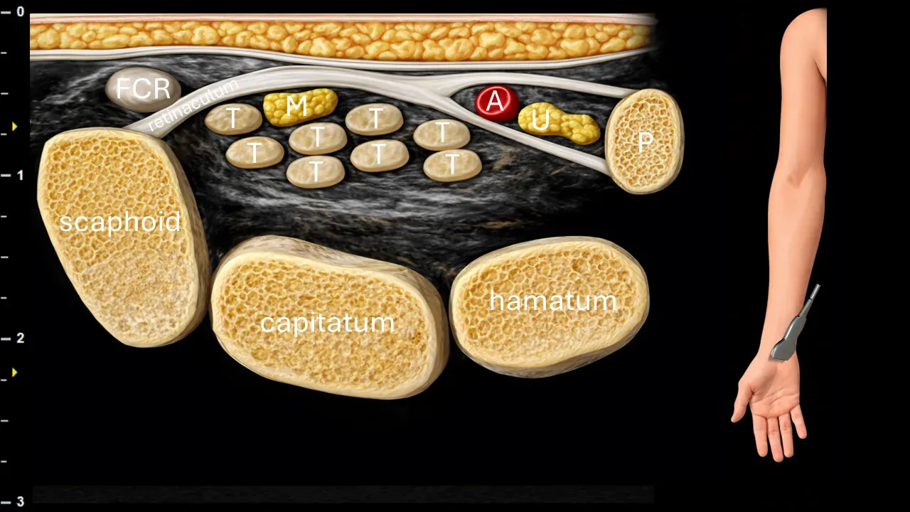
Figure 8. Carpus, transverse plane. M: n. medianus, T: flexor tendons, A: a. ulnaris, U: n. ulnaris, P: os pisiforme, FCR: tendon of m. flexor carpi radialis.
Transverse ultrasound section in the carpal region. N. medianus (M) runs beneath the flexor retinaculum in the carpal tunnel, where it is positioned superficially above the flexor tendons (T). The image also shows the tendon of m. flexor carpi radialis (FCR), a. ulnaris (A) and n. ulnaris (U) located ulnarly outside the carpal tunnel, as well as os pisiforme (P) and carpal bones, particularly os scaphoideum, os capitatum and os hamatum.
Clinical Note
The carpal tunnel area under the retinaculum flexorum represents the most common site of compression of the median nerve, which leads to the development of carpal tunnel syndrome.
2. Anterior interosseus nerve
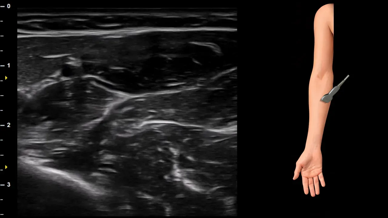
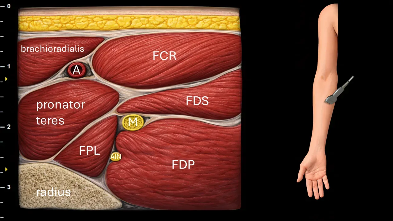
Figure 9. Forearm, transverse plane. A: a. radialis, M: n. medianus, AIN: n. interosseus anterior, FCR: m. flexor carpi radialis, FDS: m. flexor digitorum superficialis, FPL: m. flexor pollicis longus, FDP: m. flexor digitorum profundus.
Transverse ultrasound section in the forearm region. The image shows the n. medianus (M) after passing through the m. pronator teres, from whose main branch the n. interosseus anterior (AIN) arises. The AIN then descends between the m. flexor pollicis longus (FPL) and m. flexor digitorum profundus (FDP) toward the membrana interossea. The image also captures the a. radialis (A), m. flexor carpi radialis (FCR), m. flexor digitorum superficialis (FDS), m. pronator teres, and radius.
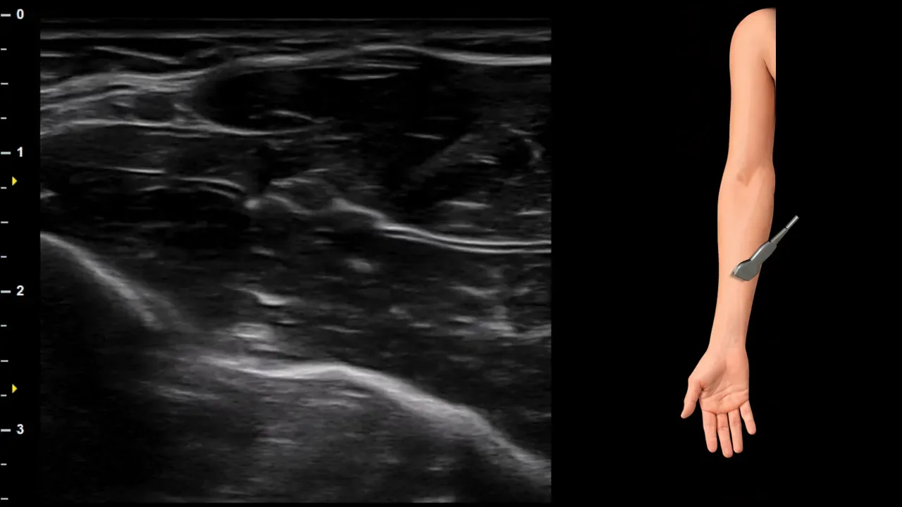
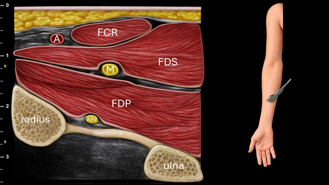
Figure 10. Forearm, transverse plane. A: a. radialis, M: n. medianus, AIN: n. interosseus anterior, FCR: m. flexor carpi radialis, FDS: m. flexor digitorum superficialis, FDP: m. flexor digitorum profundus.
Transverse ultrasound section in the forearm region. N. interosseus anterior (AIN) runs along the interosseous membrane deep beneath the m. flexor digitorum profundus (FDP) and continues distally under the m. pronator quadratus. Also visible in the image are the n. medianus (M) situated between the flexor muscles, a. radialis (A), m. flexor carpi radialis (FCR), m. flexor digitorum superficialis (FDS), radius, ulna and interosseous membrane.
3. Palmar cutaneous nerve

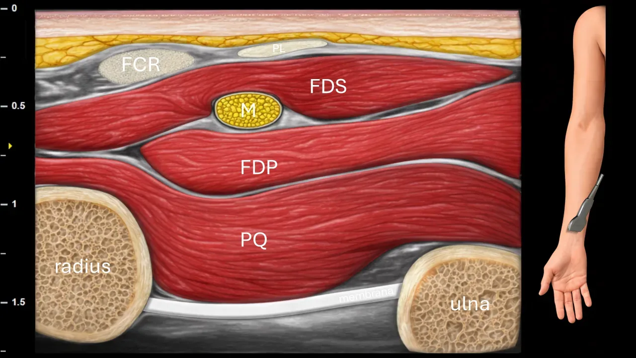
Figure 11. Antebrachium, transverse plane. M: n. medianus, FCR: m. flexor carpi radialis, PL: m. palmaris longus, FDS: m. flexor digitorum superficialis, FDP: m. flexor digitorum profundus, PQ: m. pronator quadratus.
Transverse ultrasound section in the more distal region of the forearm. N. medianus (M) is located in this projection between m. flexor digitorum superficialis (FDS) and m. flexor digitorum profundus (FDP) in the area above m. pronator quadratus (PQ). The image also shows m. flexor carpi radialis (FCR), m. palmaris longus (PL), radius, ulna and membrana interossea.
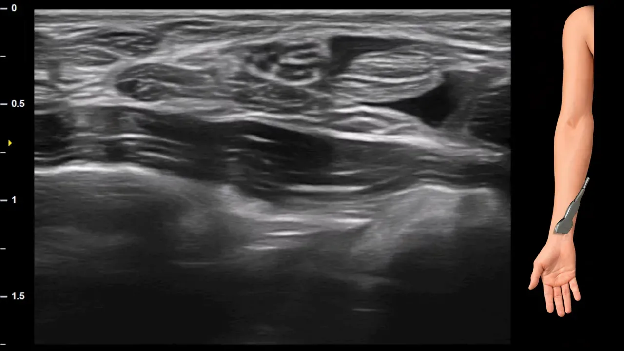
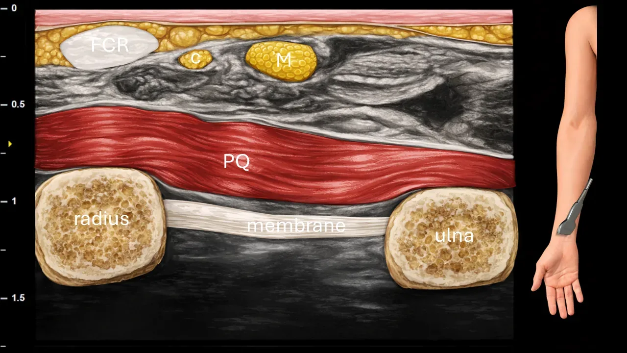
Figure 12. Antebrachium, transverse plane. M: n. medianus, c: ramus cutaneus palmaris n. mediani, FCR: tendon of m. flexor carpi radialis, PQ: m. pronator quadratus.
Transverse ultrasound section in the distal part of the forearm. Ramus cutaneus palmaris n. mediani (c) branches off at the level of m. pronator quadratus (PQ) laterally from the main trunk of n. medianus (M) and courses superficially toward the tendon of m. flexor carpi radialis (FCR). The image also shows the radius, ulna, interosseous membrane and m. pronator quadratus.
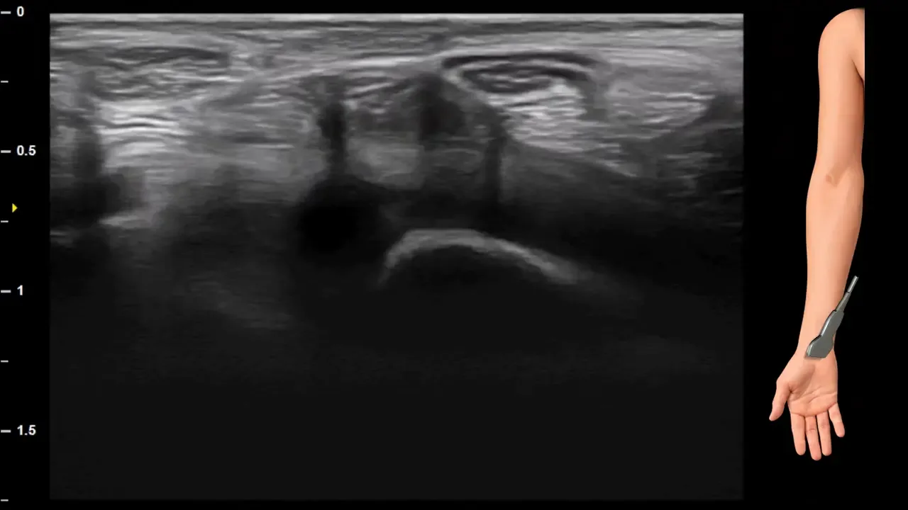
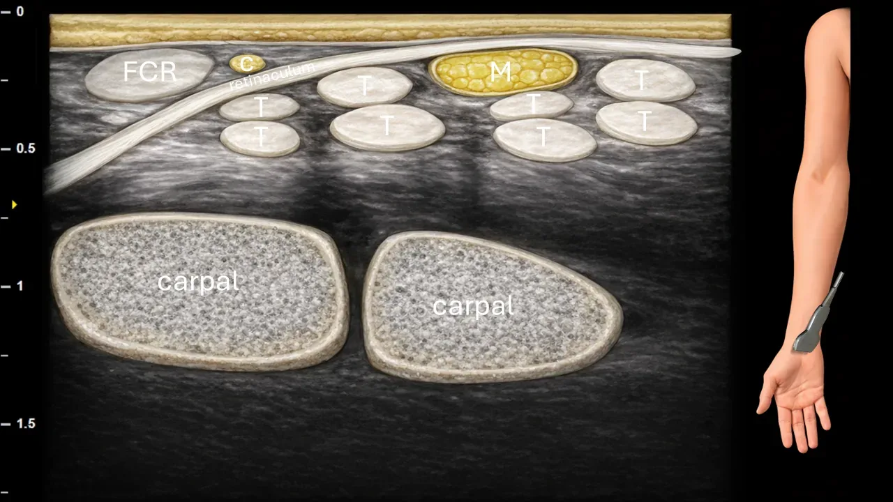
Figure 13. Carpus, transverse plane. M: median nerve, c: palmar cutaneous branch of median nerve, T: flexor tendons, FCR: flexor carpi radialis tendon.
Transverse ultrasound section in the carpal region. The median nerve (M) is located in the carpal tunnel beneath the flexor retinaculum among the flexor tendons (T). The palmar cutaneous branch of the median nerve (c) has already perforated the forearm fascia at this level and runs superficially to the flexor retinaculum toward the palm. The image also shows the flexor carpi radialis tendon (FCR) and carpal bones.
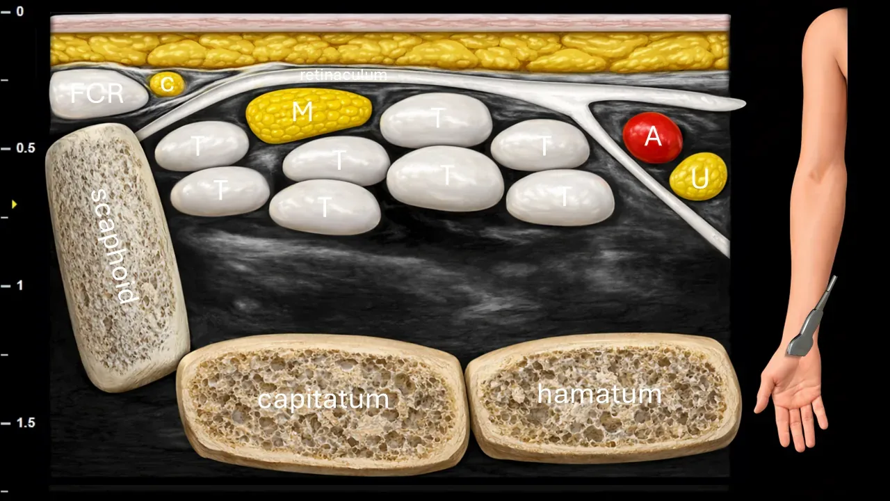
Figure 14. Carpus, transverse plane. M: n. medianus, c: ramus cutaneus palmaris n. mediani, T: flexor tendons, A: a. ulnaris, U: n. ulnaris, FCR: tendon of m. flexor carpi radialis.
Transverse ultrasound section in the carpal region. N. medianus (M) is located in the carpal tunnel beneath the retinaculum flexorum between the flexor tendons (T). N. ulnaris (U) together with a. ulnaris (A) runs ulnarly from the carpal tunnel in Guyon's canal. Ramus cutaneus palmaris n. mediani (c) is at this level located superficially to the retinaculum flexorum. The image also shows the tendon of m. flexor carpi radialis (FCR) and carpal bones, particularly os scaphoideum, os capitatum and os hamatum.
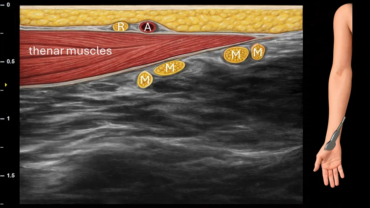
Figure 15. Hand, transverse plane. M: terminal branches of n. medianus, R: ramus recurrens n. mediani, A: artery in the thenar region.
Transverse ultrasound section in the palm region. The image shows the terminal branches of n. medianus (M) after exiting the carpal tunnel and their further branching in the palm. Superficially in the thenar region, the ramus recurrens n. mediani (R) is captured, which courses toward the thenar muscles. The image also shows the thenar muscles and a small arterial branch (A) in their vicinity.
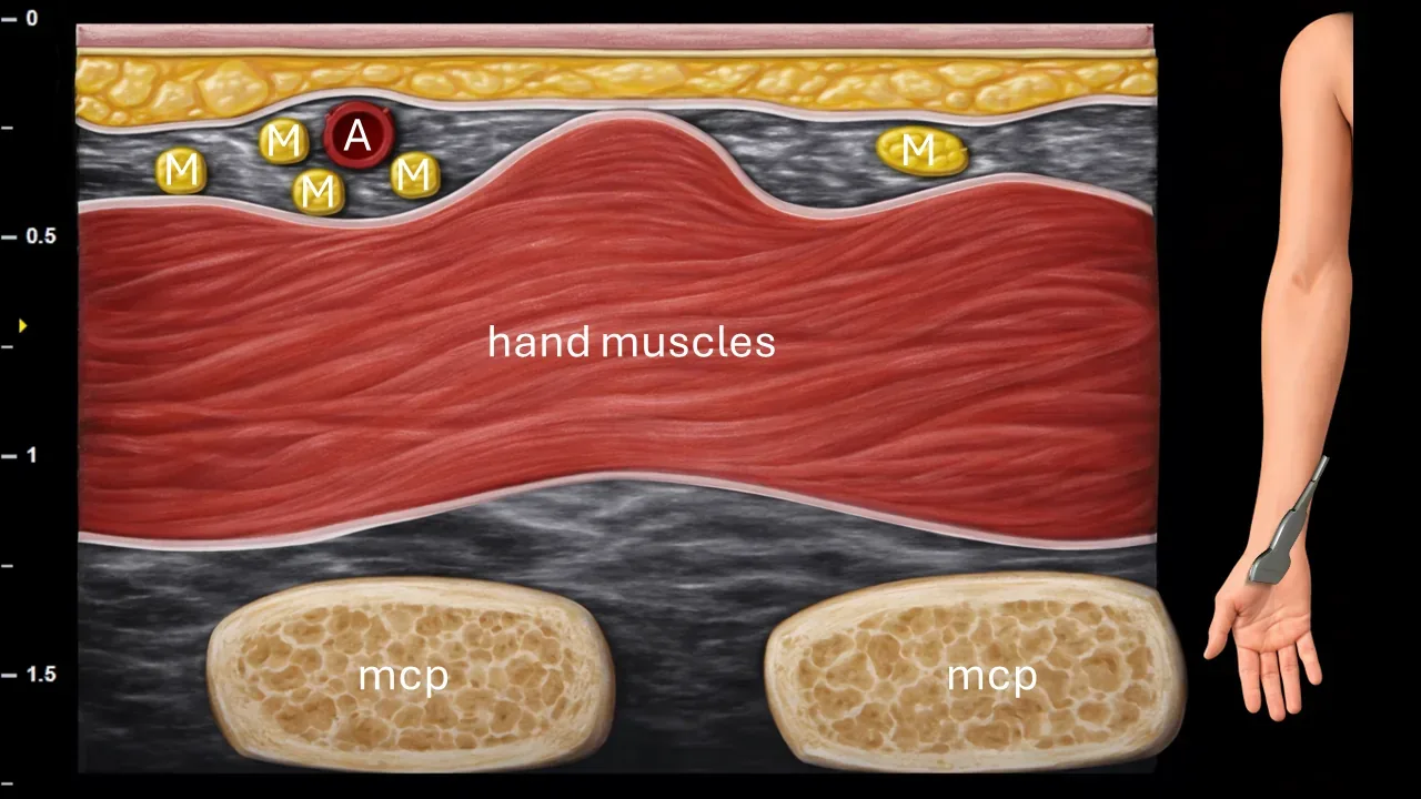
Figure 16. Hand, transverse plane. M: terminal branches of median nerve, A: arteries in the palm region.
Transverse ultrasound section in the palm region at a more distal level. The image shows terminal branches of the median nerve (M) after their further branching distally in the palm. The recurrent branch of the median nerve at this level approaches the other terminal branches of the median nerve. The image also captures small arterial branches (A), hand muscles, and contours of the metacarpophalangeal joints (MCP).