Anatomy
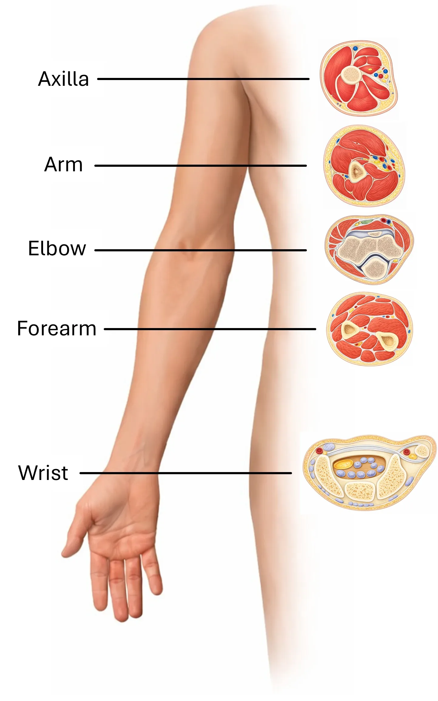
N. radialis arises from the fasciculus posterior plexus brachialis, most commonly from roots C5–C8, sometimes with contribution from T1. It is the main nerve of the posterior compartment of the arm and forearm. It provides motor innervation to the extensors of the elbow, wrist, and fingers, and sensory innervation to the posterior aspect of the arm, forearm, and radial portion of the dorsum of the hand. For sonography, its characteristic transition from the posterior aspect of the arm to the anterior aspect and its terminal division in the elbow region into ramus superficialis and ramus profundus are important. Cross-sectional anatomy is key to understanding the sonographic image, as ultrasound displays structures in individual sections.
Axilla
In the axilla, the n. radialis lies behind the a. axillaris and leaves the plexus as its strong posterior terminal branch. Already in this area, it gives off branches to the m. triceps brachii and also a sensory branch to the posterior side of the arm.
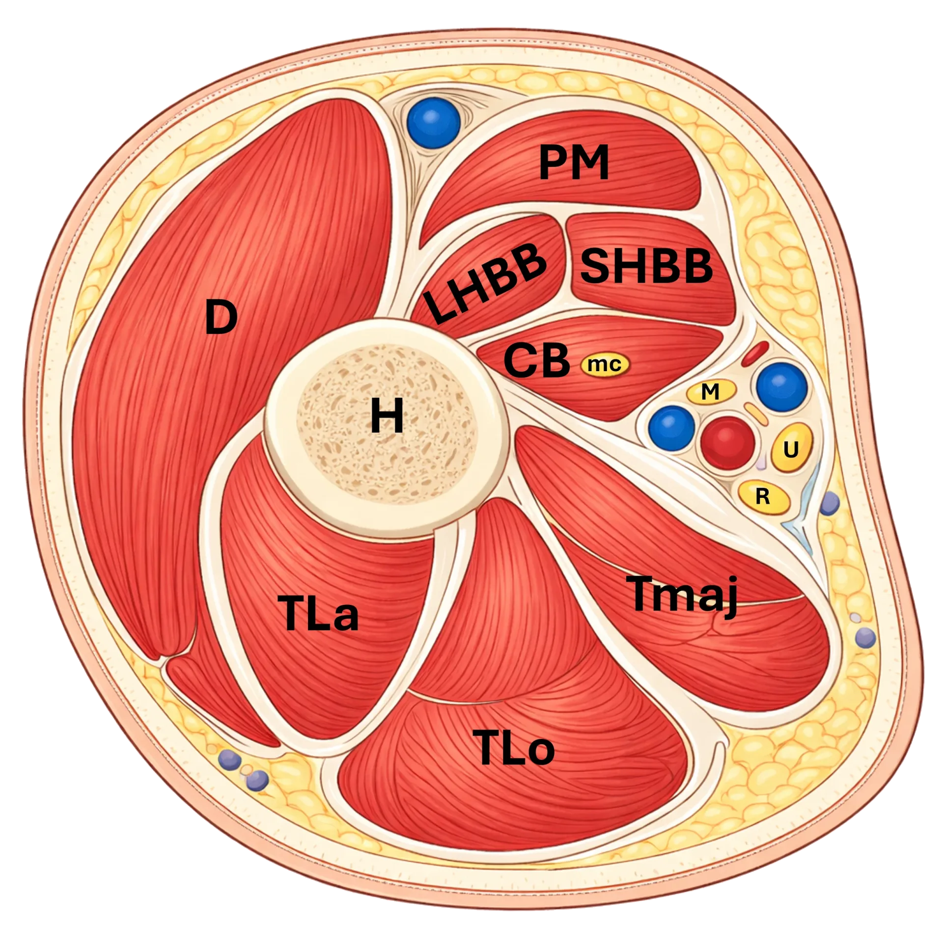
Figure 1: Axillary cross-section. D – m. deltoideus, PM – m. pectoralis major, LHBB – caput longum m. biceps brachii, SHBB – caput breve m. biceps brachii, CB – m. coracobrachialis, mc – n. musculocutaneus, M – n. medianus, U – n. ulnaris, R – n. radialis, Tmaj – m. teres major, TLa – caput laterale m. triceps brachii, TLo – caput longum m. triceps brachii, H – humerus
Arm
In the arm, the n. radialis passes posteriorly through the triangular interval and enters the posterior compartment of the arm. It then runs in the sulcus n. radialis on the humerus together with the a. profunda brachii, between the heads of the m. triceps brachii. In the middle part of the arm, it wraps around the humerus spirally and subsequently pierces the septum intermusculare brachii laterale, thereby returning to the anterior aspect of the arm. Distally, it then lies between the m. brachialis and m. brachioradialis.
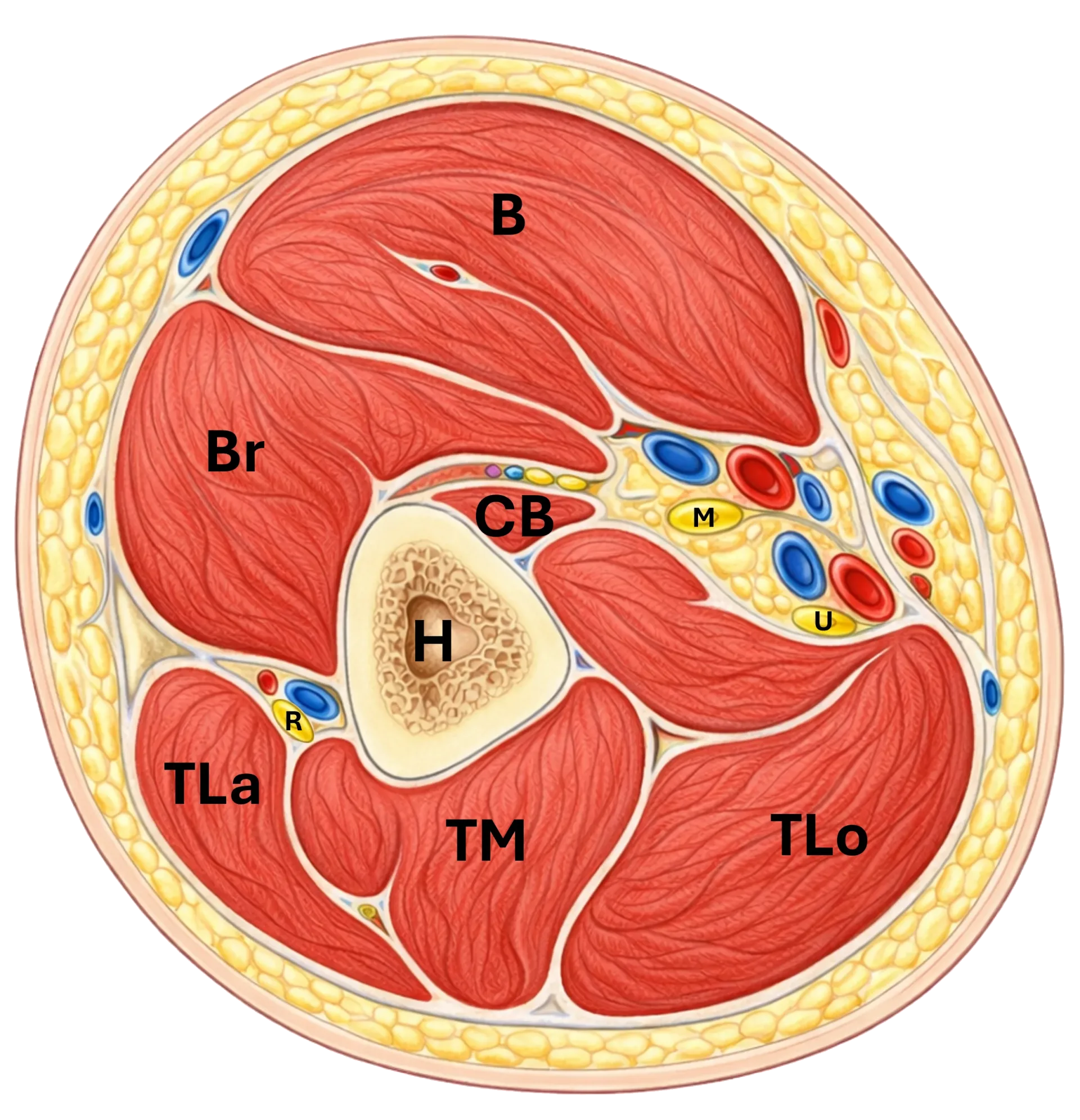
Figure 2: Cross-section of the arm. B – m. biceps brachii, Br – m. brachialis, CB – m. coracobrachialis, H – humerus, TLa – caput laterale m. triceps brachii, TM – caput mediale m. triceps brachii, TLo – caput longum m. triceps brachii, R – n. radialis, M – n. medianus, U – n. ulnaris
Elbow
In the elbow region, the n. radialis lies in the groove between m. brachialis and m. brachioradialis. Here it divides into:
ramus superficialis
ramus profundus
Motor branches also originate in this area for m. brachioradialis, m. extensor carpi radialis longus and often also for m. extensor carpi radialis brevis. The bifurcation area of the n. radialis is crucial for sonographic differentiation of the superficial sensory and deep motor branches.
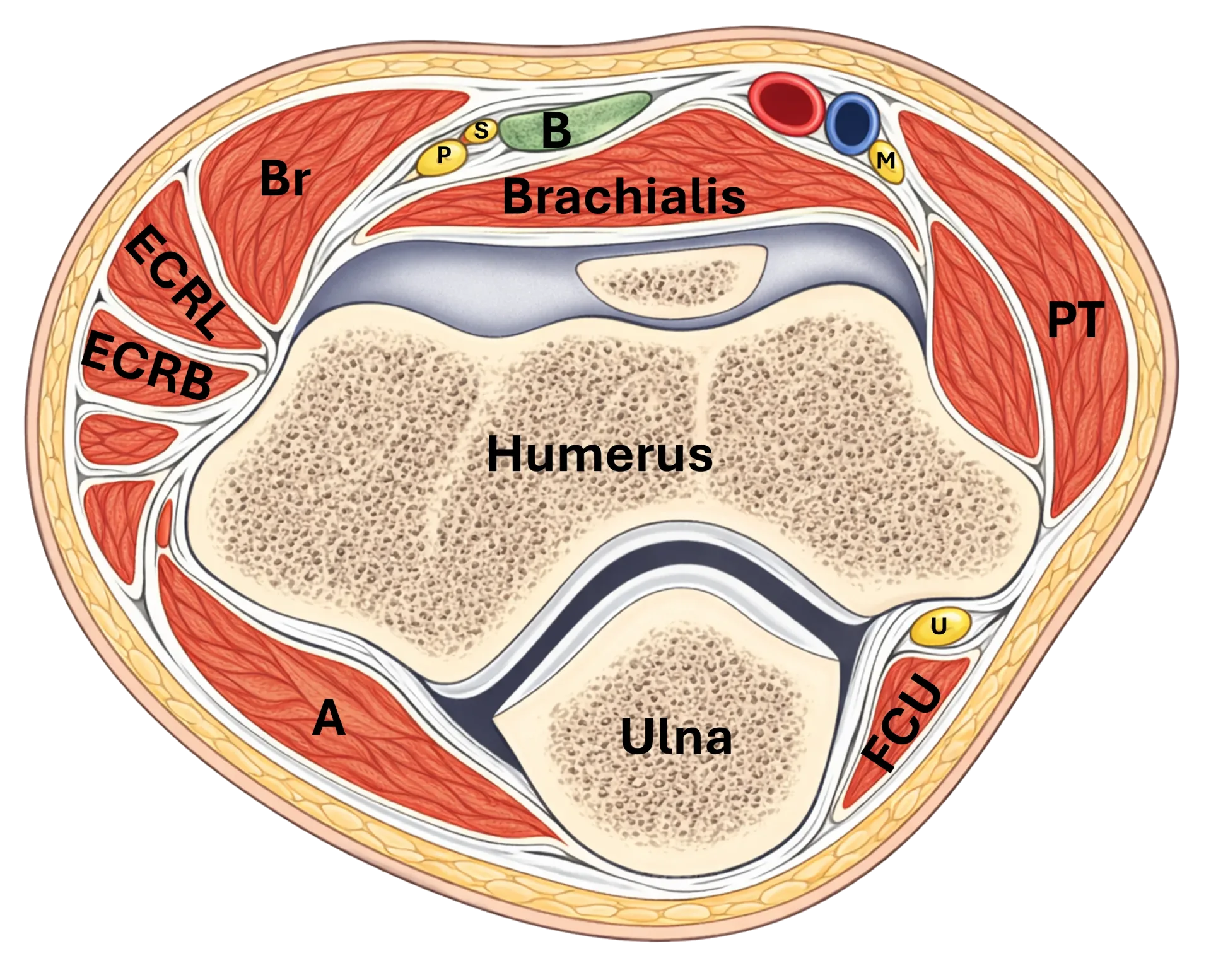
Figure 3: Cross-section of the elbow. Br – m. brachioradialis, ECRL – m. extensor carpi radialis longus, ECRB – m. extensor carpi radialis brevis, B – tendon of m. biceps brachii, S – r. superficialis n. radialis, P – r. profundus n. radialis (n. interosseus posterior), M – n. medianus, PT – m. pronator teres, U – n. ulnaris, FCU – m. flexor carpi ulnaris, A – m. anconeus
Forearm
Ramus superficialis continues in the forearm under m. brachioradialis, lateral to the radial artery. In the distal part of the forearm, it pierces the fascia between m. brachioradialis and m. extensor carpi radialis longus and becomes subcutaneous.
Ramus profundus enters the supinator tunnel under the arcade of Frohse and after passing through m. supinator continues as n. interosseus posterior (PIN). This runs in the posterior compartment of the forearm between the superficial and deep layers of extensors and innervates most of the wrist and finger extensors. Distally, PIN runs deeper along the membrana interossea and terminates at the dorsal capsule of the wrist.
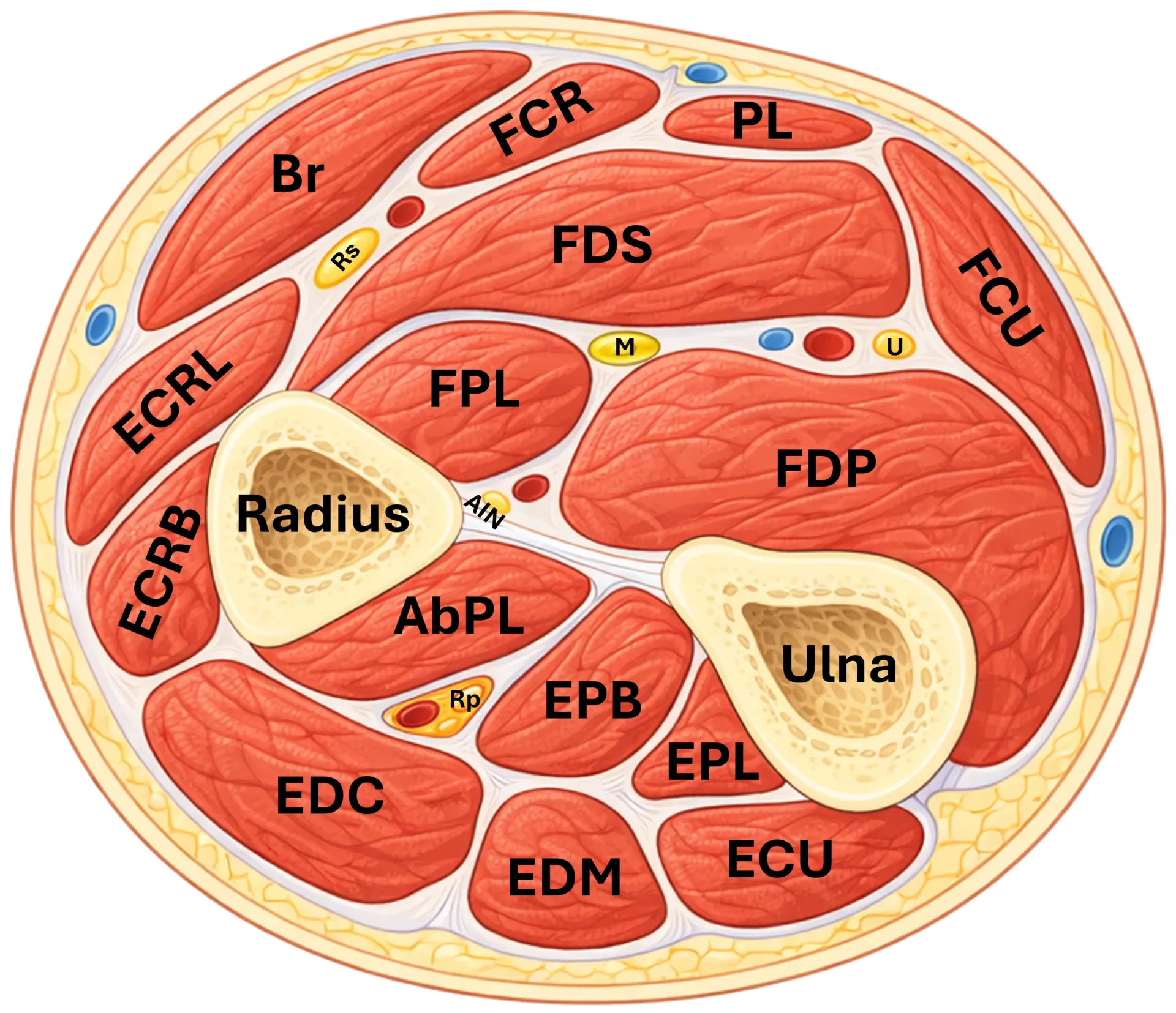
Figure 4: Cross-section of the forearm. Br – m. brachioradialis, ECRL – m. extensor carpi radialis longus, ECRB – m. extensor carpi radialis brevis, EDC – m. extensor digitorum communis, EDM – m. extensor digiti minimi, ECU – m. extensor carpi ulnaris, AbPL – m. abductor pollicis longus, EPB – m. extensor pollicis brevis, EPL – m. extensor pollicis longus, FCR – m. flexor carpi radialis, PL – m. palmaris longus, FDS – m. flexor digitorum superficialis, FPL – m. flexor pollicis longus, FDP – m. flexor digitorum profundus, FCU – m. flexor carpi ulnaris, M – n. medianus, U (nerve) – n. ulnaris, Rs – r. superficialis n. radialis, Rp – r. profundus n. radialis (n. interosseus posterior), AIN – n. interosseus anterior, R – radius, U (bone) – ulna
Wrist
In the wrist region, the ramus superficialis n. radialis is of primary clinical significance. After emerging from beneath the fascia, it continues to the dorsoradial side of the wrist and into the area of the anatomical snuffbox, where it runs in close proximity to the tendons of the 1st and 2nd dorsal compartments. In this area, it may be crossed by the v. cephalica. The nerve provides sensory innervation to the radial portion of the dorsum of the hand and the dorsal side of the thumb, index finger, and part of the middle finger.
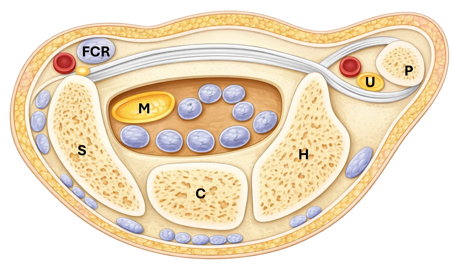
Figure 5: Cross-section of the wrist. FCR – m. flexor carpi radialis, M – n. medianus, U – n. ulnaris, P – os pisiforme, H – os hamatum, C – os capitatum, S – os scaphoideum
Practical landmarks for ultrasound
in axilla: behind a. axillaris
in brachium: in sulcus n. radialis together with a. profunda brachii
distally on arm: through septum intermusculare laterale anteriorly
in cubita: between m. brachialis and m. brachioradialis
on forearm:
ramus profundus under arcade of Frohse into supinator tunnel
ramus superficialis under m. brachioradialis
in wrist: ramus superficialis on dorsoradial side, in region of fossa tabatier
Schalten Sie die vollständige Health Library frei
Voller Zugriff auf Scan-Protokolle, Anatomie und klinische Referenzen. Jederzeit kündbar.
- Alle Protokolle und Anatomiereferenzen
- Originale Ultraschall-Illustrationen und Video-Demonstrationen
- Synchronisierung zwischen Mobilgerät und Web