Ultrasound examination
Examination Protocol
Nervus ulnaris throughout the entire upper extremity
- Axilla
- Brachium
- Cubitum
- Forearm
- Carpus
Dorsal cutaneous branch
- Distance from the ulnar nerve
- Course around the ulna
Terminal branches
- Guyon's canal
- Terminal branches
Interaktive Funktion, verfügbar in der App
1. Ulnar nerve throughout the entire upper extremity
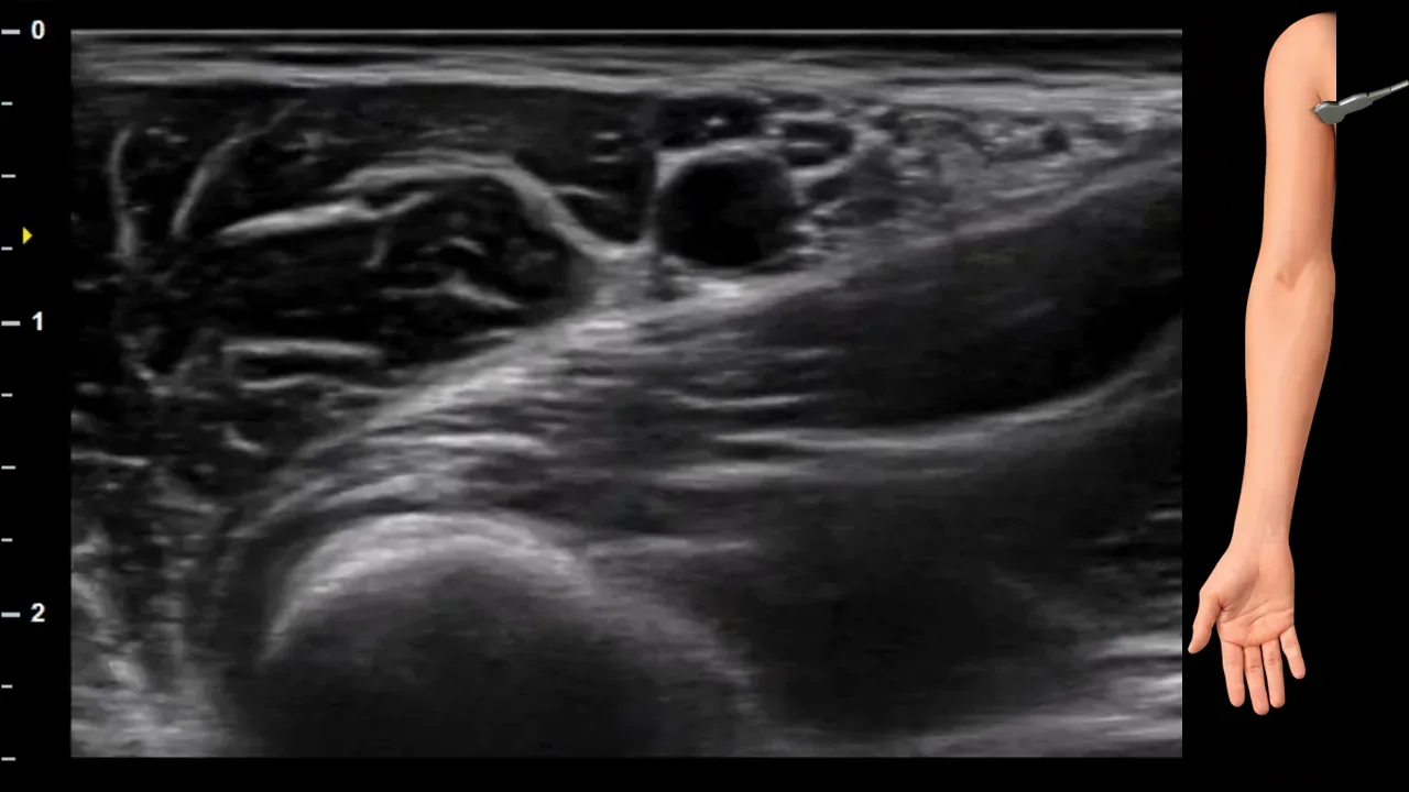
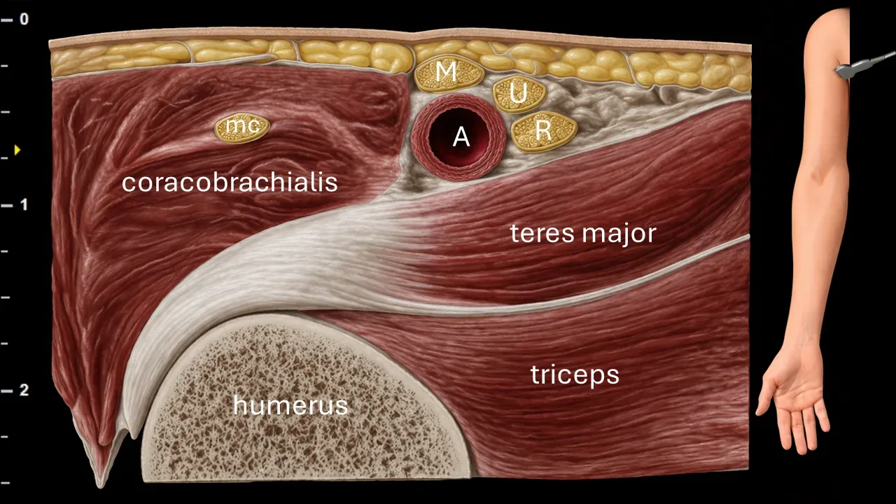
Figure 1. Axilla, transverse plane. A: axillary artery, M: median nerve, U: ulnar nerve, R: radial nerve, mc: musculocutaneous nerve
Transverse ultrasound section of the axilla. The image shows the axillary artery (A), which represents the main landmark structure for identifying nerve elements of the brachial plexus. The ulnar nerve (U) is positioned medially and dorsally to the axillary artery in this projection. Near the vessel, the median nerve (M), radial nerve (R), and musculocutaneous nerve (mc) are also visible. The image also captures surrounding muscle structures, particularly the coracobrachialis muscle, teres major muscle, and triceps brachii muscle, as well as the contour of the humerus.
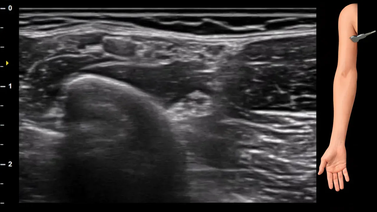
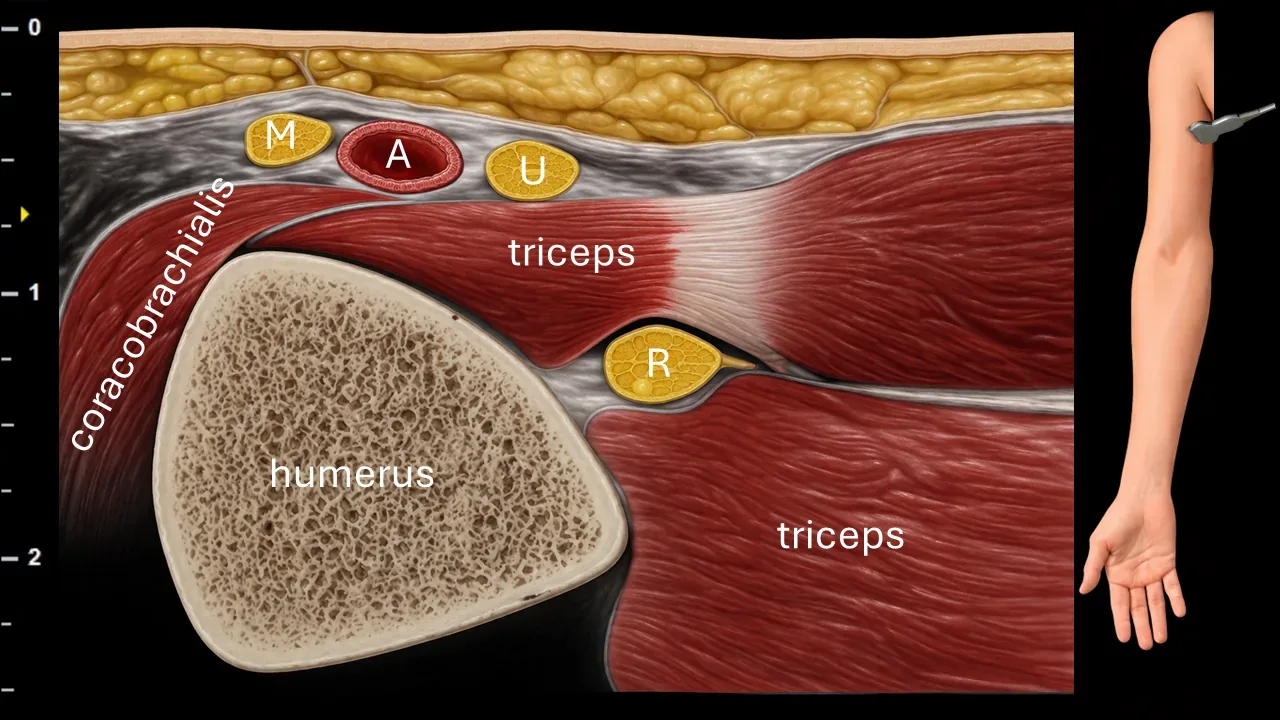
Figure 2. Brachium, transverse plane. A: artery, M: n. medianus, U: n. ulnaris, R: n. radialis.
Transverse ultrasound section of the arm showing the n. ulnaris (U), which is positioned medially and superficially in this projection, adjacent to the medial head of m. triceps brachii. Near the vascular structure, the n. medianus (M) can also be identified, and deeper at the humerus the n. radialis (R). The image also captures surrounding muscle structures, particularly m. coracobrachialis and m. triceps brachii, and the contour of the humerus.
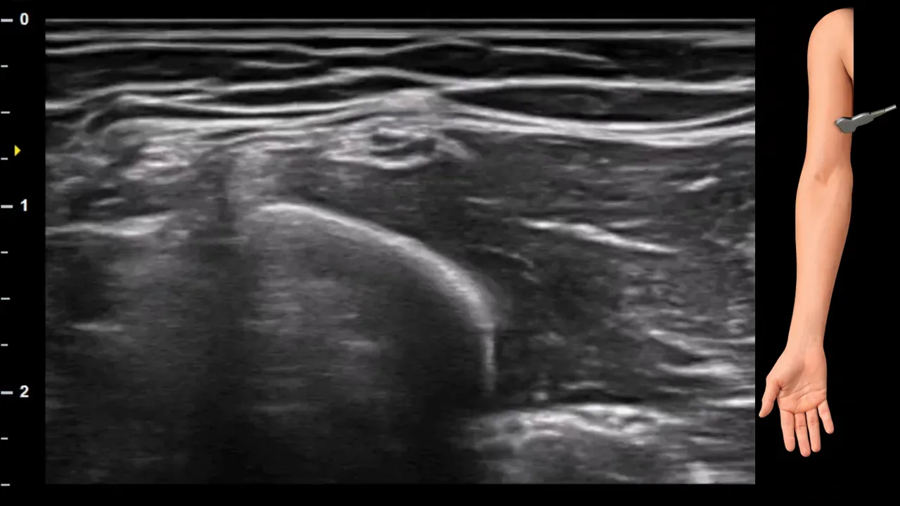
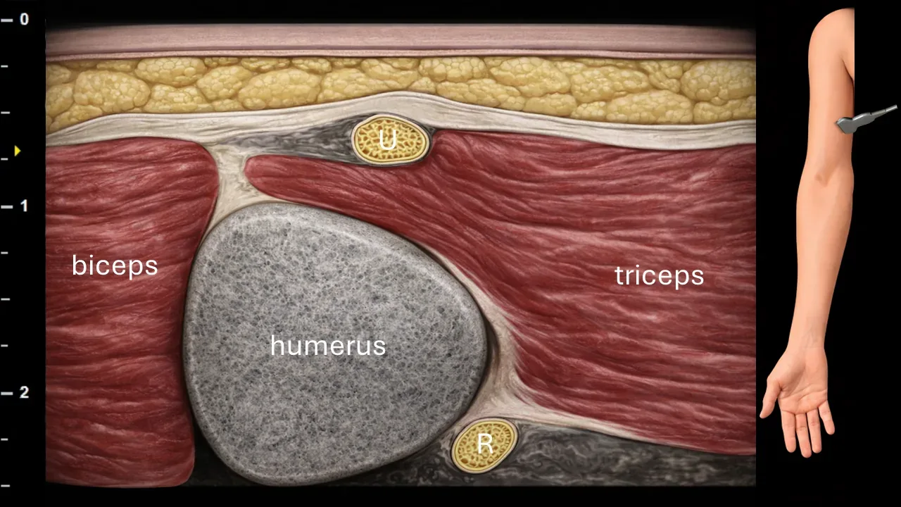
Figure 3. Brachium, transverse plane. U: ulnar nerve, R: radial nerve.
Transverse ultrasound section of the arm at the level where the ulnar nerve (U) turns dorsally and perforates the medial intermuscular septum. In this projection, the nerve is located superficially at the septum between the muscular structures of the arm. The image shows surrounding anatomical structures, particularly the biceps brachii, triceps brachii, contour of the humerus, and the deeper radial nerve (R).
Clinical Note
The area of perforation of the medial intermuscular septum represents a possible site of n. ulnaris compression, either by the septum itself or by the arcade of Struthers.
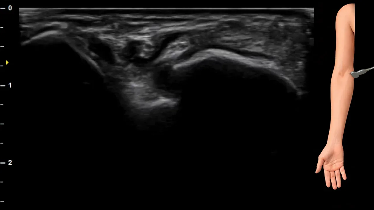
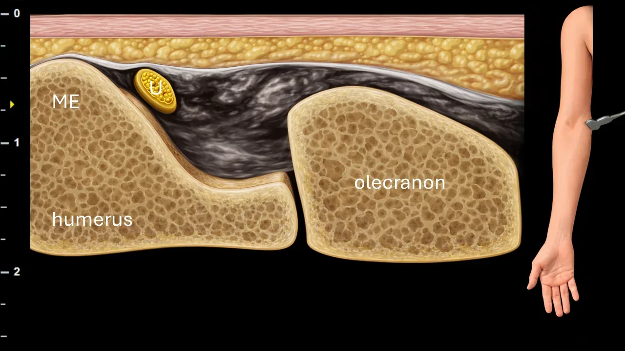
Figure 4. Elbow, transverse plane. U: ulnar nerve, ME: medial epicondyle of humerus.
Transverse ultrasound section in the elbow region. The ulnar nerve (U) runs dorsally behind the medial epicondyle of the humerus (ME) in the ulnar nerve groove area. The image also shows bony landmark structures, particularly the distal humerus and olecranon.
Clinical Note
In this area, you can dynamically examine the stability of the n. ulnaris, particularly to assess whether it snaps over the medial epicondyle of the humerus during elbow flexion.
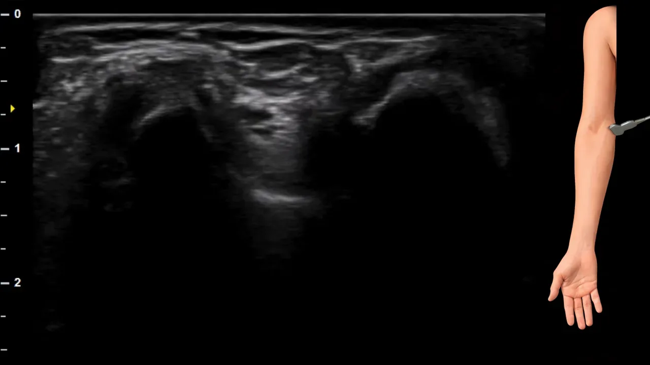
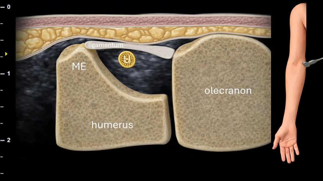
Figure 5. Elbow, transverse plane. U: ulnar nerve, ME: medial epicondyle of humerus.
Transverse ultrasound section in the area of the cubital tunnel. The ulnar nerve (U) is positioned between the medial epicondyle of humerus (ME) and the olecranon, where it enters the cubital tunnel. The roof of the tunnel is formed by Osborne's ligament, under which the nerve passes. The image also shows bony landmark structures, particularly the distal humerus and olecranon.
Clinical Note
In this area, the ulnar nerve may be compressed by Osborne's ligament, which represents one of the typical compression sites in cubital tunnel syndrome.
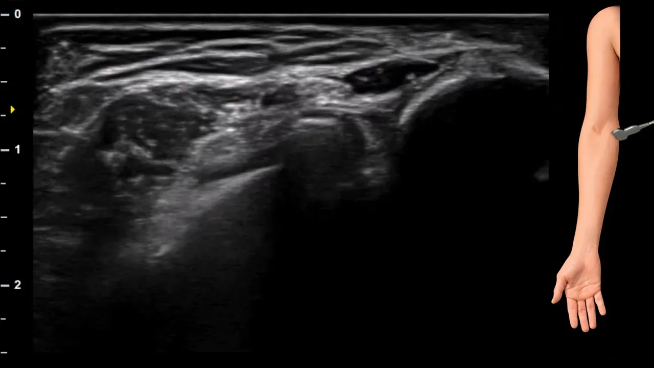
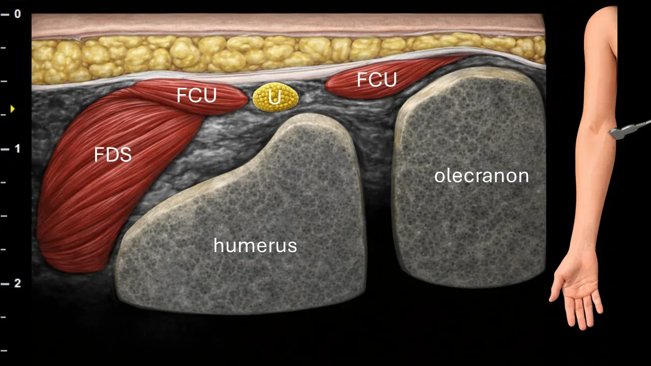
Figure 6. Cubitum, transverse plane. U: n. ulnaris, FCU: m. flexor carpi ulnaris, FDS: m. flexor digitorum superficialis.
Transverse ultrasound section in the elbow region distal to the cubital tunnel. N. ulnaris (U) runs here beneath the ligamentum arcuatum, which represents a fascial aponeurotic arch between the humeral and ulnar heads of m. flexor carpi ulnaris (FCU). In this projection, the nerve is located between both heads of m. flexor carpi ulnaris, adjacent to the flexor musculature of the forearm. The image also shows m. flexor digitorum superficialis (FDS) and bony landmark structures, particularly the distal humerus and olecranon.
Clinical Note
The area beneath the ligamentum arcuatum, between the heads of the m. flexor carpi ulnaris, represents another possible site of n. ulnaris compression in cubital tunnel syndrome.
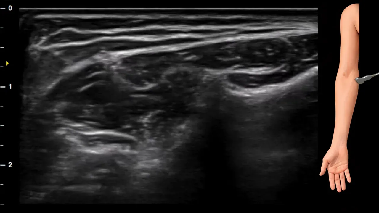
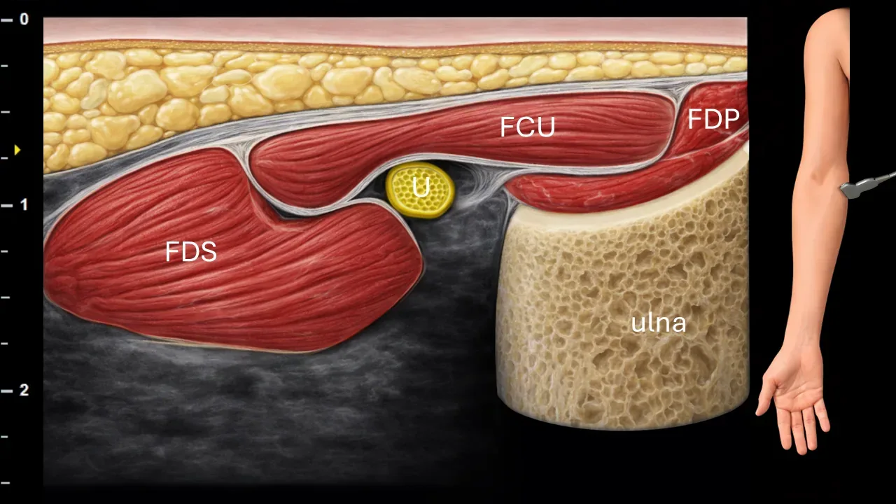
Figure 7. Antebrachium, transverse plane. U: n. ulnaris, FCU: m. flexor carpi ulnaris, FDS: m. flexor digitorum superficialis, FDP: m. flexor digitorum profundus.
Transverse ultrasound section of the proximal forearm. N. ulnaris (U) is located in this projection between the heads of m. flexor carpi ulnaris (FCU), during its transition from the cubital tunnel area into the forearm. The image also shows surrounding flexor muscle structures, particularly m. flexor digitorum superficialis (FDS) and m. flexor digitorum profundus (FDP), as well as the bony contour of the ulna.
Clinical Note
In this area, the ulnar nerve may be compressed when exiting the cubital tunnel, particularly in connection with the tight passage between the heads of the flexor carpi ulnaris muscle.
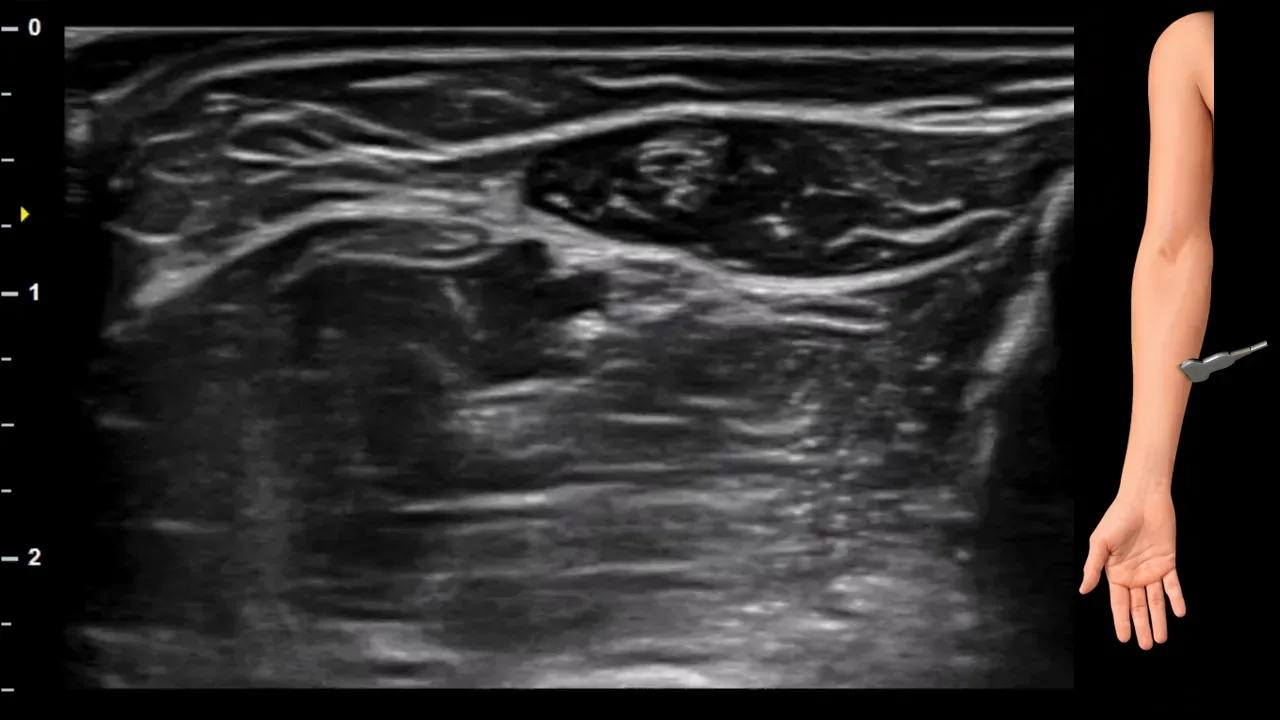
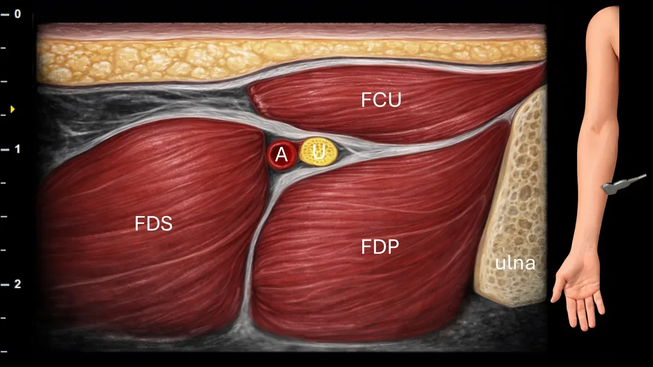
Figure 8. Antebrachium, transverse plane. U: ulnar nerve, A: ulnar artery, FCU: flexor carpi ulnaris muscle, FDS: flexor digitorum superficialis muscle, FDP: flexor digitorum profundus muscle.
Transverse ultrasound section of the forearm. The ulnar nerve (U) in this projection is located between the flexor digitorum superficialis muscle (FDS) and the flexor digitorum profundus muscle (FDP). In the more distal levels of the forearm, the nerve is accompanied by the ulnar artery (A), which represents an important landmark structure for its identification. Superficial to the neurovascular bundle, the flexor carpi ulnaris muscle (FCU) is visible. The bony contour of the ulna is also captured in the image.
Clinical Note
In this area, during ultrasound examination, it is advisable to monitor the relationship between the n. ulnaris and a. ulnaris, because the vessel facilitates orientation in the more distal part of the forearm and helps to precisely localize the course of the nerve.
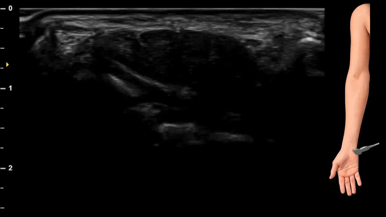
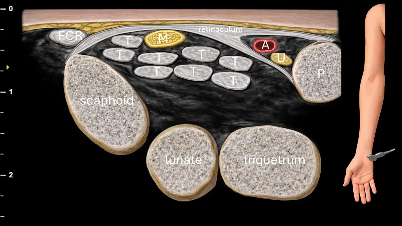
Figure 9. Carpus, transverse plane. U: ulnar nerve, A: ulnar artery, M: median nerve, T: flexor tendons, FCR: flexor carpi radialis tendon, P: os pisiforme.
Transverse ultrasound section in the wrist area. The ulnar nerve (U) is located in this projection in Guyon's canal, where it runs in close proximity to the ulnar artery (A) between os pisiforme (P) and superficial connective tissue structures. In the more radial portion of the image, the median nerve (M) is visible, located beneath the flexor retinaculum in the carpal tunnel, surrounded by flexor tendons (T). Also captured in the image are bony structures of the carpus, particularly os scaphoideum, os lunatum and os triquetrum, as well as the flexor carpi radialis tendon (FCR).
Clinical Note
Guyon's canal represents a significant potential site of ulnar nerve compression in the wrist region. Ultrasound enables assessment of the canal lumen, the relationship of the nerve to the ulnar artery, and possible compressive causes, such as ganglion, hypertrophy of surrounding structures, or post-traumatic changes.
2. Dorsal cutaneous branch
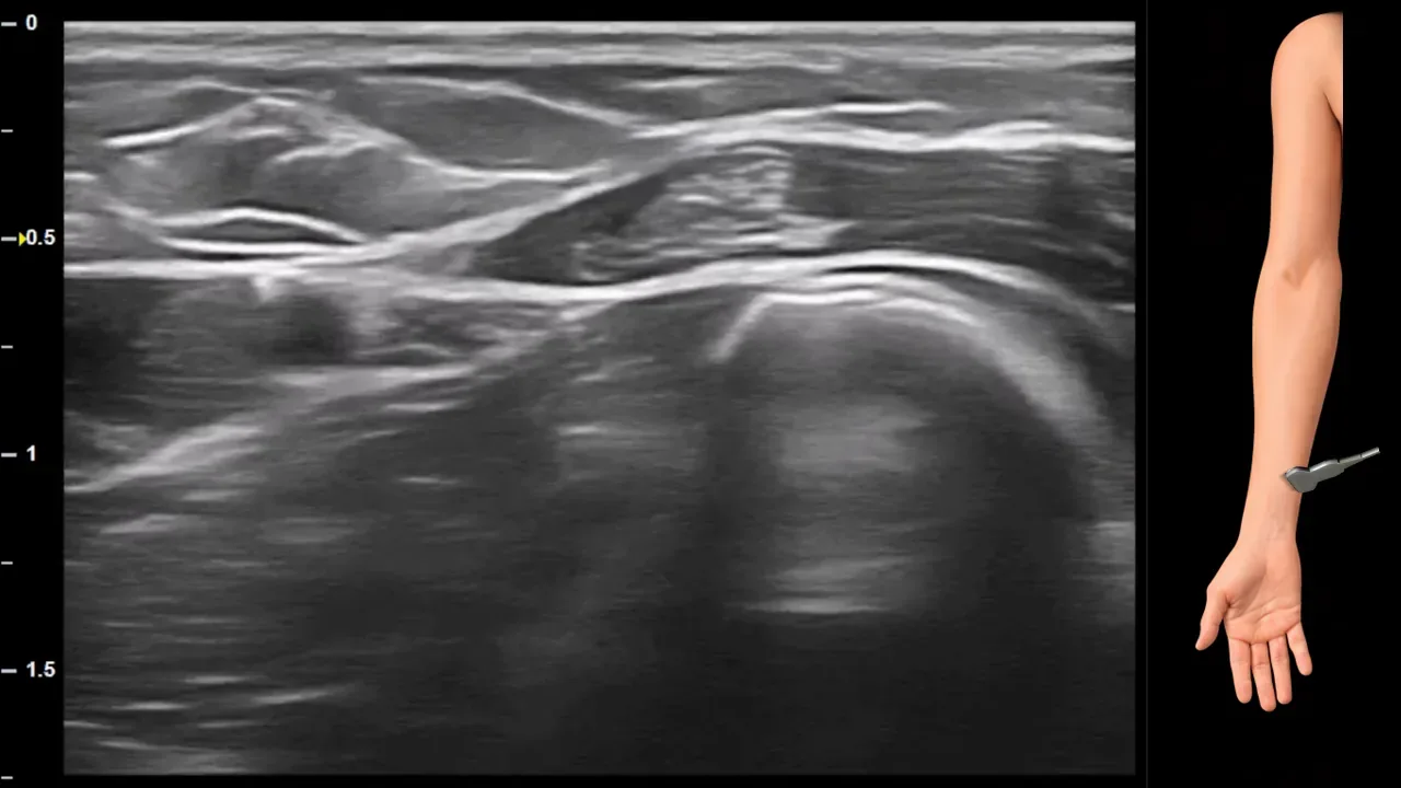
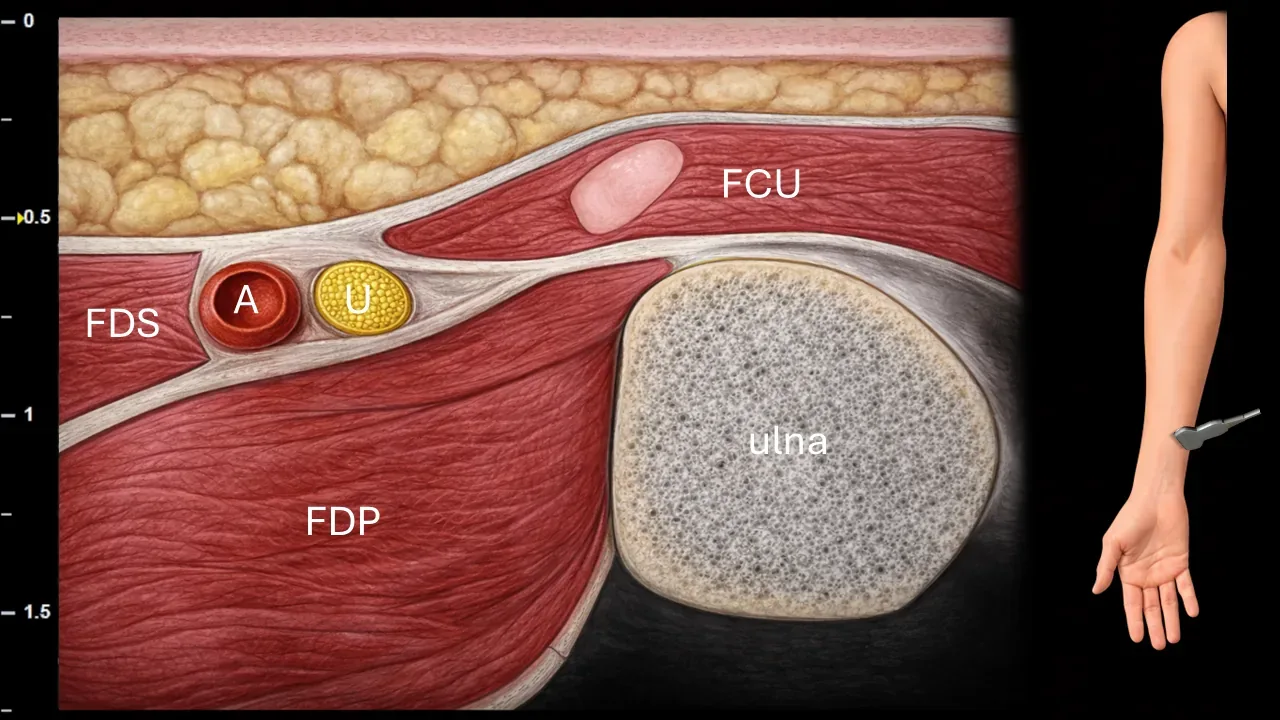
Figure 10. Distal third of the forearm, transverse plane. U: n. ulnaris, A: a. ulnaris, FCU: m. flexor carpi ulnaris, FDS: m. flexor digitorum superficialis, FDP: m. flexor digitorum profundus.
Transverse ultrasound section in the area of the distal third of the forearm. The n. ulnaris (U) runs here in close proximity to the a. ulnaris (A) between the surrounding flexor muscles. Superficially and ulnarly, the m. flexor carpi ulnaris (FCU) is visible, deeper lies the m. flexor digitorum profundus (FDP) and more radially the m. flexor digitorum superficialis (FDS). The image also shows the bony contour of the ulna.
Clinical Note
At this level, ultrasound can be used to locate the branch point of the ramus cutaneus dorsalis n. ulnaris, which typically separates from the main trunk in the distal forearm. This area is therefore suitable for its targeted identification and subsequent sonographic tracking.
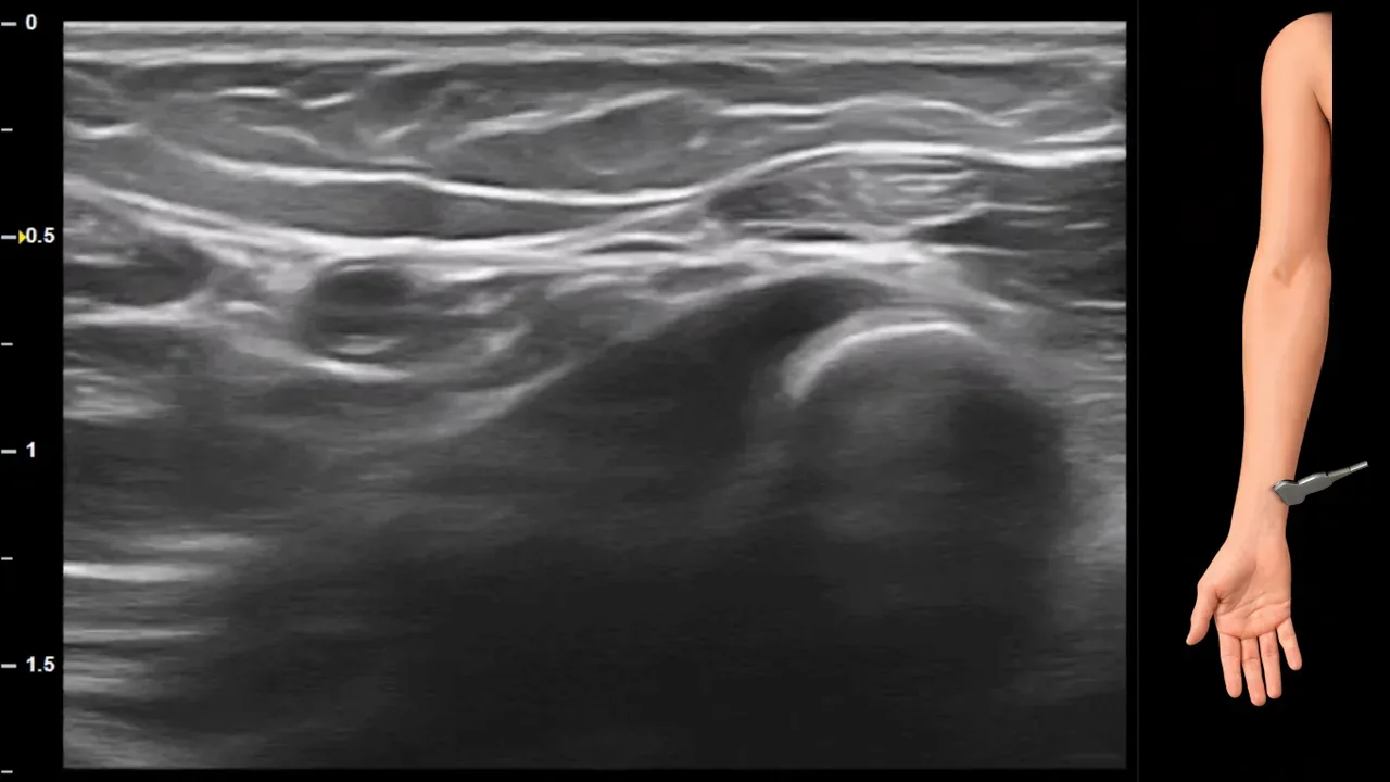
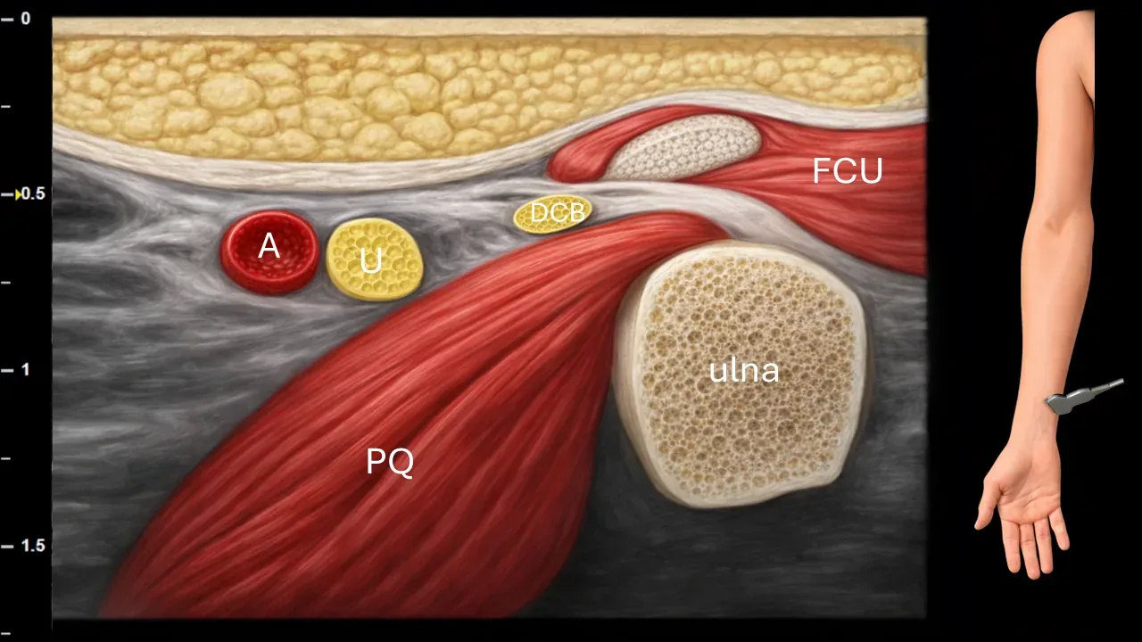
Figure 11. Distal third of the forearm, transverse plane. U: n. ulnaris, A: a. ulnaris, DCB: ramus cutaneus dorsalis n. ulnaris, FCU: m. flexor carpi ulnaris, PQ: m. pronator quadratus.
Transverse ultrasound section in the area of the distal third of the forearm. N. ulnaris (U) runs here in close proximity to a. ulnaris (A). From its trunk at this level branches the dorsal cutaneous branch (DCB, ramus cutaneus dorsalis n. ulnaris), which turns dorsoulnarly and heads toward the subcutaneous tissue. The image also shows surrounding anatomical structures, particularly m. flexor carpi ulnaris (FCU), the deeper m. pronator quadratus (PQ) and the bony contour of the ulna.
Clinical Note
Sonographic identification of the ramus cutaneus dorsalis n. ulnaris is important in evaluating injuries to small sensory branches as well as for preoperative orientation, because this branch can be a source of local sensory complaints on the dorsoulnar side of the hand.
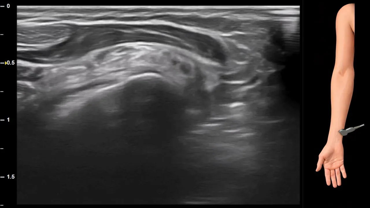
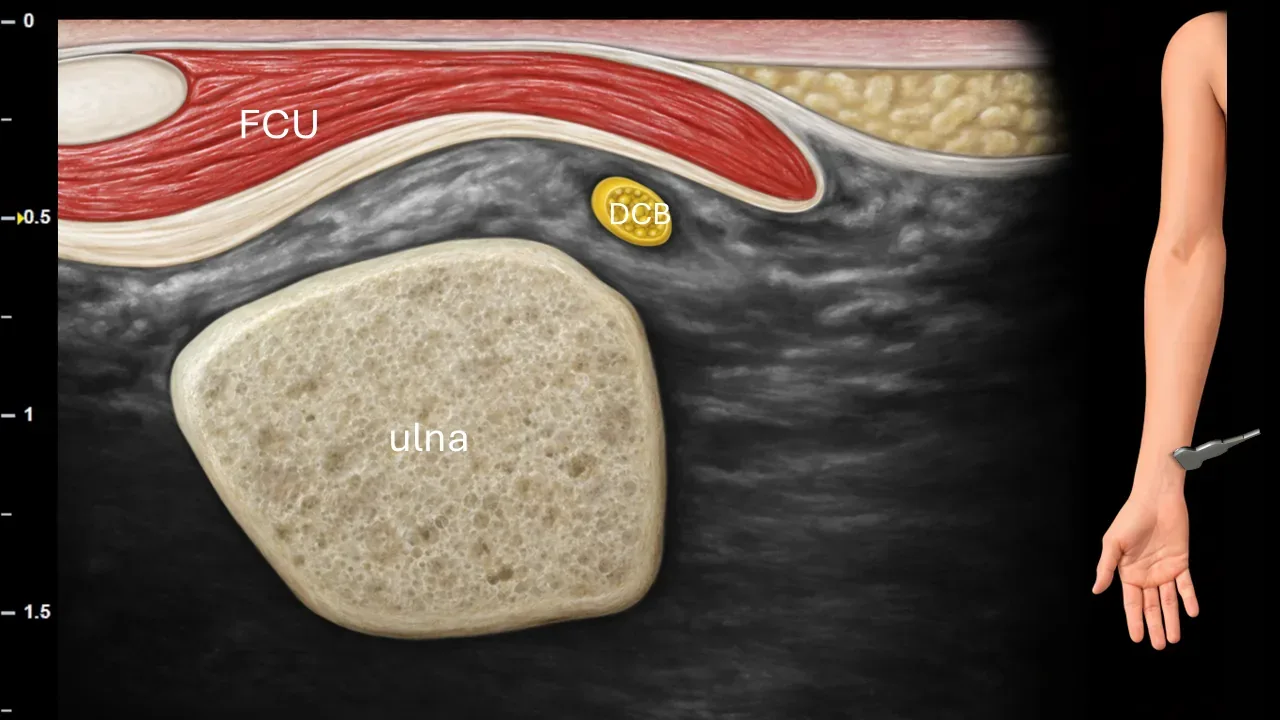
Figure 12. Distal third of the forearm, transverse plane. DCB: ramus cutaneus dorsalis n. ulnaris, FCU: m. flexor carpi ulnaris.
Transverse ultrasound section in the region of the distal third of the forearm. The dorsal cutaneous branch of the ulnar nerve (DCB) separates from the main trunk at this level and curves dorsally around the ulna, heading toward the subcutaneous tissue on the dorsoulnar aspect of the forearm and hand. Also visible in the image are the m. flexor carpi ulnaris (FCU) and the bony contour of the ulna.
3. Terminal branches
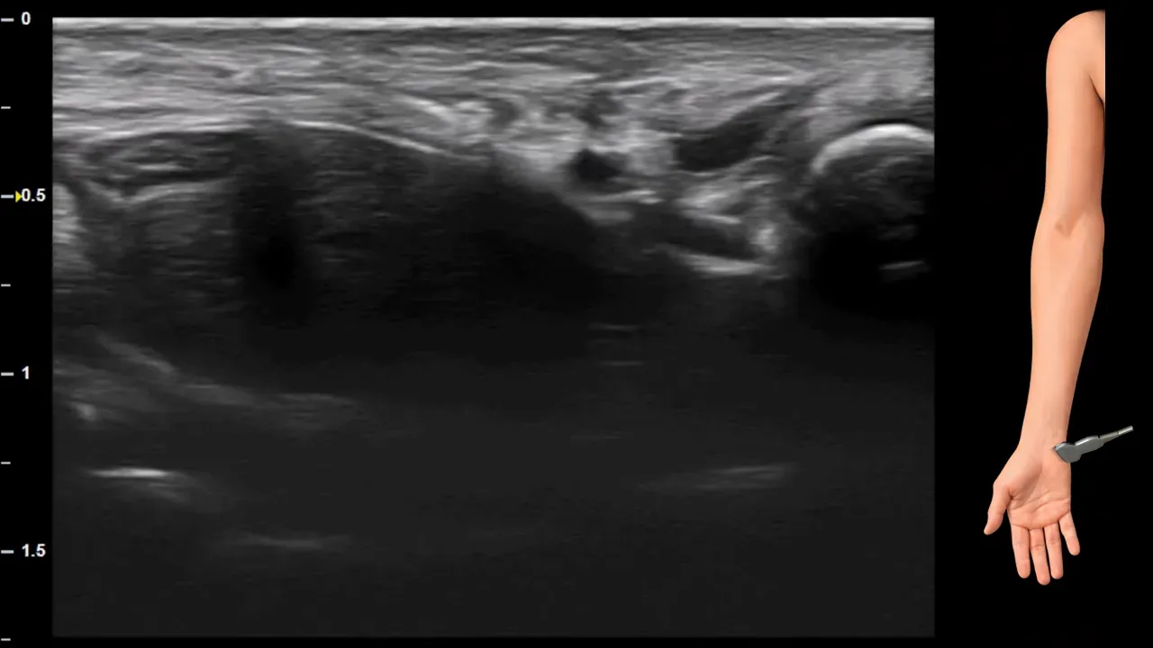
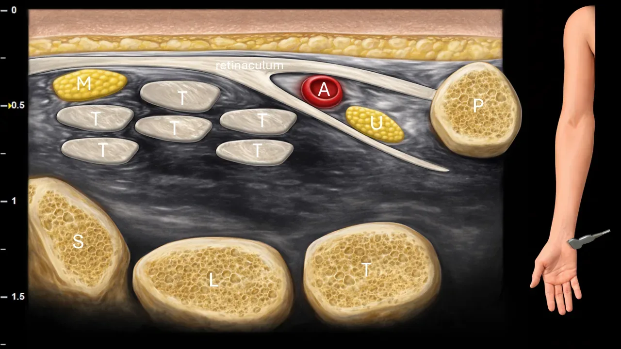
Figure 13. Carpus, transverse plane. U: ulnar nerve, A: ulnar artery, M: median nerve, T: flexor tendons, P: pisiform bone, S: scaphoid bone, L: lunate bone, T: triquetral bone.
Transverse ultrasound section in the wrist area. The ulnar nerve (U) is located in Guyon's canal in close proximity to the ulnar artery (A) at the pisiform bone (P). This projection is suitable as an initial orientation section for tracking the terminal branching of the ulnar nerve in the area of Guyon's canal, where the nerve further divides into its superficial sensory and deep motor branches. In the radial part of the image, the median nerve (M) is visible, located beneath the flexor retinaculum in the carpal tunnel, surrounded by flexor tendons. The image also captures bony structures of the carpus, particularly the scaphoid bone (S), lunate bone (L) and triquetral bone (T).
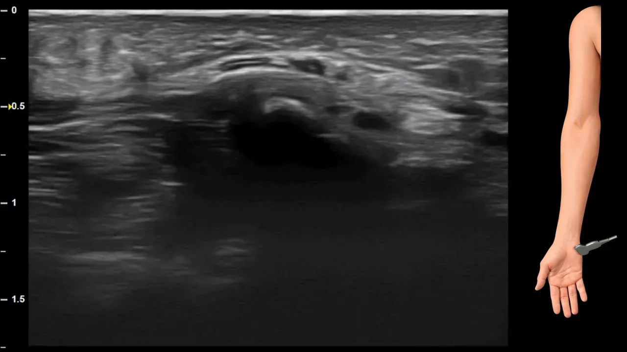
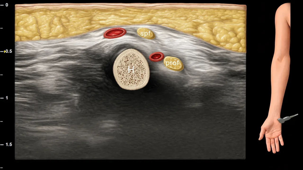
Figure 14. Carpus, transverse plane. H: hamulus ossis hamati, spf: superficial sensory branch of ulnar nerve, prof: deep motor branch of ulnar nerve.
Transverse ultrasound section in the wrist area at the level of hamulus ossis hamati (H). At this level, terminal branching of the ulnar nerve occurs into the superficial sensory branch (spf) and deep motor branch (prof). The superficial branch is located more superficially, while the deep branch runs deeper in close relation to the hook of hamate area. Vascular structures accompanying the individual nerve branches are also visible in the image.
Schalten Sie die vollständige Health Library frei
Voller Zugriff auf Scan-Protokolle, Anatomie und klinische Referenzen. Jederzeit kündbar.
- Alle Protokolle und Anatomiereferenzen
- Originale Ultraschall-Illustrationen und Video-Demonstrationen
- Synchronisierung zwischen Mobilgerät und Web