 MSK Academy
3. Laterale Ansicht
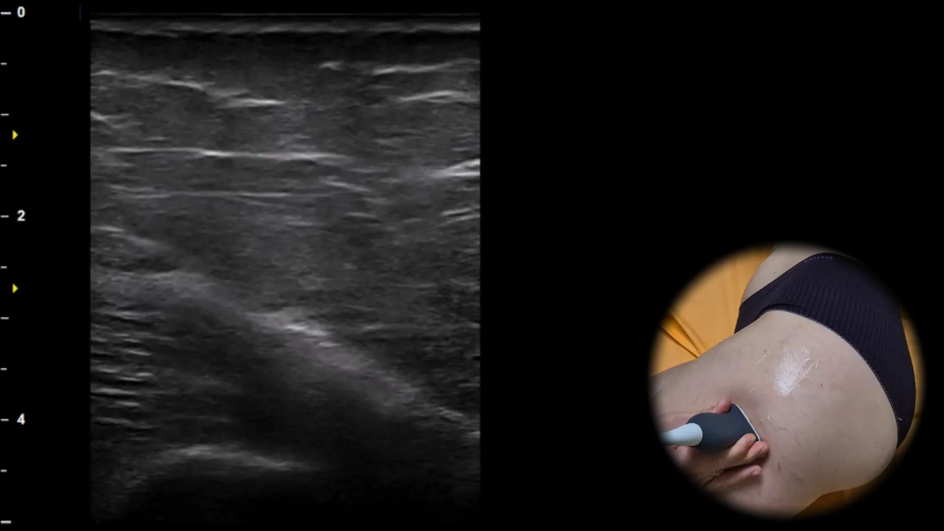
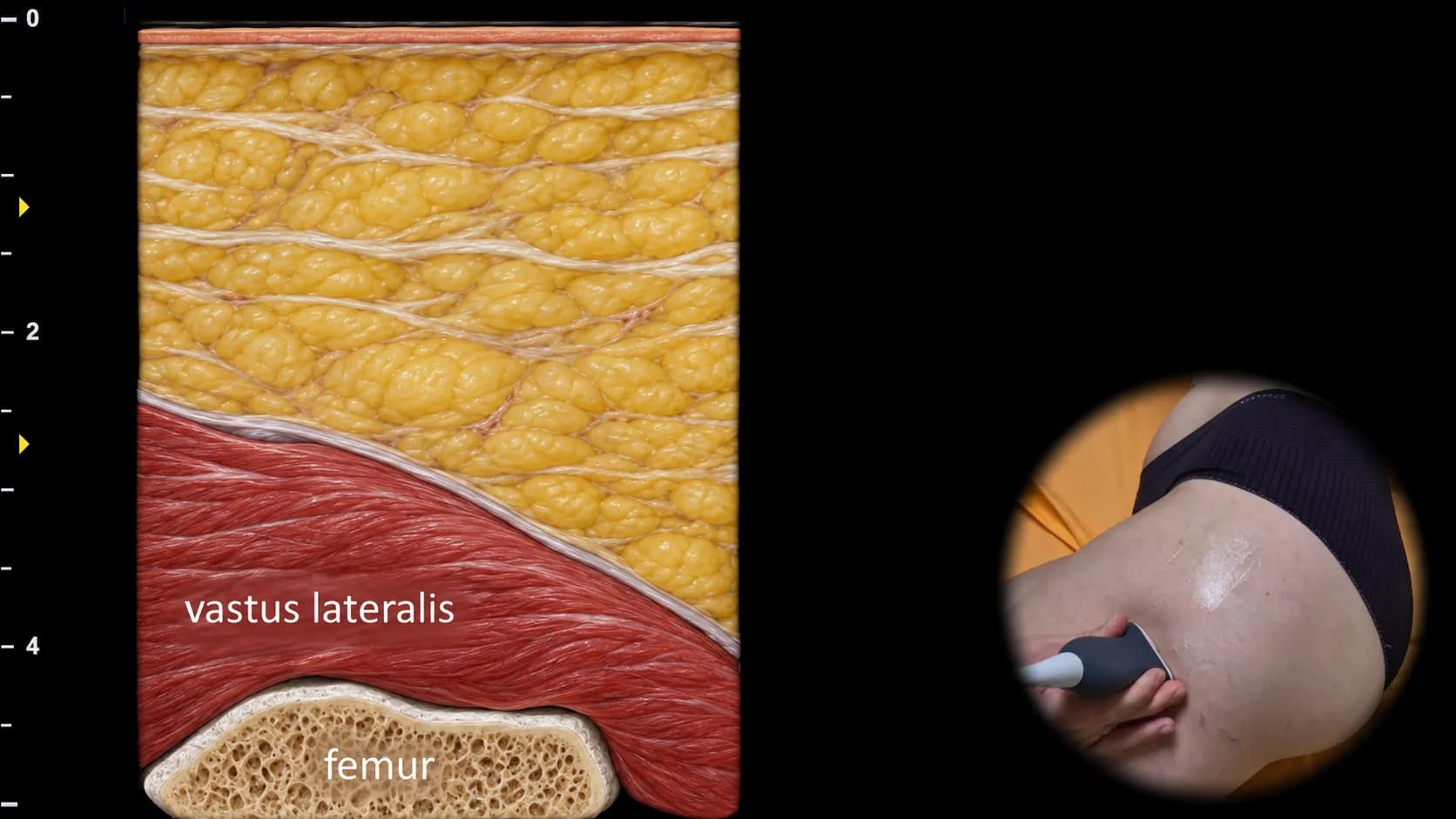
Abbildung 1. Laterale Ansicht, Transversalebene.
Transversaler Ultraschallschnitt des lateralen Anteils des Oberschenkels auf distaler Höhe, wobei der m. vastus lateralis oberflächlich als echoarme Muskelschicht mit feinen inneren fibrösen Bändern sichtbar ist. Tiefer wird die kortikale Kontur des Femurs als durchgehende, gekrümmte echoreiche Linie mit dorsaler Schallschattenbildung dargestellt, die den wichtigsten knöchernen Orientierungspunkt dieser Projektion bildet. Diese Ansicht dient primär der Identifikation des Femurs und als Referenzausgangsebene für seine proximale Nachverfolgung in Richtung des Trochanter major.
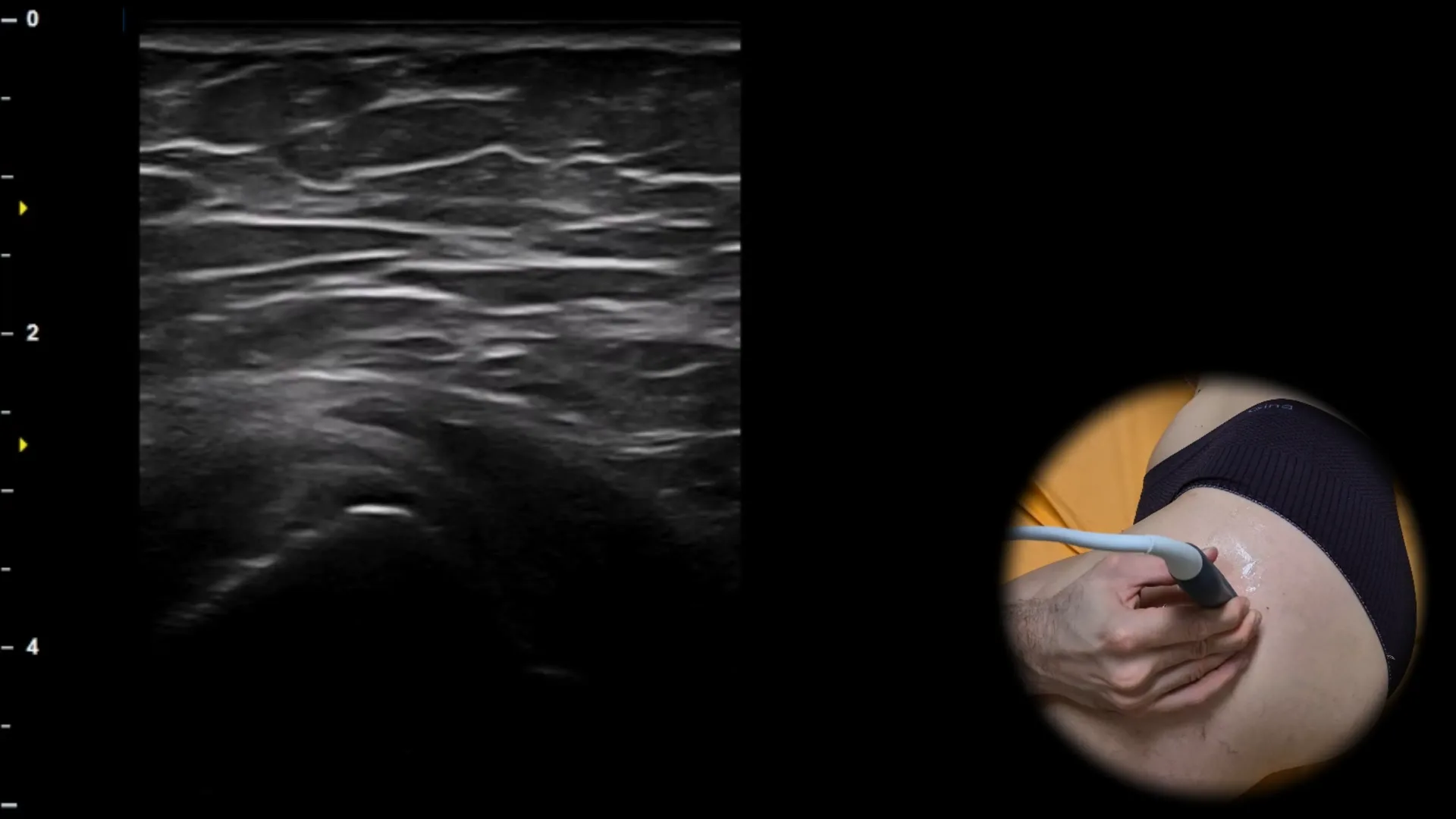
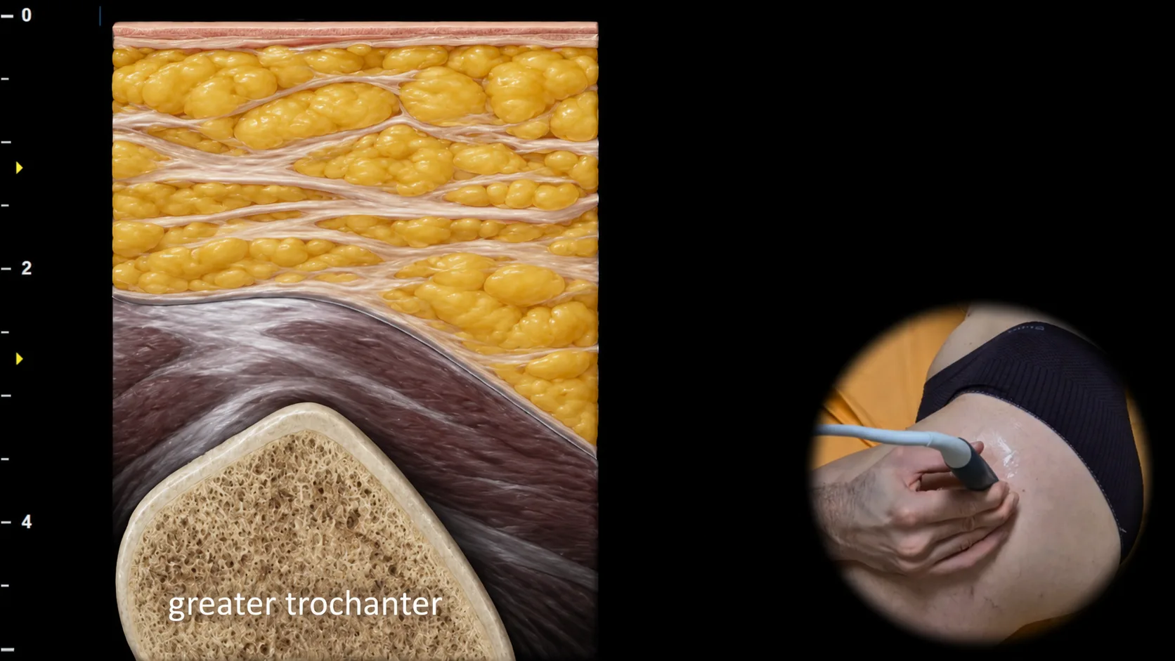
Abbildung 2. Laterale Ansicht, Transversalebene.
Transversaler Ultraschallschnitt durch den lateralen Teil der Hüftregion bei proximaler Verschiebung des Schallkopfs entlang des Femurs. Auf dieser Höhe geht die ursprünglich runde Form des Femurs in eine ausgeprägtere dreieckige bis pyramidenförmige Knochenkontur über, die dem Bereich des Trochanter major entspricht und bei der sonografischen Untersuchung der lateralen Hüfte eine wichtige Landmarke darstellt. Die korrekte Identifizierung des Trochanter major ist für die anschließende Beurteilung der umgebenden Sehnenansätze und Bursae in dieser Region von entscheidender Bedeutung.
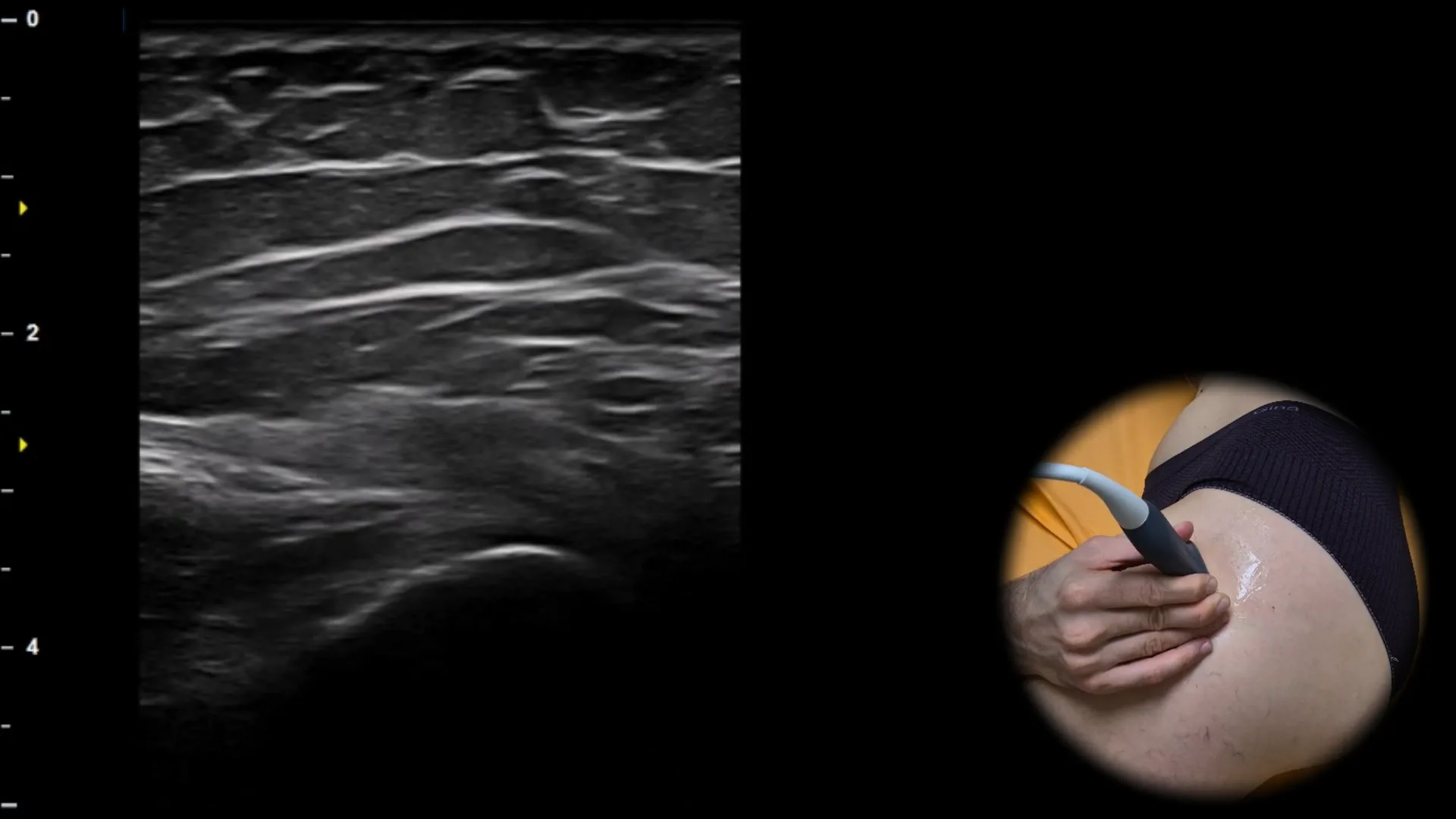
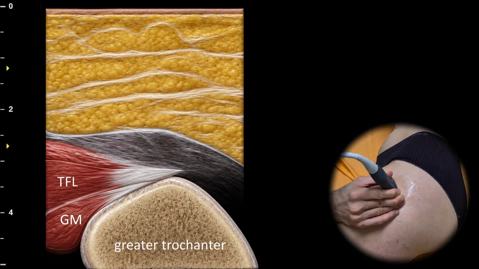
Abbildung 3. Laterale Ansicht, Schrägebene. TFL: m. tensor fasciae latae, GM: Sehne des m. gluteus minimus
Schräger sonografischer Schnitt durch den lateralen Teil der Hüftregion mit Fokus auf die anteriore Facette des Trochanter major. Oberflächlich und lateral ist der m. tensor fasciae latae (TFL) sichtbar, unter dem die Sehne des m. gluteus minimus (GM) verläuft, die an der anterioren Facette des Trochanter major ansetzt und als klar abgegrenztes fibrilläres hyperechogenes Band erscheint. Diese Projektion ist für die Beurteilung des Ansatzes der Sehne des m. gluteus minimus und für den Nachweis einer Tendinopathie oder partieller Rupturen der Glutealsehnen bei Patienten mit lateralen Hüftschmerzen unerlässlich.
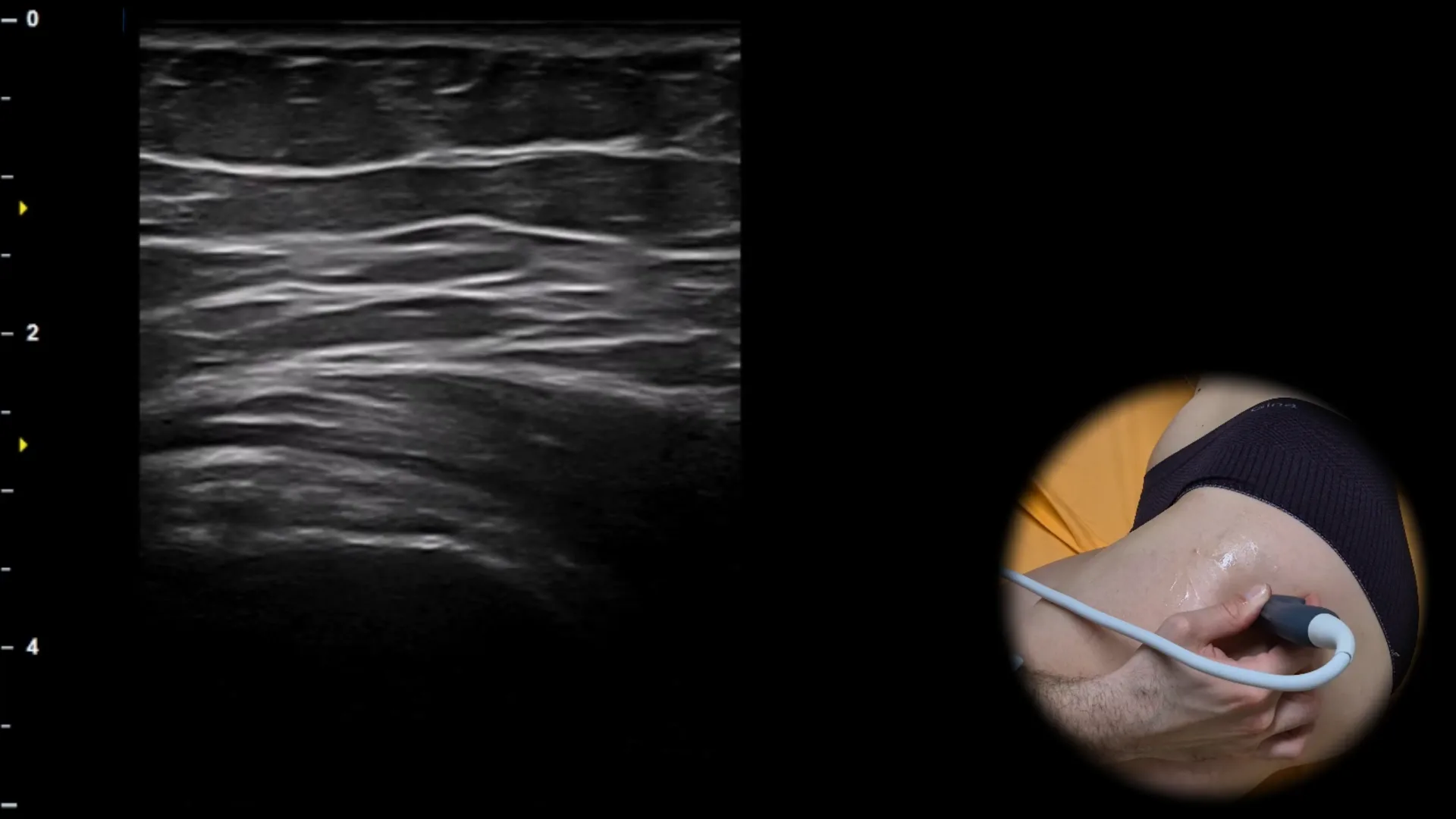
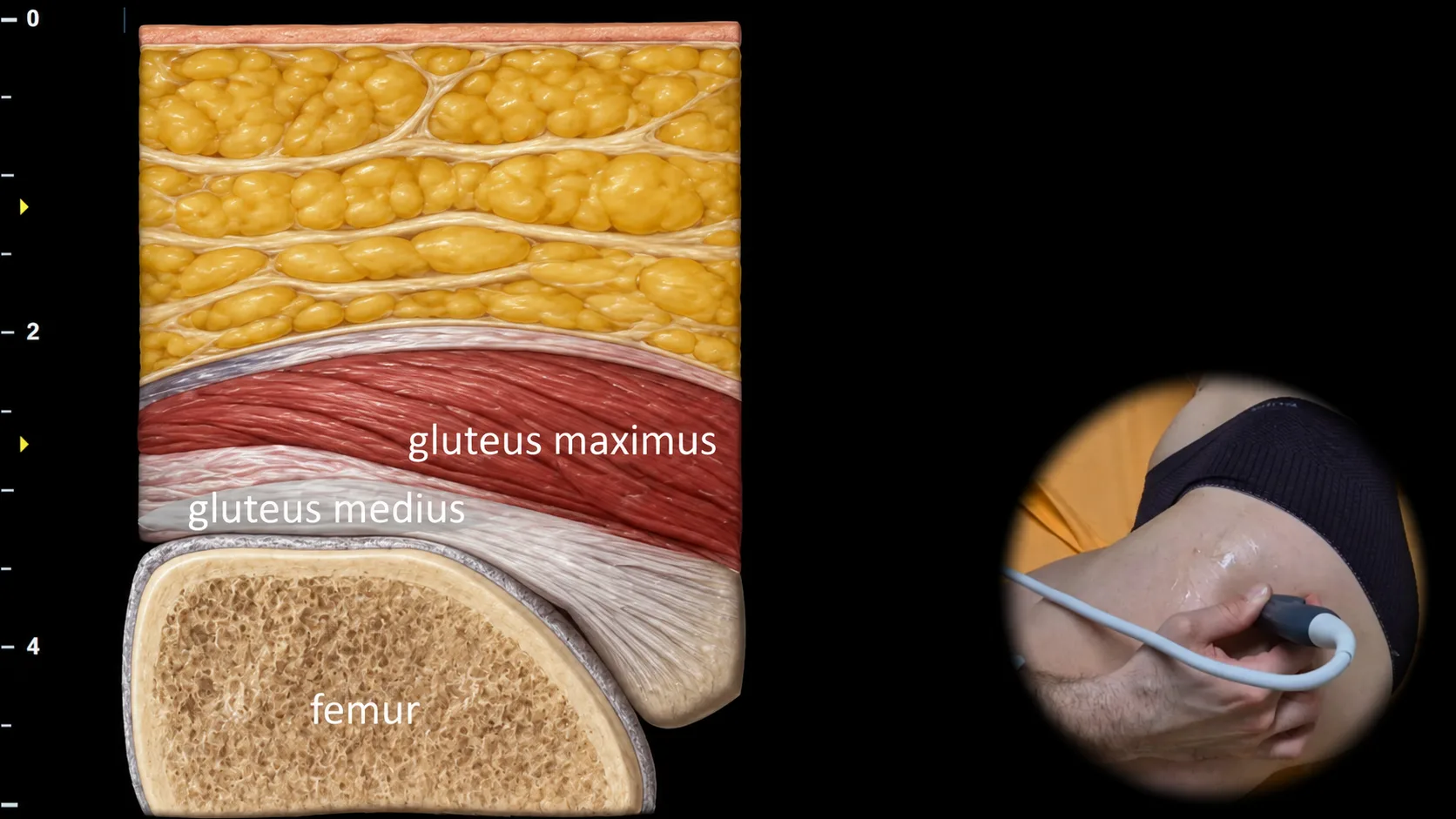
Abbildung 4. Laterale Ansicht, schräge Ebene.
Schräger sonografischer Schnitt durch den lateralen Anteil der Hüftregion mit Darstellung des Bereichs des Trochanter major. Oberflächlich ist der M. gluteus maximus sichtbar, darunter die an die laterale Facette des Trochanter major angrenzende Sehne des M. gluteus medius; in der Tiefe ist die knöcherne Kontur des proximalen Femurs als wichtigster Orientierungspunkt dieser Projektion dargestellt. Diese Projektion ist für die Beurteilung des Ansatzes des M. gluteus medius und für den Nachweis von Tendinopathien, Partialrupturen und Veränderungen der peritrochantären Bursen bei Patienten mit lateralen Hüftschmerzen essenziell.
Zugang zur gesamten Bibliothek
Voller Zugriff auf Scan-Protokolle, Anatomie und klinische Referenzen. Jederzeit kündbar.
- Alle Protokolle und Anatomiereferenzen
- Originale Ultraschall-Illustrationen und Video-Demonstrationen
- Synchronisierung zwischen Mobilgerät und Web