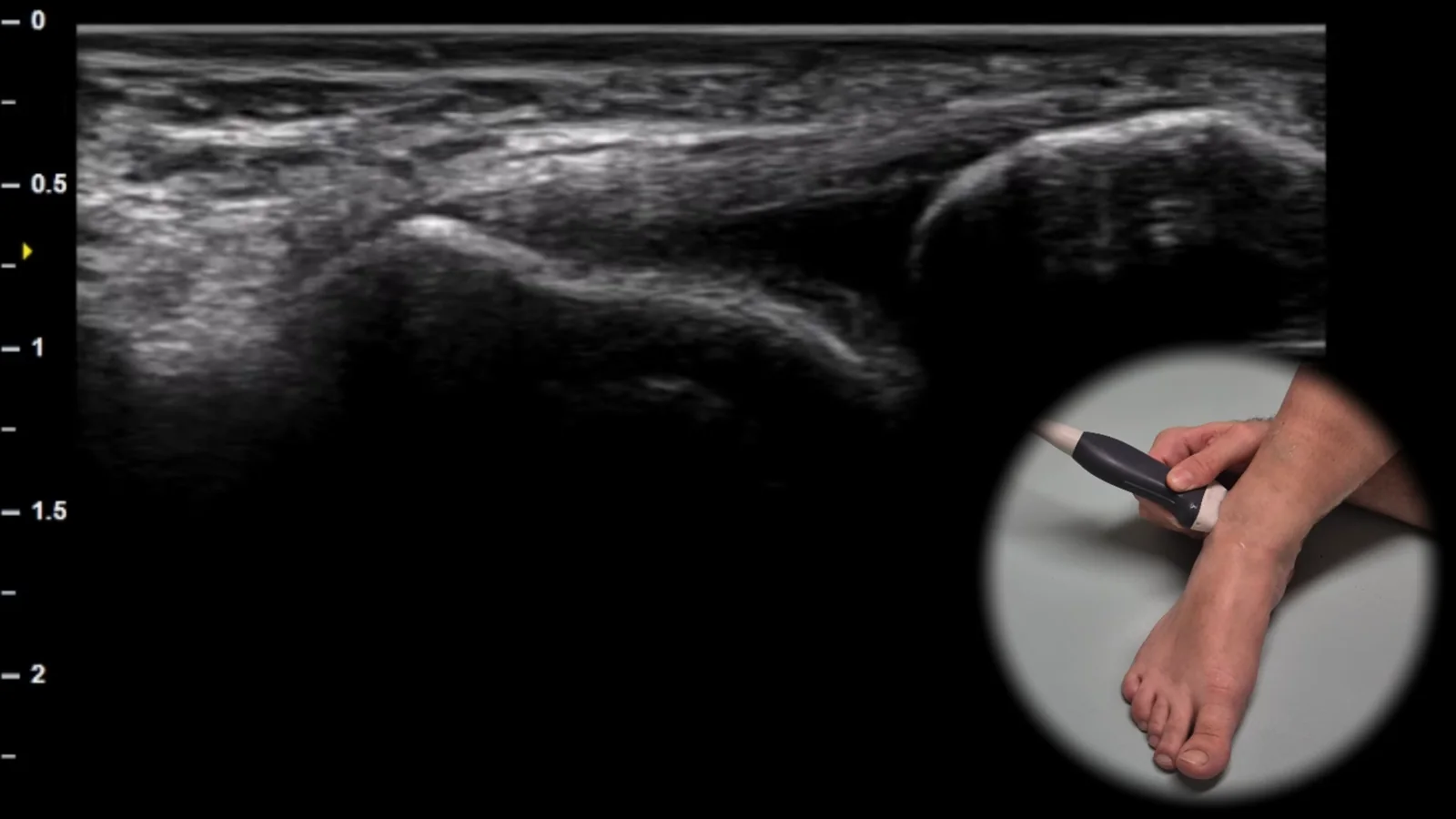
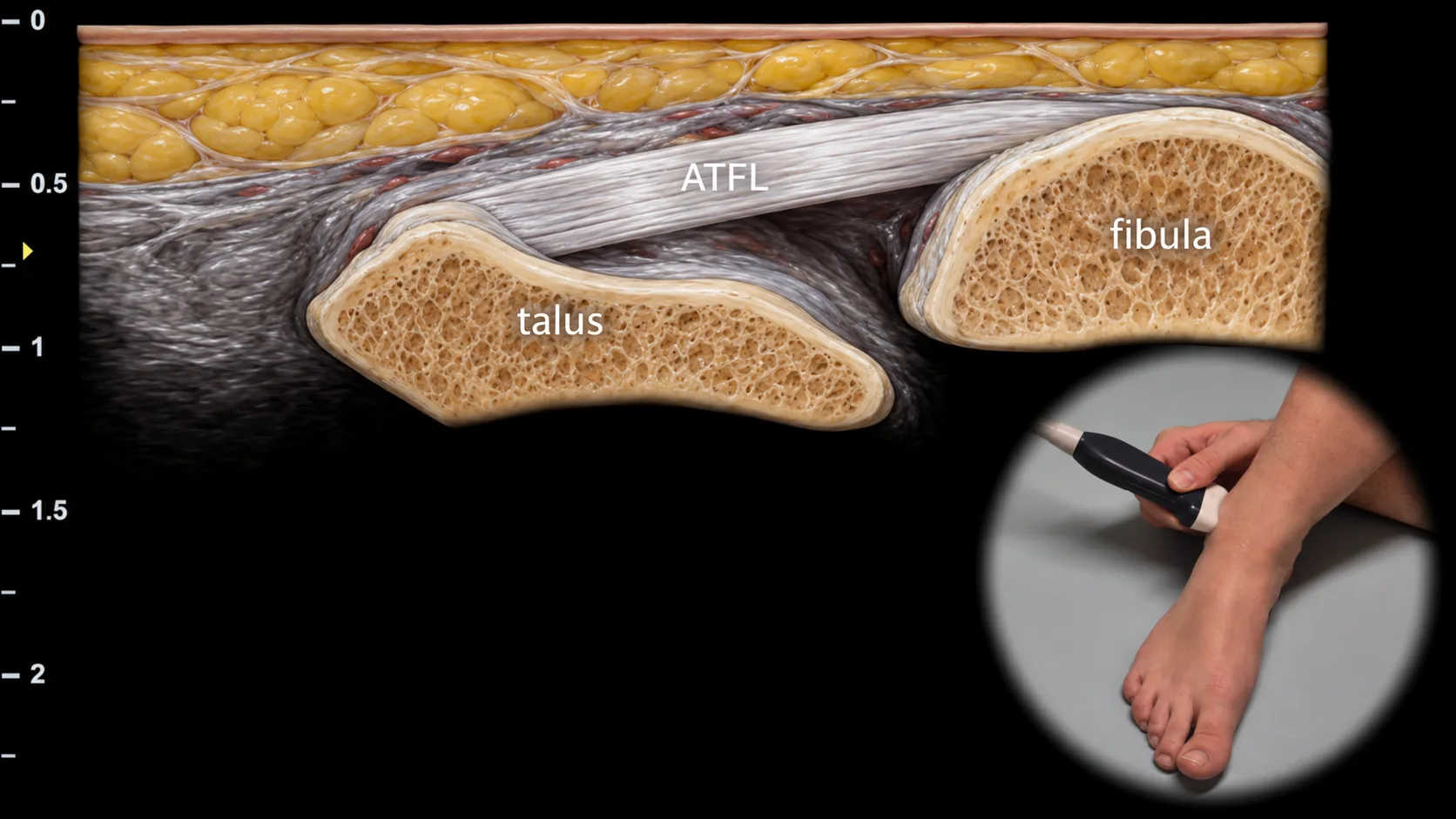
Figure 1. Lateral view, transverse plane. ATFL: ligamentum talofibulare anterius
Transverse ultrasound section of the lateral part of the ankle showing the ligamentum talofibulare anterius (ATFL) as a thin hyperechoic fibrillar structure running between the anterior border of the fibula and the lateral part of the talar neck. The fibula and talus form the main bony landmarks of this projection, with the ligament bridging the anterior part of the lateral ankle complex. This projection is essential for evaluating the continuity, echotexture, and tension of the ATFL when lateral ankle sprain, partial rupture, or complete lesion is suspected.
 MSK Academy