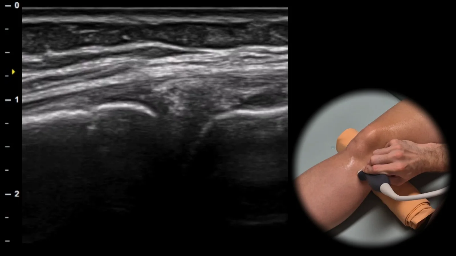
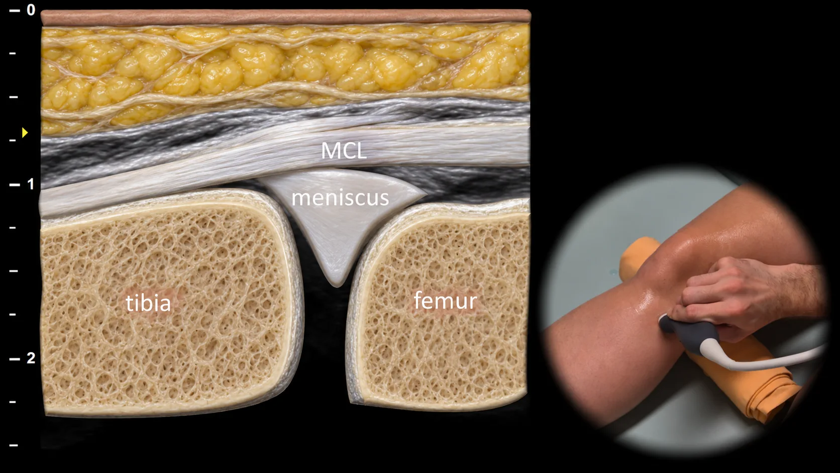
Figure 1. Medial view, frontal plane. MCL: medial collateral ligament
Longitudinal ultrasound section of the medial side of the knee showing the medial collateral ligament (MCL) as a thin, well-defined hyperechoic fibrillar band running between the femur and tibia. At the level of the joint space, the medial meniscus is visible beneath the ligament as a triangular structure positioned between the bony contours of the femur and tibia, which appear as hyperechoic cortical lines with dorsal acoustic shadowing. This projection is essential for assessing MCL integrity, detecting medial meniscus extrusion, and evaluating effusion or capsular abnormalities in the medial recess.
 MSK Academy