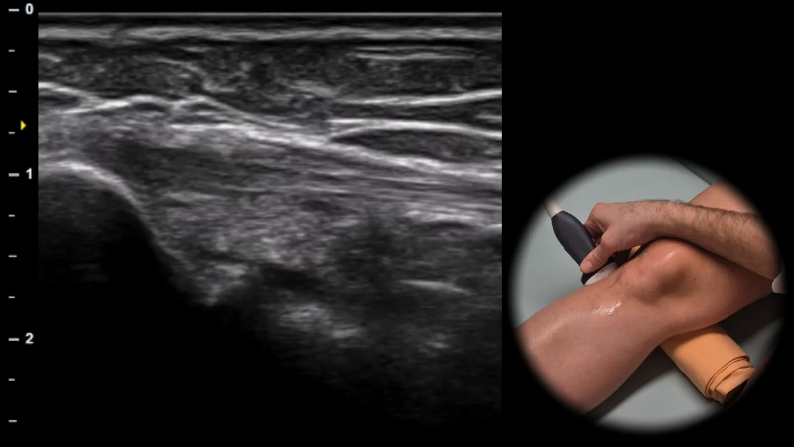
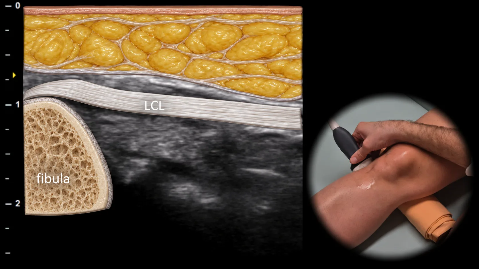
Figure 1. Lateral view, frontal plane. LCL: lateral collateral ligament
Longitudinal ultrasound section of the lateral side of the knee showing the lateral collateral ligament (LCL) as a thin hyperechoic fibrillar structure running from the lateral epicondyle of the femur to its insertion on the fibular head. The fibula is visible in this projection as the main bony landmark with a hyperechoic cortical line and posterior acoustic shadowing. This projection is essential for assessing LCL integrity and for detecting enthesopathy, partial ruptures, or avulsion injuries in the area of the fibular insertion.
 MSK Academy