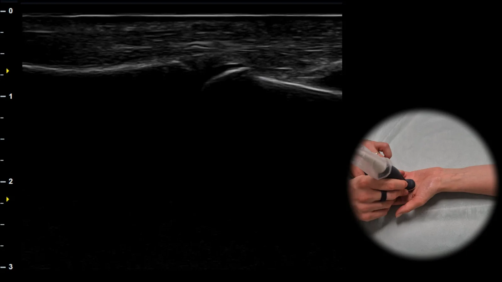
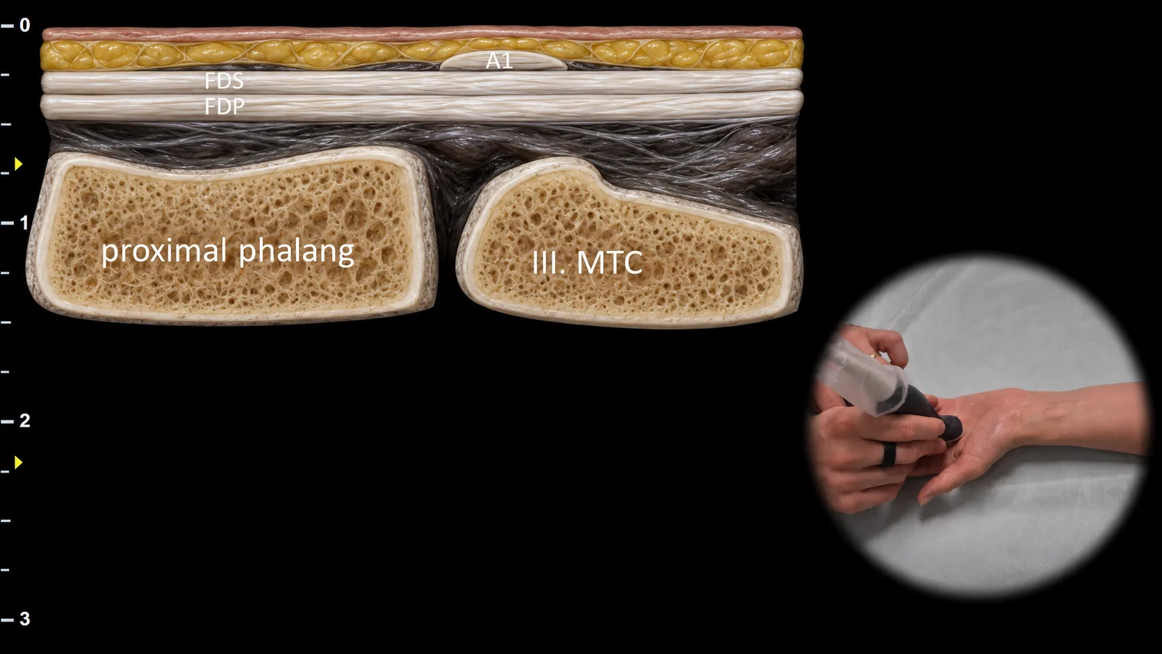
Figure 1. View of the MCP joint, sagittal plane. A1: A1 pulley, FDS: flexor digitorum superficialis tendon, FDP: flexor digitorum profundus tendon, III. MTC: third metacarpal
Longitudinal ultrasound section through the metacarpophalangeal joint region showing the finger flexor apparatus at the level of the A1 pulley. The A1 pulley is visible superficially above the flexor tendons, beneath which run the flexor digitorum superficialis (FDS) and flexor digitorum profundus (FDP) tendons, while the head of the third metacarpal (III. MTC) serves as the bony landmark. This projection is important for evaluating the relationship of the flexor tendons to the A1 pulley and during dynamic examination allows assessment of their gliding, possible pulley thickening, and signs of stenosing tenosynovitis.
 MSK Academy