Patellar tendon - partial rupture
Written by Jakub Jacisko, MD, PhDUpdated
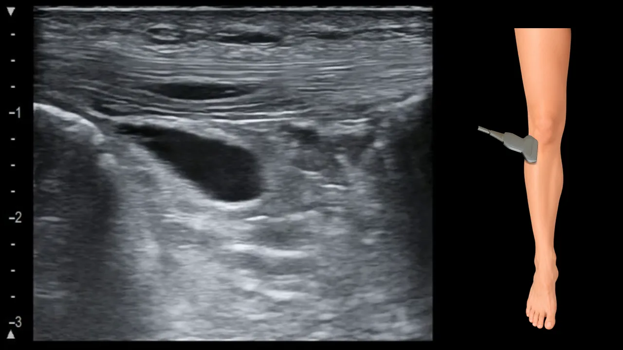

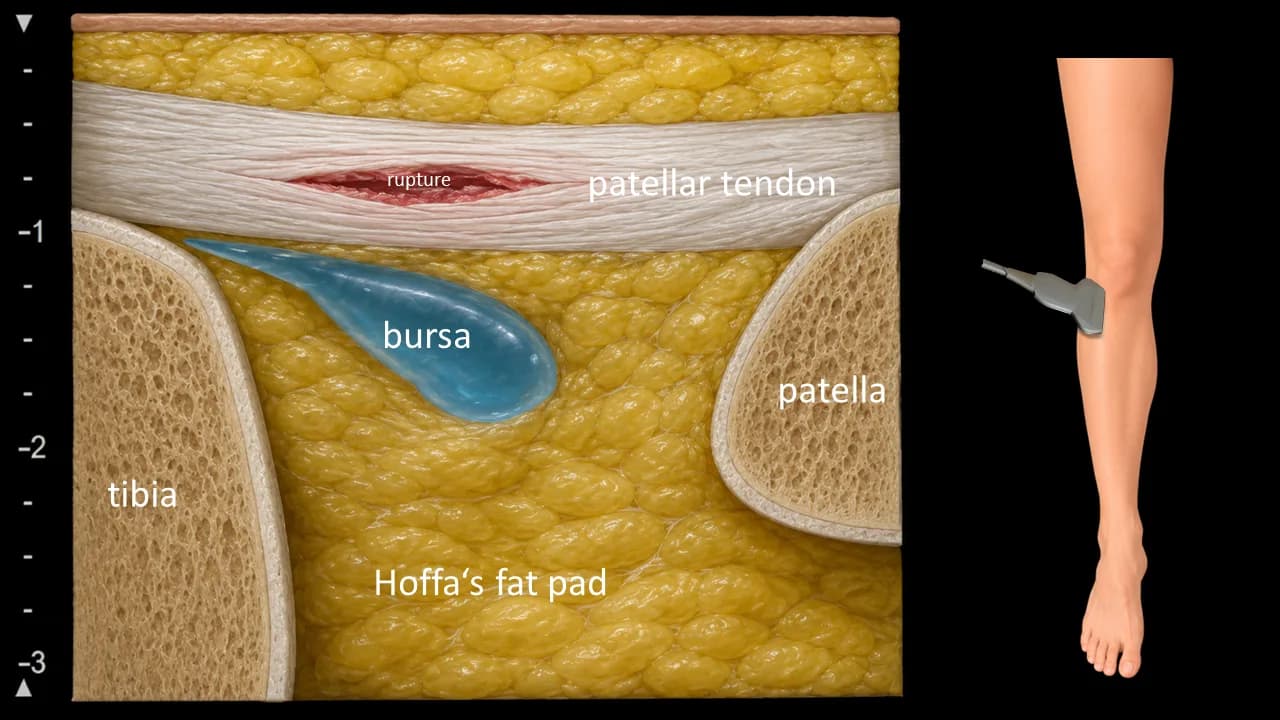
Figure 1. Ventral view, sagittal plane, infrapatellar.
Partial tear of the patellar ligament, presenting as a focal hypoechoic defect with thickening of the surrounding tendon fibers. Increased fluid can also be observed beneath the ligament within the deep infrapatellar bursa.
Clinical Note
A partial tear may be highly focal; therefore, the patellar ligament should be carefully examined throughout its entire extent in both the mediolateral and craniocaudal directions.


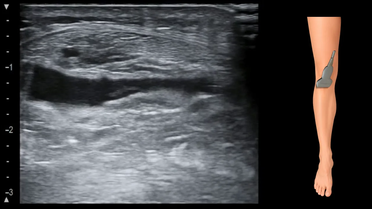
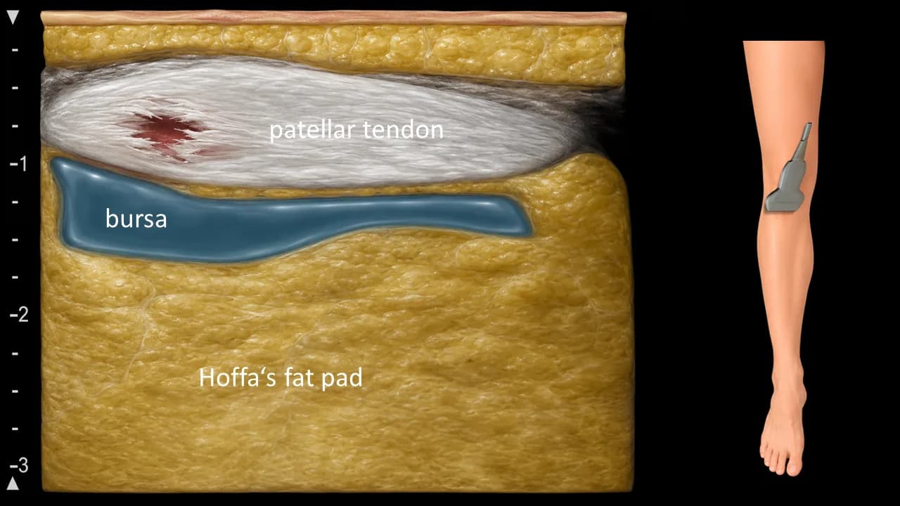
Figure 2. Ventral view, transverse plane, infrapatellar.
Partial tear of the patellar ligament, presenting as a focal hypoechoic defect with thickening of the surrounding tendon fibers. Increased fluid can also be observed beneath the ligament within the deep infrapatellar bursa.