September 2015
EURO-MUSCULUS/USPRM. Basic scanning protocols for hip
Authors: L Özçakar, M Kara, K V Chang, N Akkaya, C Y Hung, F Tok, C H Wu, A B Çarli, M Y Hsiao, L Tekin, T G Wang, A M Ulaşlı, W S Chen, M De Muynck
Journal: European Journal of Physical and Rehabilitation Medicine
Published: September 2015
This article presents a practical ultrasound protocol for examining the hip. Its main strength is the clear step-by-step approach to patient and probe positioning, making it useful for learning a standardized hip scan.
Interesting highlights:
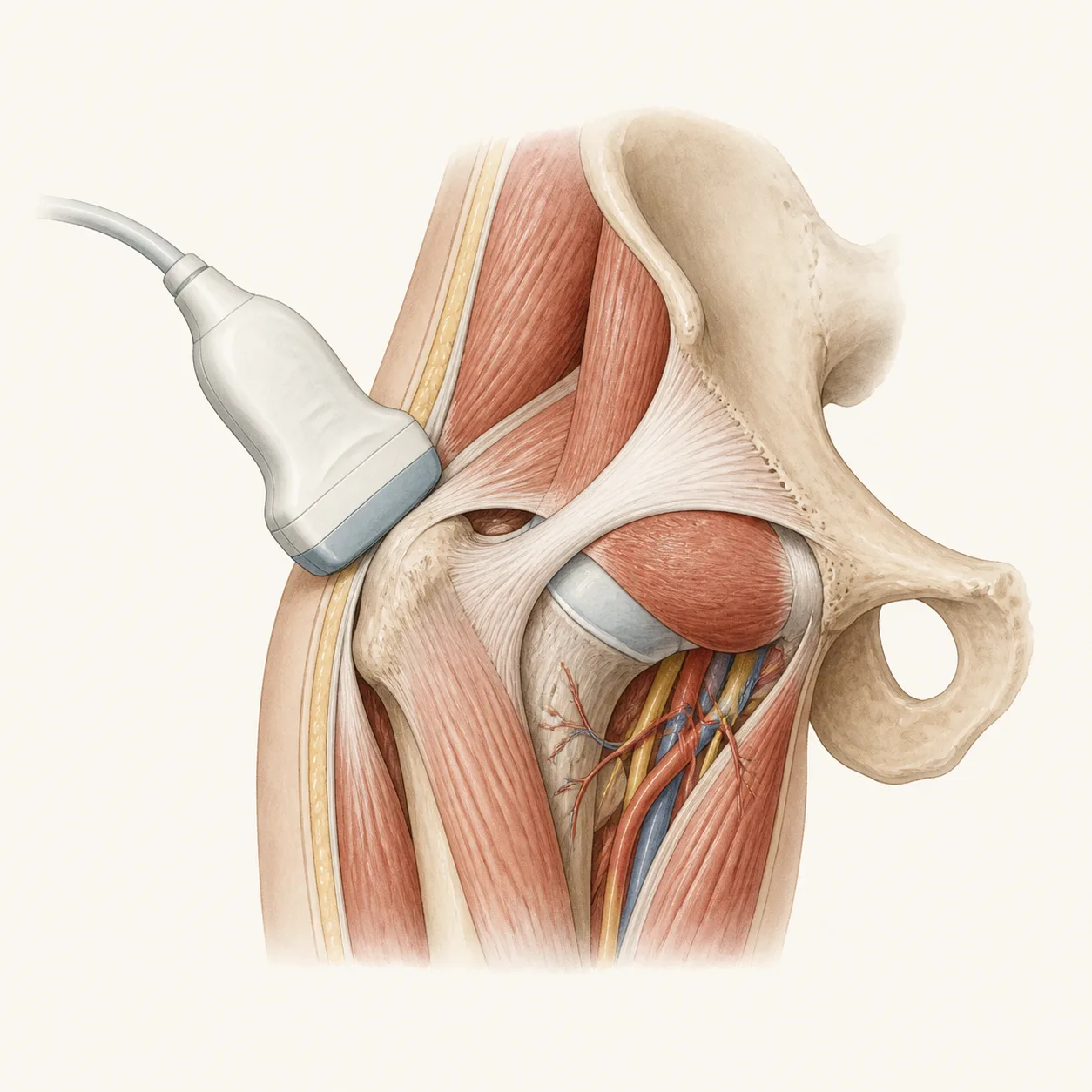
The protocol shows how to assess the anterior hip joint, including the acetabulum, labrum, femoral head and neck, iliopsoas tendon, and possible joint fluid.
It covers important medial and lateral structures, including the adductor muscles, tensor fascia lata, gluteus medius and minimus tendons, greater trochanter, and bursae.
It also includes posterior hip scanning, especially the sciatic nerve, gluteal muscles, hamstrings, and ischial tuberosity.
Most useful for:
MSK ultrasound beginners, rehabilitation physicians, sports medicine clinicians, radiologists, orthopedic physicians, and anyone learning a structured approach to hip ultrasound.