July 2015
EURO-MUSCULUS/USPRM Basic Scanning Protocols for shoulder
Authors: L Özçakar, M Kara, K V Chang, L Tekin, C Y Hung, A M Ulaülı, C H Wu, F Tok, M Y Hsiao, N Akkaya, T G Wang, A B Çarli, W S Chen, M De Muynck
Journal: European Journal of Physical and Rehabilitation Medicine
Published: July 2015
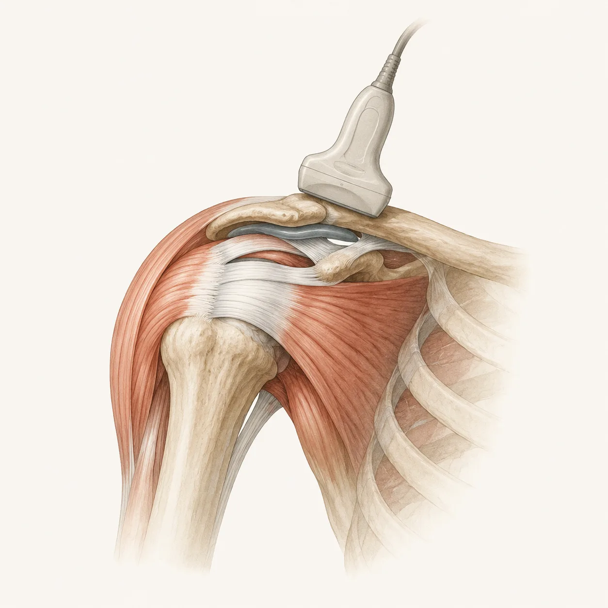
This article presents a practical ultrasound protocol for examining the shoulder. Its main strength is the clear, step-by-step guidance on patient and probe positioning, making it useful for learning a standardized shoulder scan.
Interesting highlights:
Dynamic shoulder positioning helps improve tendon visualization, especially the subscapularis and infraspinatus.
The protocol shows how to assess the rotator cuff, long head of the biceps, subdeltoid bursa, glenohumeral joint, and acromioclavicular joint.
It also includes important positions such as the Crass and modified Crass views for better rotator cuff evaluation.
Most useful for:
MSK ultrasound beginners, rehabilitation physicians, sports medicine clinicians, radiologists, and anyone learning a structured approach to shoulder ultrasound.