MUSCLES
Sonoanatomy
In the context of US imaging, myofibers appear hypoechoic compared to adjacent connective and nervous structures, while fibroadipose tissues (perimysium and epimysium) are hyperechogenic.
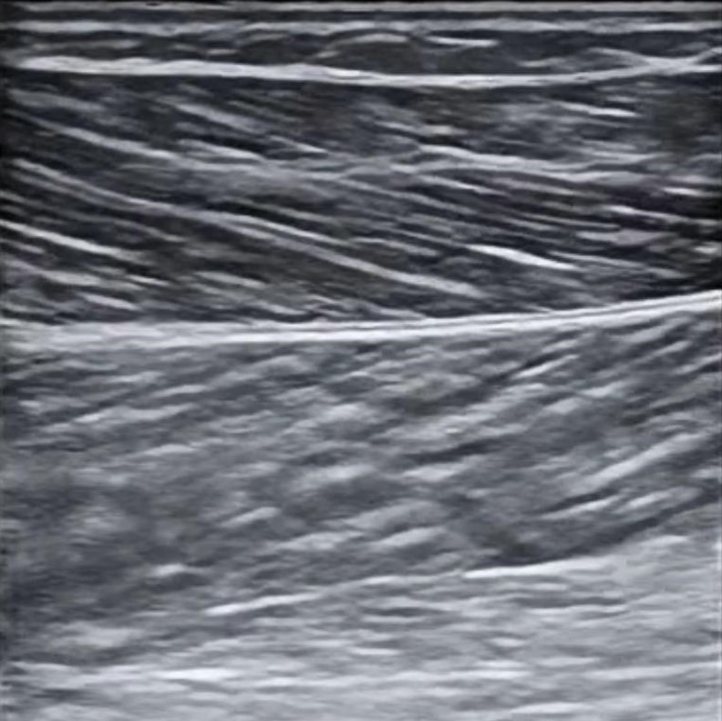
In the transverse axis, the tissue appears to be formed by a background hypoechoic compound with hyperechoic dots inside (“starry sky”). Longitudinal axis images of muscle tissue show an alternation of parallel hypo/hyperechoic bands (“feather” or “veins on a leaf”) with a variable orientation depending on pennation angle.
Different orientation of muscle fibers with respect to the US beam might cause artifacts of anisotropy, especially in long axis evaluation.
Transverse – Starry sky
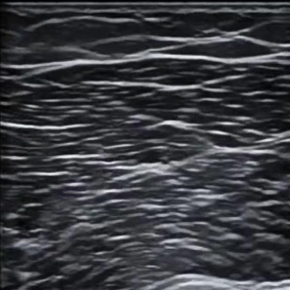
Longitudinal – Feather
Pathologies
Strain
Definition: Strain is a muscle injuries that involve stretching or elongation. However, its definition is not well-established and there is inconsistency in how it is used to describe different types of muscle injuries.
US appearence: x
Tear
Definition: Injury to muscle fibres or internal aponeuroses.
US appearence: Variable depending on the severity of the injury. This can range from an increase echogenicity in a muscle that is still intact, to partial or complete disrupting of the muscle or its connection to tendons, which may be accompanied by bleeding of varying echogenity. Doppler imaging may show an increase in blood flow in the affected area.
Contusion
Definition: Muscle injury with or without haematoma most commonly after blunt trauma.
US appearence: Mixed echogenicity of area of disrupted muscle fibers. This area can appear very bright (hyperechoic) when the injury is recent, but over time may become completely dark (anechoic) as it heals. Additionally, there may be a mass effect caused by a hematoma forming in the area of the injury, and Doppler imaging may show increased blood flow in the affected region.
Myositis
In construction
Myositis ossificans
In construction
Pyomyositis
In construction
Fatty infiltration
In construction
Mnemonics and metaphors
Videos
Articles
