
Want to confidently perform musculoskeletal ultrasound exams? This course is perfect for beginners, guiding you step-by-step through essential skills needed to examine joints, muscles, tendons, and more.
After completing the course you will be able to:
💡How to understand and interpret ultrasound images
🎯Proper use of probes and positioning techniques
🎛️Knobology: buttons and machine settings explained
💪Key anatomy: muscles, tendons, nerves, vessels, and bones
🗝️10 essential structures in the upper and lower extremities
📝Practical tips and memory aids for easy learning
Course Features:
📽️High-Quality Video Tutorials
📚Downloadable PDF Guide
⏱️Learn at Your Own Pace
Who Is This Course For?
👨⚕️Physical medicine & rehabilitation specialists
🤸Physiotherapists
🦴Orthopedic doctors
🧠Neurologists
☢️Radiologists
🩺GPs
👨🎓Medical students
💉Other healthcare professionals
Knobology
Knobology is a term used in ultrasonography to refer to the knowledge and understanding of the various knobs, buttons, and controls on an ultrasound machine. It is an important aspect of ultrasonography because the operator must be familiar with the various controls on the machine in order to properly adjust the settings and obtain high-quality images.
Knobology involves understanding the purpose and function of each control on the machine, as well as how to properly use them to adjust the gain, depth, focus, and other parameters of the ultrasound image. It also involves understanding how to troubleshoot any issues that may arise with the machine and how to properly maintain and care for it.
Knobology is an essential part of training for anyone who wants to become an ultrasonographer, and it is something that is continuously developed and refined throughout a career in the field.

The probe button is used to select and change the type of the ultrasound probe being used (linear, konvex or hockey stick probe).

Freeze button on ultrasound machine pauses image display for closer examination or still image capture. Press to freeze and press again to unfreeze.

The gain button amplifies ultrasound signals for clearer images. It can brighten or reduce noise depending on the operator’s adjustments.

The store button is used to save the ultrasound images or clips onto the internal memory or external storage media, allowing for later review or documentation.
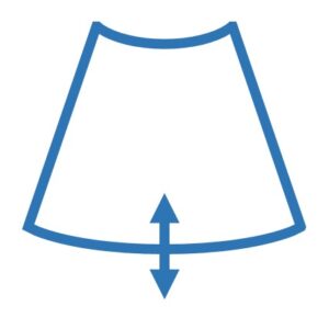
The depth control is used to adjust the depth of the image being displayed, which is useful for examining specific structures at different depths.

The focus control is used to adjust the depth at which the ultrasound beam is most sharply focused, which affects the quality and clarity of the resulting image.

The frequency button adjusts ultrasound waves’ frequency emitted by the transducer to determine depth of penetration and image detail.

The zoom button is used to magnify or enlarge the image on the display screen to better visualize and examine specific structures of interest.

The measure button is used to take measurements (distnce, circumference, cross section area) of structuresbvisible on the ultrasound image.

The dual-screen button allows for two separate images to be viewed side-by-side on the same screen, which can be useful for comparing structures.

The print button is used to print a hard copy of the ultrasound image or report, which can be used for documentation or keeping the record.

Color Doppler is used to visualize and assess the direction and speed of blood flow in real-time, providing diagnostic information about vascular structures.

Power Doppler is used to detect the presence of blood flow in real-time, particularly in low-flow situations where color Doppler may be unable to detect flow.

The shear wave elastography button is used in to measure the stiffness or elasticity of tissue to diagnose muscle or tendon injuries.

The freeze button is a control on an ultrasound machine that allows the operator to pause the real-time display of the image on the screen. It is typically used to take a closer look at a specific area of the image or to record or print a still image. To use the freeze button, the operator simply presses it, and the image will be frozen on the screen. To unfreeze the image and resume real-time display, the operator can press the freeze button again.

The gain button or gain control allows the operator to amplify the amount of signal that is being received by the ultrasound transducer. Adjusting the gain can be useful when trying to improve the clarity and quality of the ultrasound image. For example, if the image is too dark or noisy, the operator can increase the gain to make the image brighter and clearer. On the other hand, if the image is too bright or has too much noise, the operator can decrease the gain to reduce the signal strength and improve the image quality.
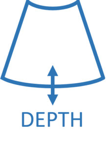
The depth control in ultrasonography is used to adjust the distance that the ultrasound waves travel through the body before being received by the transducer. It is a knob or button on the control panel of the machine that allows the operator to adjust the depth of the image being displayed. Adjusting the depth can be useful for examining specific areas of the body or visualizing structures at different depths within the body.

The zoom button in ultrasonography is a control on the machine that is used to magnify or enlarge the image on the display screen. It is typically located on the control panel of the machine and is marked with an icon or label indicating its function. To use the zoom button, the operator presses it to magnify the image, and can adjust the level of magnification using the zoom control or by pressing the zoom button again to toggle between different magnification levels.


