Want to confidently perform musculoskeletal ultrasound exams? This course is perfect for beginners, guiding you step-by-step through essential skills needed to examine joints, muscles, tendons, and more.
After completing the course you will be able to: 💡How to understand and interpret ultrasound images 🎯Proper use of probes and positioning techniques 🎛️Knobology: buttons and machine settings explained 💪Key anatomy: muscles, tendons, nerves, vessels, and bones 🗝️10 essential structures in the upper and lower extremities 📝Practical tips and memory aids for easy learning
Course Features: 📽️High-Quality Video Tutorials 📚Downloadable PDF Guide ⏱️Learn at Your Own Pace
Who Is This Course For? 👨⚕️Physical medicine & rehabilitation specialists 🤸Physiotherapists 🦴Orthopedic doctors 🧠Neurologists ☢️Radiologists 🩺GPs 👨🎓Medical students 💉Other healthcare professionals
PROBES
Introduction
Using ultrasound probe is like driving a car.
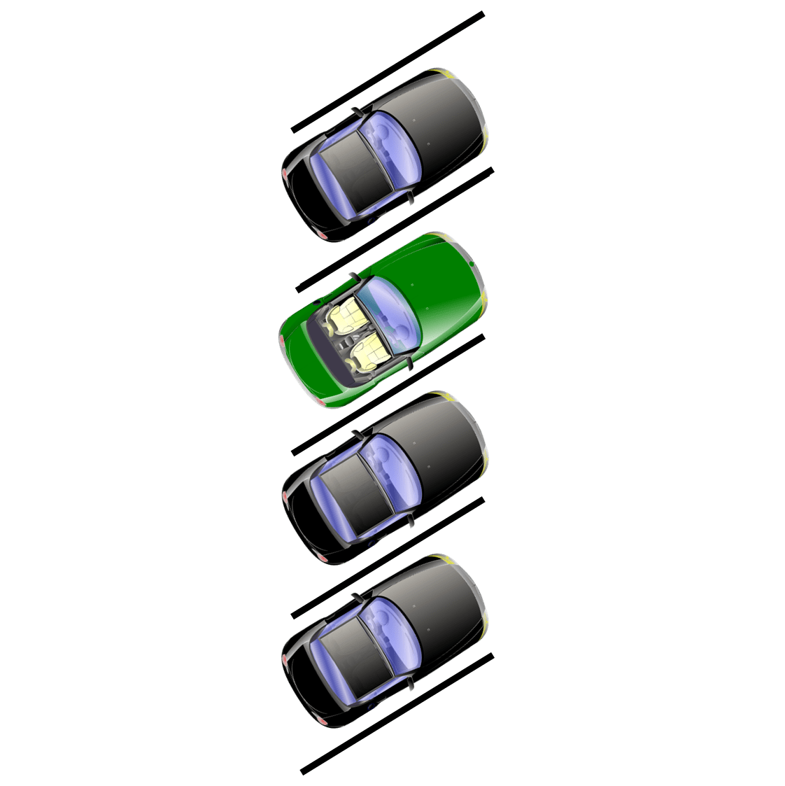
If you want to be able to use a car, you have to know how to choose the right vehicle, how to drive (perform movements) the car and last but not least how to park the car.
The same rules apply for musculoskeletal ultrasound probe: you need to know how to choose, move and position the probe.
In the following lines you can find a manual designed as an easy and entertaining way to learn about probes and how to use them in musculoskeletal ultrasonography.
Content
1. “The right vehicle” – Probe types A. Convex B. Linear C. Hockey-stick 2. “Driving” – Probe movements A. Slide B. Rock (heel-toe) C. Sweep D. Fan E. Compression F. Decopmression G. Helicopter H. Wiper 3. “Parking” – Probe positioning on the patient A. Transversal B. Longitudinal C. Oblique
1. “The right vehicle” – Probe types
Introduction
There are multiple types of vehicles. Small, big, fast, slow, new, old, etc. Vehicle preference depends on the purpose of use. Formula 1 can not drive through the forest as well as tractor can not win a car racing competition.
Analogically there are multiple types of ultrasound probes/transducers. They vary in their size, shape, crystals configuration, image and utility. Crucial for successful ultrasound examination is to choose appropriate probe for planned examination.
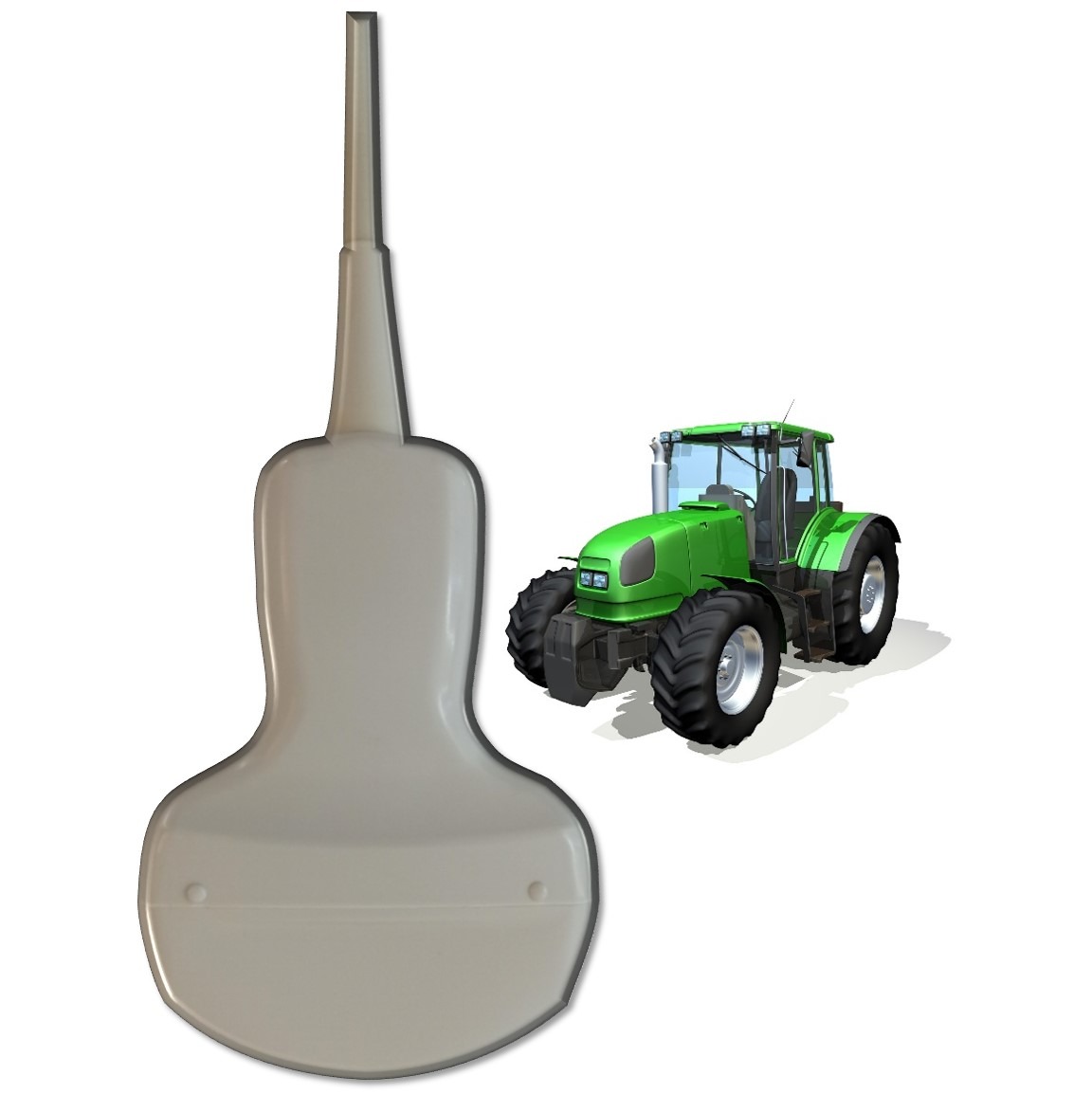
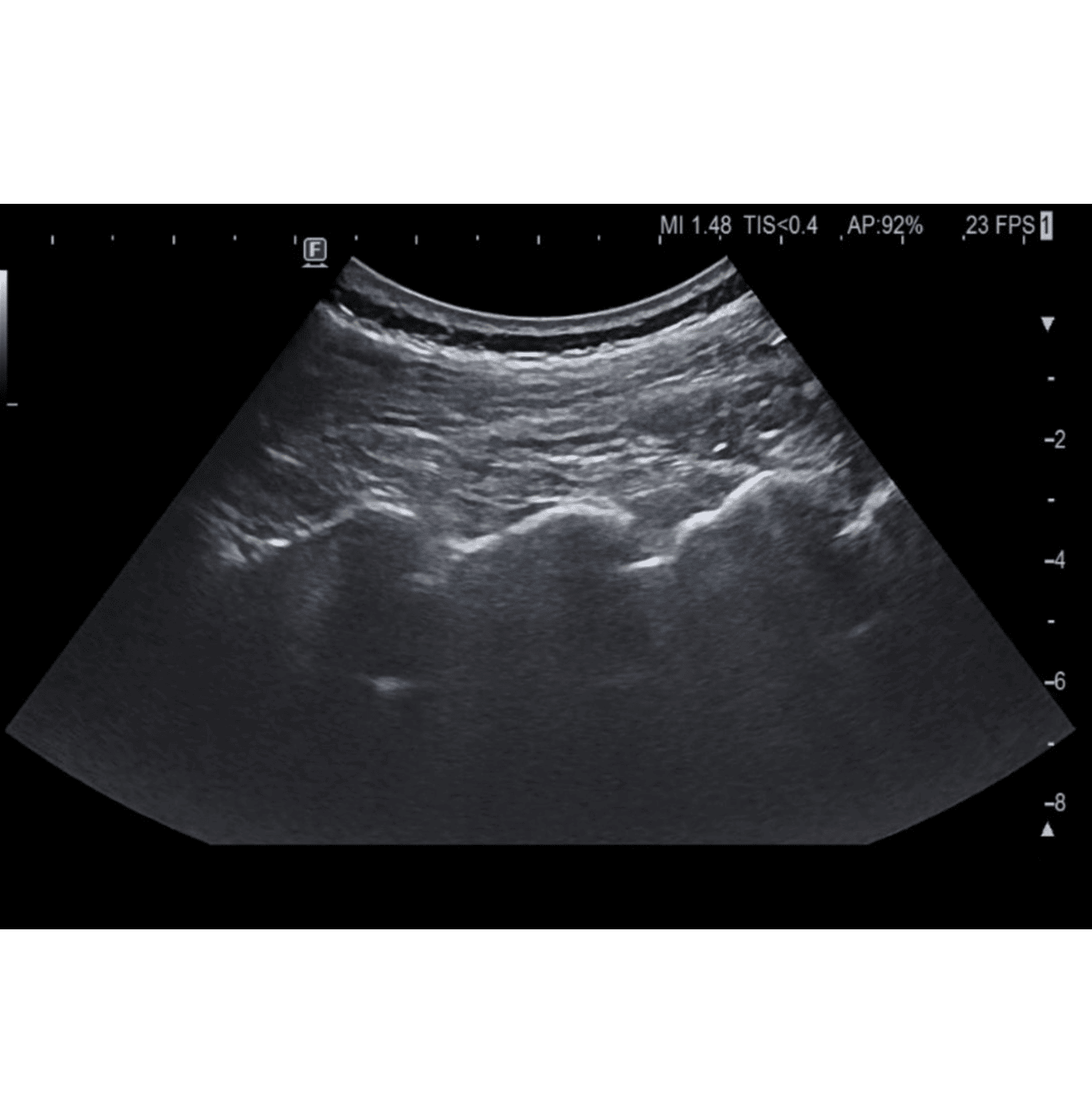
A. Convex
Tractor is a vehicle that moves slowly but can operate in difficult, rough terrain with deep holes and hills.
Convex probe produces ultrasound with low frequency and therefore can be used mainly for depicting deep structures such as hip or spine muscles and joints.
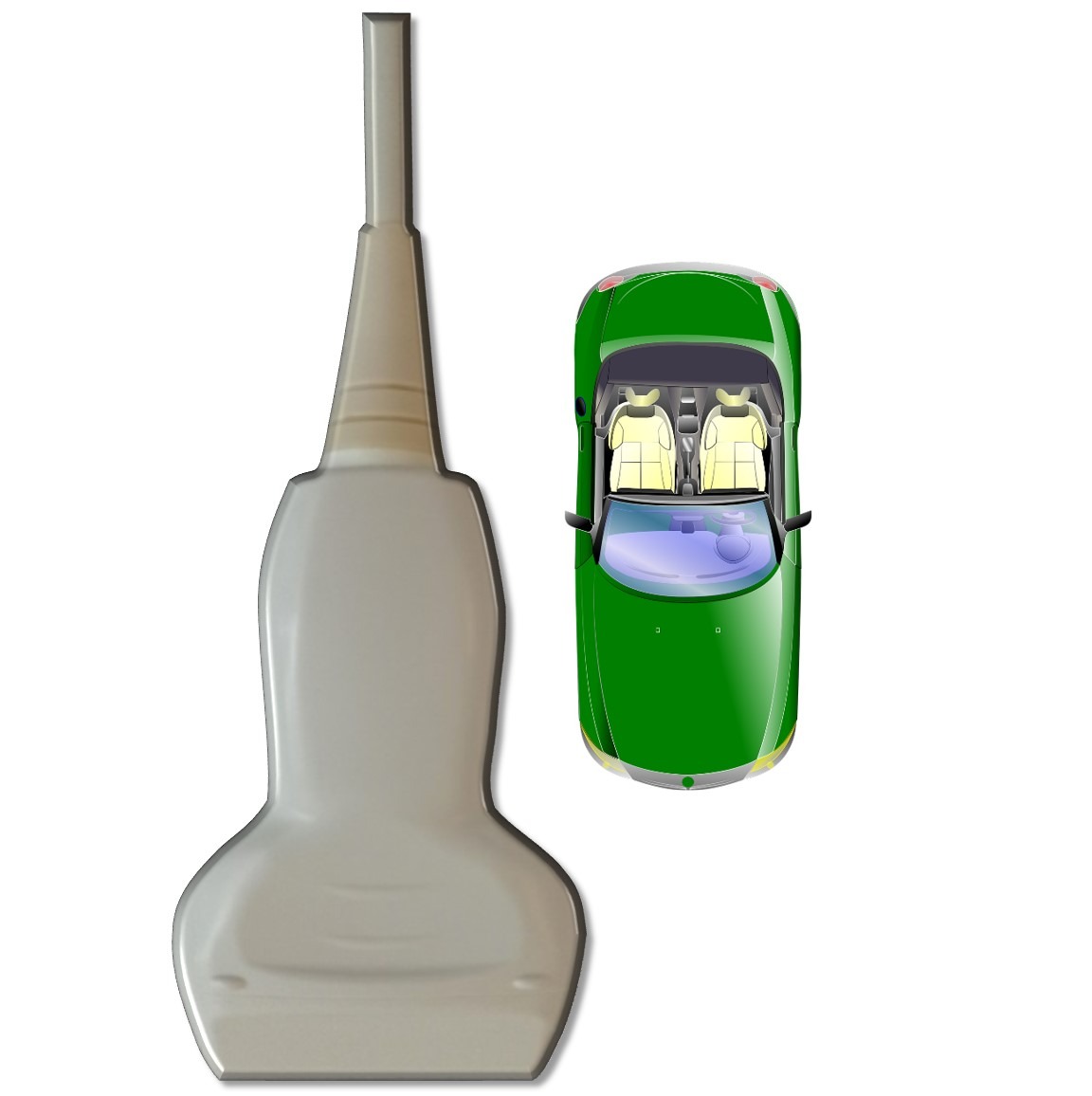
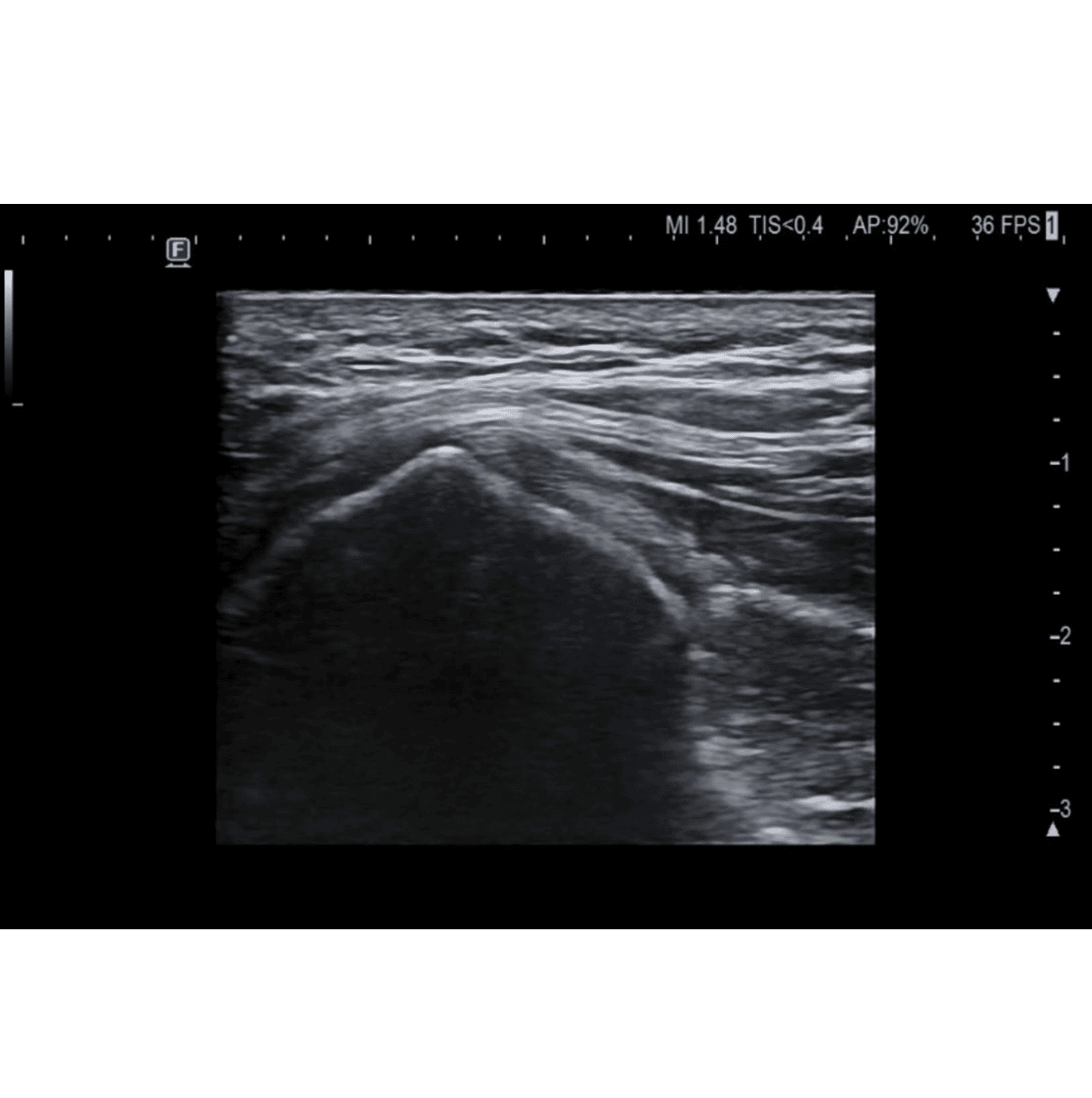
B. Linear
A “gold standard” vehicle is a car. It is the most common one and brings reasonable balance between speed and usage.
Linear probe is the “gold standard” in musculoskeletal ultrasound probes. It works on moderate frequencies and can be used for depicting most of the structures.
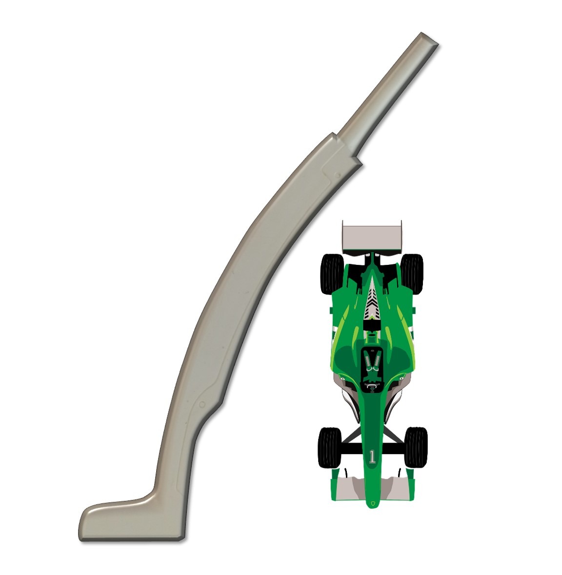
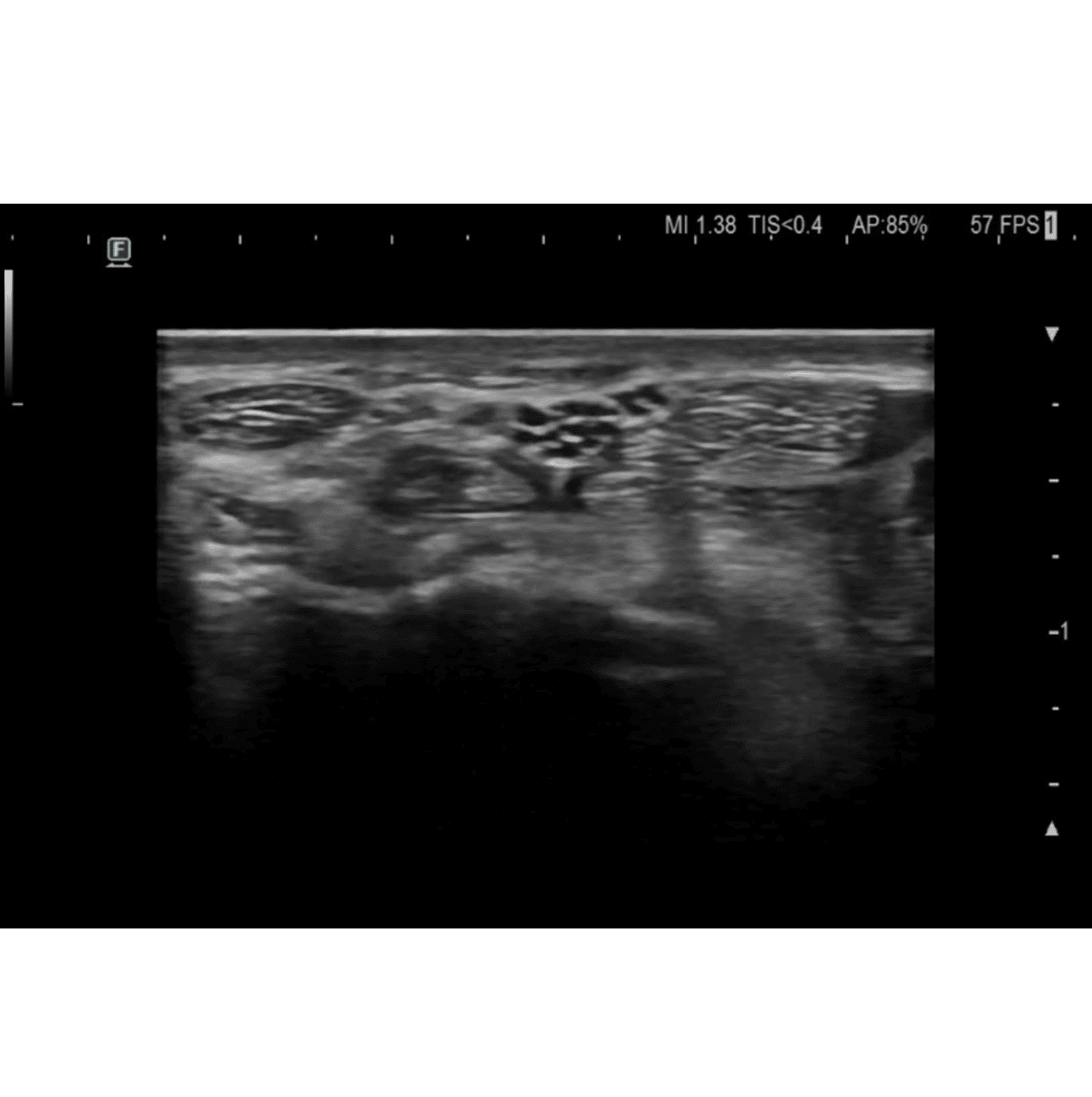
C. Hockey stick
The fastest vehicle is the formula 1. It can reach incredible speed but can be driven only on high quality surface.
A hockey stick probe works on the highest frequencies. Therefore the quality of the image is really great but can be used for structers close to the surface only.
2. “Driving” – Probe movements
Introduction
Driving a car requires a combination of controlling speed and turning the steering wheel. Handling these two modalities are essential to move the car.
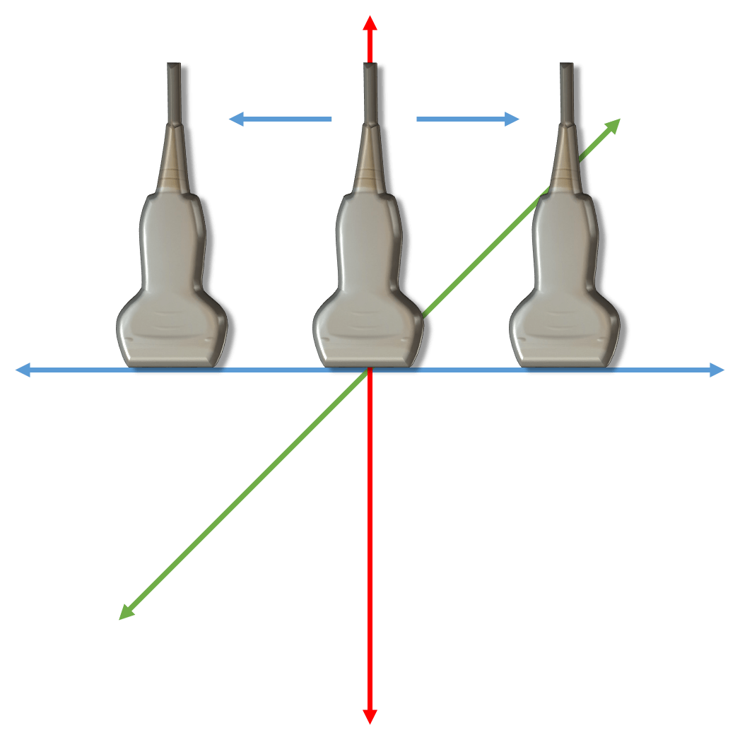
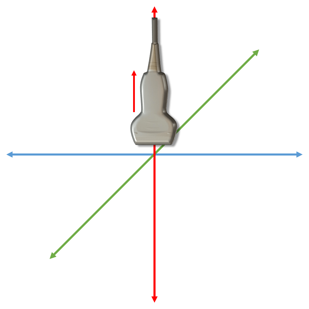
For successful ultrasound examination it is essential to know the basic ultrasound probe movement possibilities in three dimensional space. Every single one is used for another purpose. The basic movements are depicted below on the x, y, z axis (x – blue – side to side; y – green – back and forward; z – red – up and down). Every other probe movement is just a combination of the basic ones.
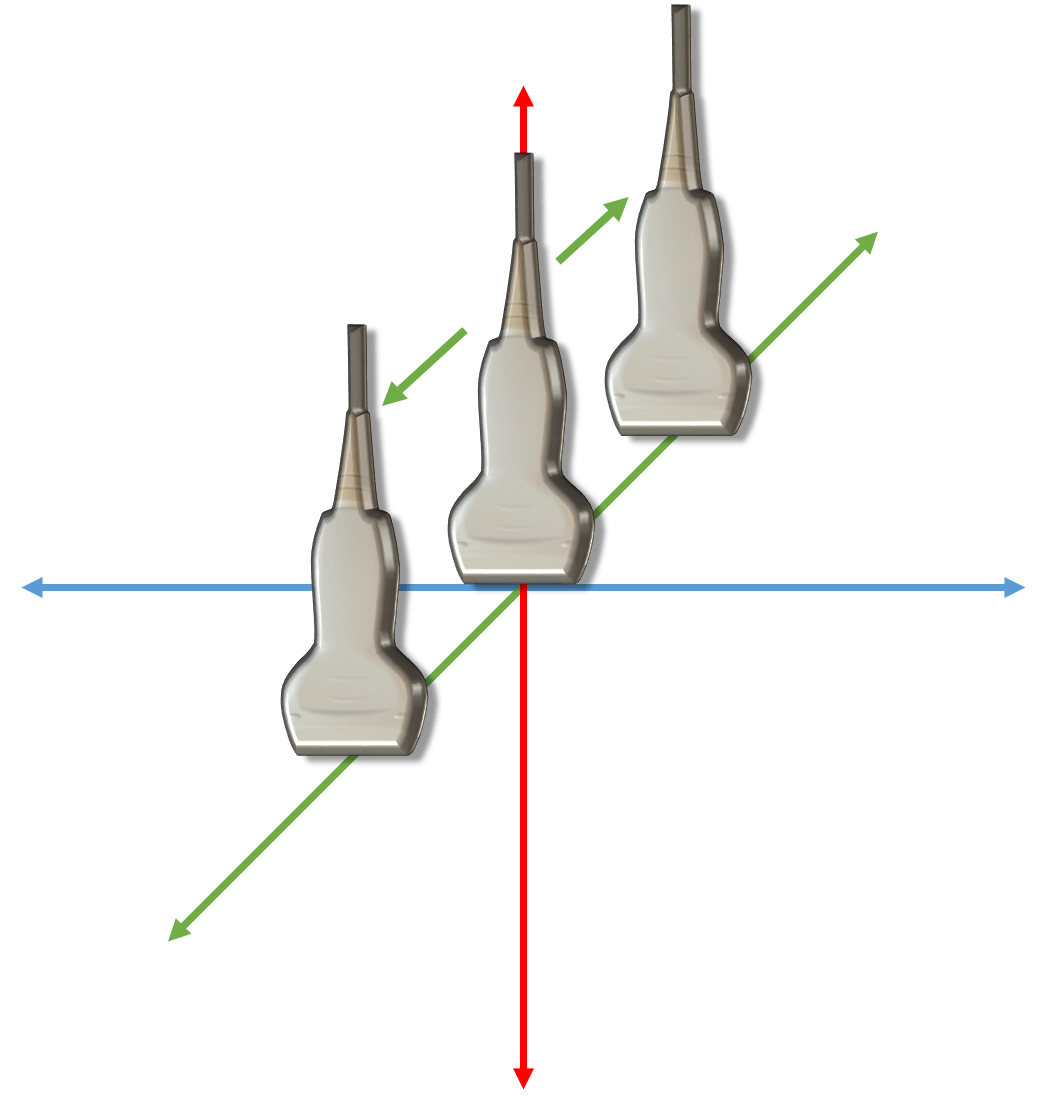
A. Slide
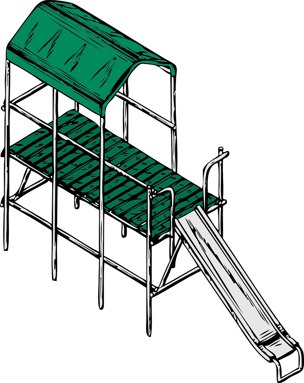
Long time before beeing able to get the driving licence as children peolple often imagine driving a car while going down the slide.
Slide is also the first movement with the probe. It is performed by moving the probe in it’s long axis.
Examples: looking for a structure, following a structure such as nerve or tendon, etc.
B. Rock (heel-toe)
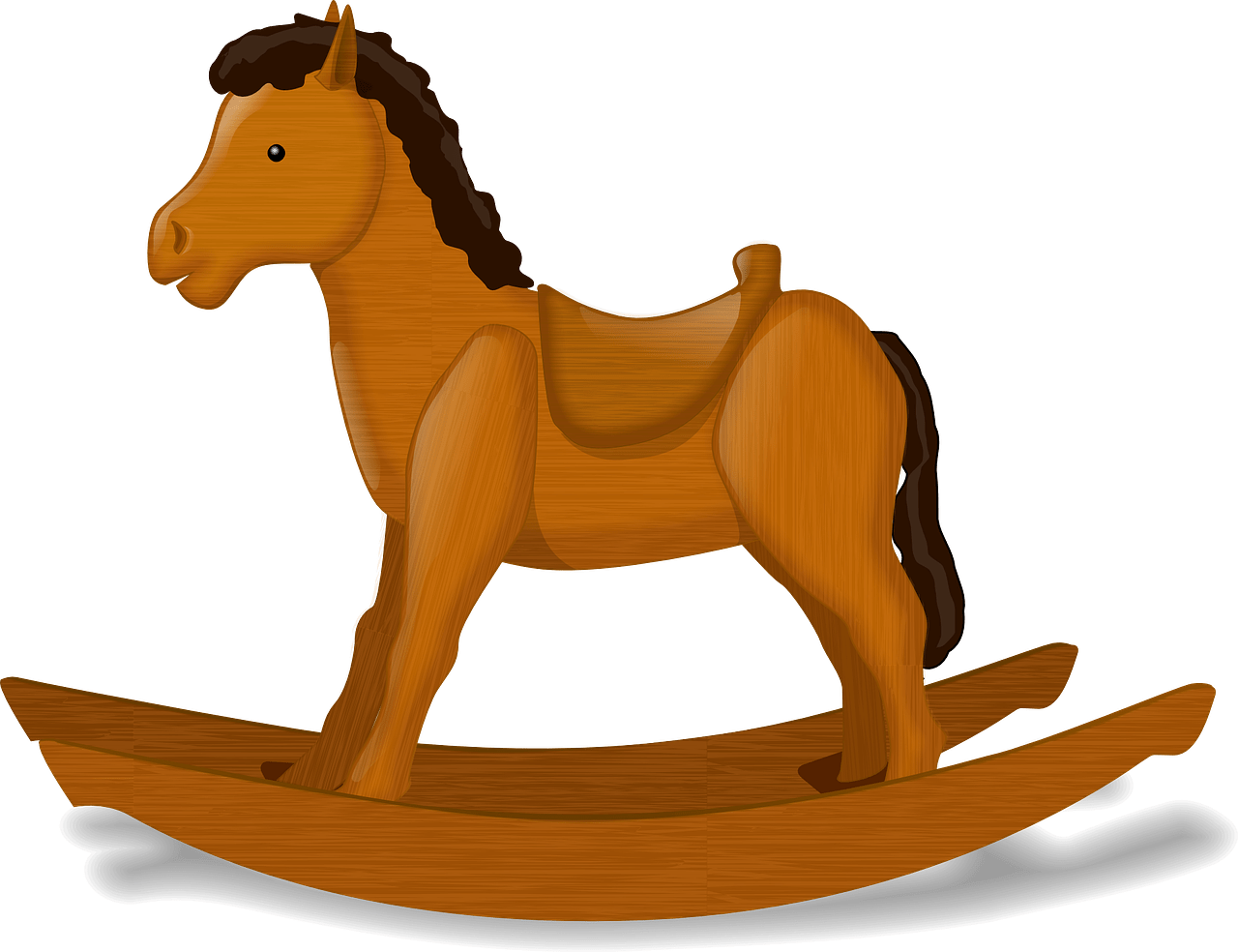
Another example of preparing for driving a car from the childhood is the rocking horse.
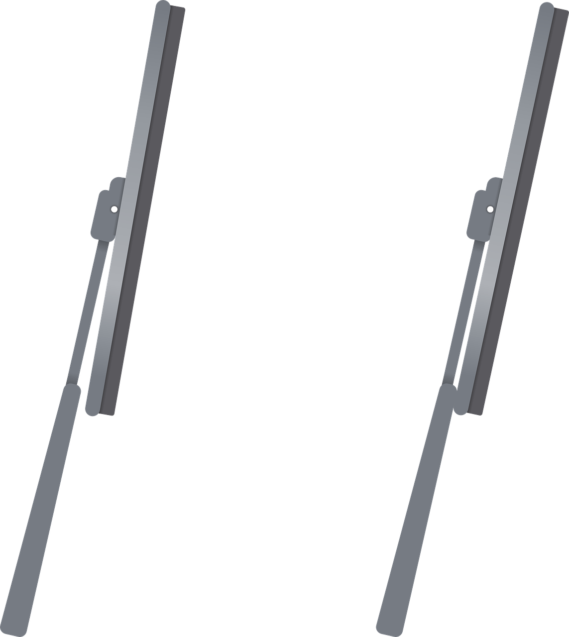
Rock is the second movement that can be performed with the probe. It is performed by tilting the probe in the long axis.
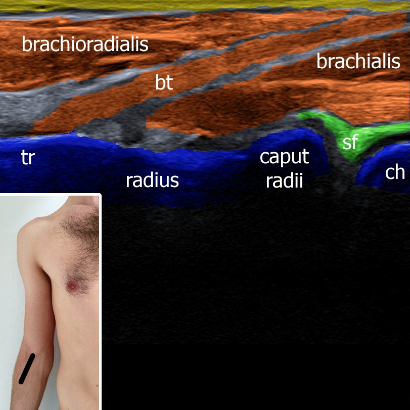
Examples: changing the angle of insolation in order to avoid anisotropy artifact e.g. in tendons, distal biceps tendon, Achilles tendon.
C. Sweep
When the winter hides the car under a blanket of snow people use a broom to sweep the snow off the car.
Sweep movement is the third movement and is perfomed by moving the probe in its short axis.
Examples: looking for a structure, following structure, etc.
D. Fan
In the ancient times when there was no air conditioning in the car people had to use a fan to cool them down.
Fan is the fourth movement and is performed by tilting the probe in the short axis.
Examples: searching for a median nerve in cubital canal, searching for a needle.
E. Compression
Driving a car can be dangerous. In the case of a car crash the cars get compressed.
Compression is the fifth movement and is performed by pushing the probe agianst the skin of the patient.
Examples: sonopalpation, pain provocation, evaluate compressibility of tissue or veins.
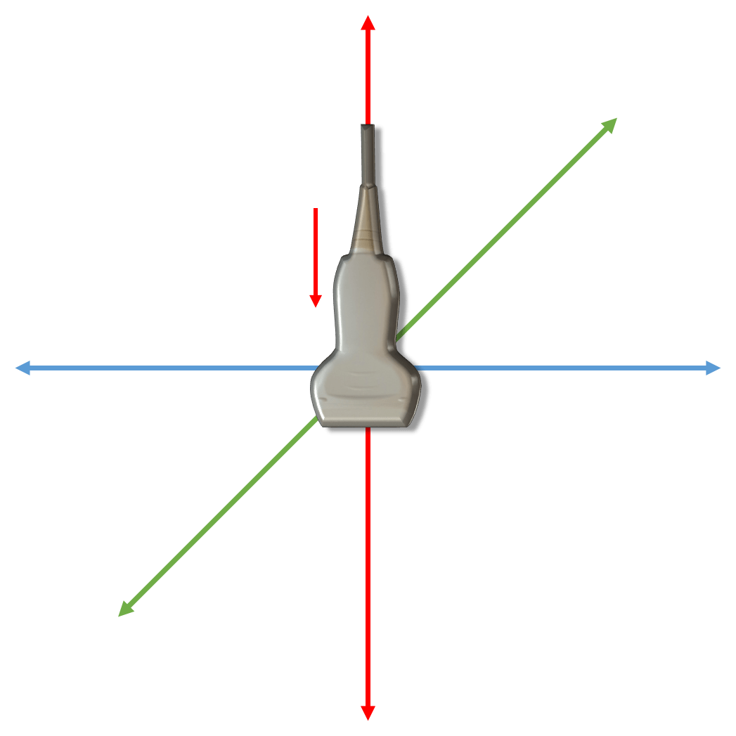
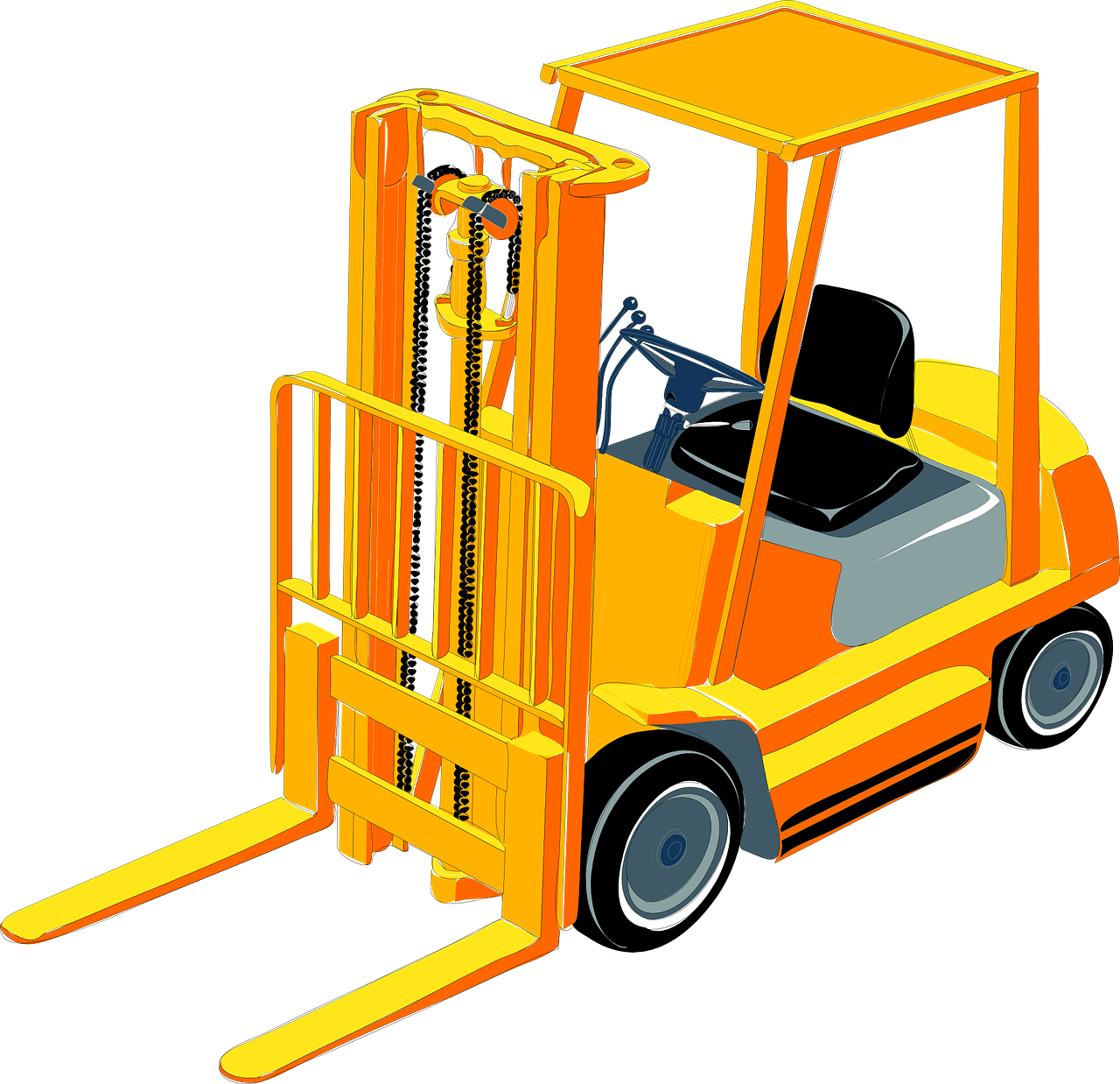
F. Lift
When dealing with a heavy subject this vehicle can come really handy: a forklift truck.
Lift is the sixth movement and is performed by releasing the pressure on the probe.
Examples: dynamic examination of the ulnar nerve stability in ulnar sulcus.
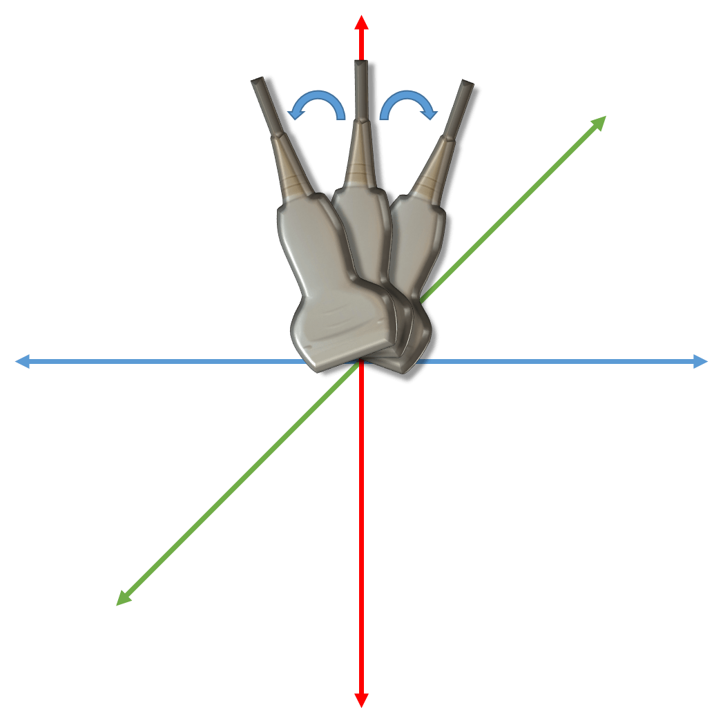
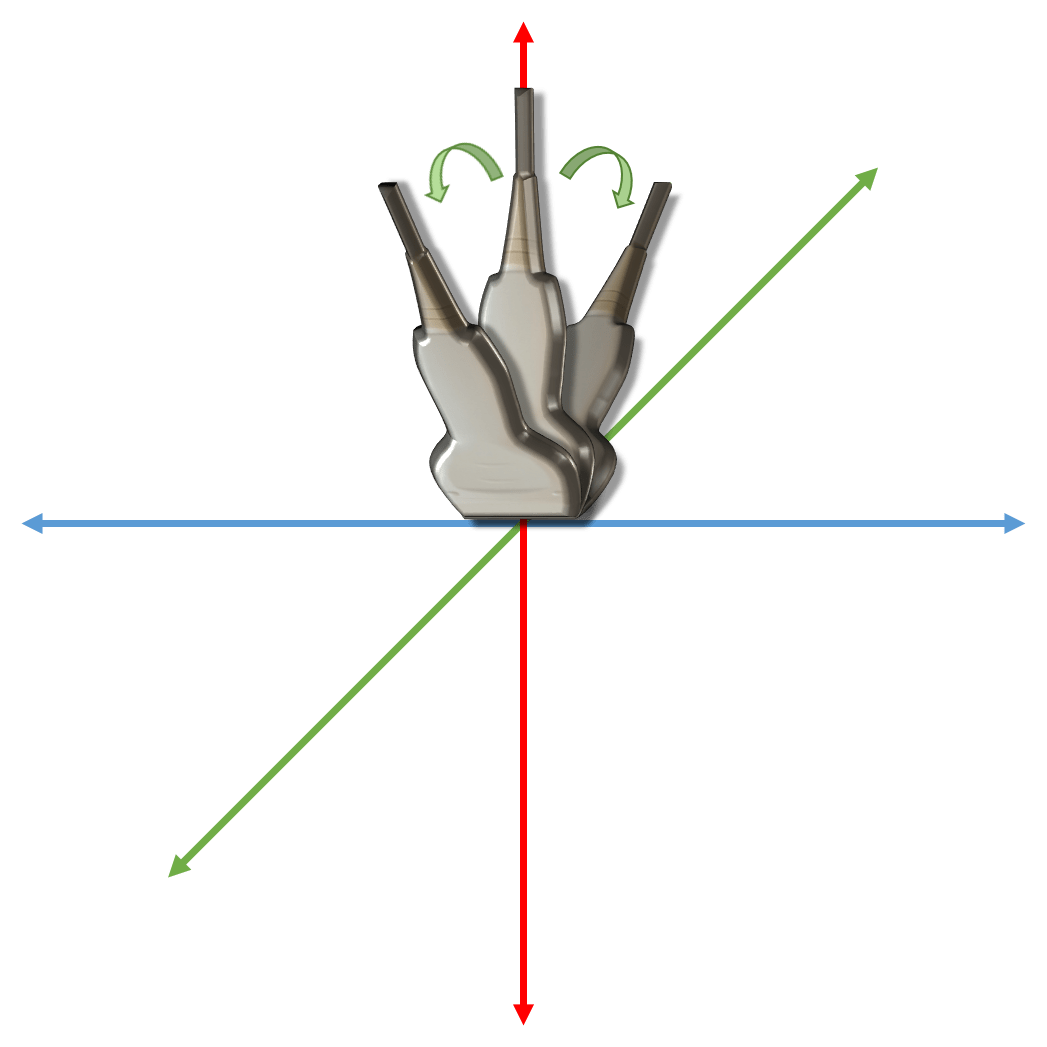
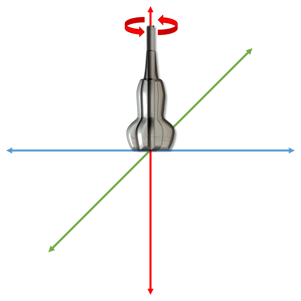
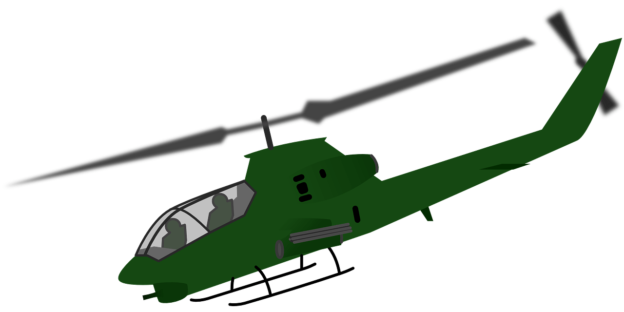
G. Helicopter
Sometimes, when the car is not fast enough, another means of transport can be used, for example: helicopter.
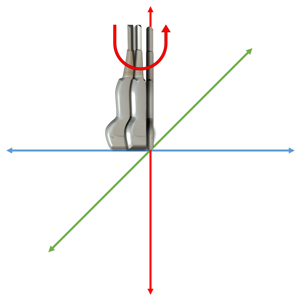
Helicopter is also the seventh movement and is performed by rotating the probe. The center of rotation is in the middle of the probe.
Examples: changing the view of a structer from longitudinal to transversal and vica versa, e.g. biceps tendon, median nerve.
H. Wiper
An usefull equipment to get rid of water from the windshield are wipers.
Wiper is the eight and the last of the movements and is performed by rotating the prebe with the center of the rotation in one end of the long axis of the probe.
Examples: to scan triangul structers such as subscapularis muscle.
3. “Parking” – Probe positioning on the patient
G. Helicopter
Sometimes, when the car is not fast enough, another means of transport can be used, for example: helicopter.
Helicopter is also the seventh movement and is performed by rotating the probe. The center of rotation is in the middle of the probe.
Examples: changing the view of a structer from longitudinal to transversal and vica versa, e.g. biceps tendon, median nerve.
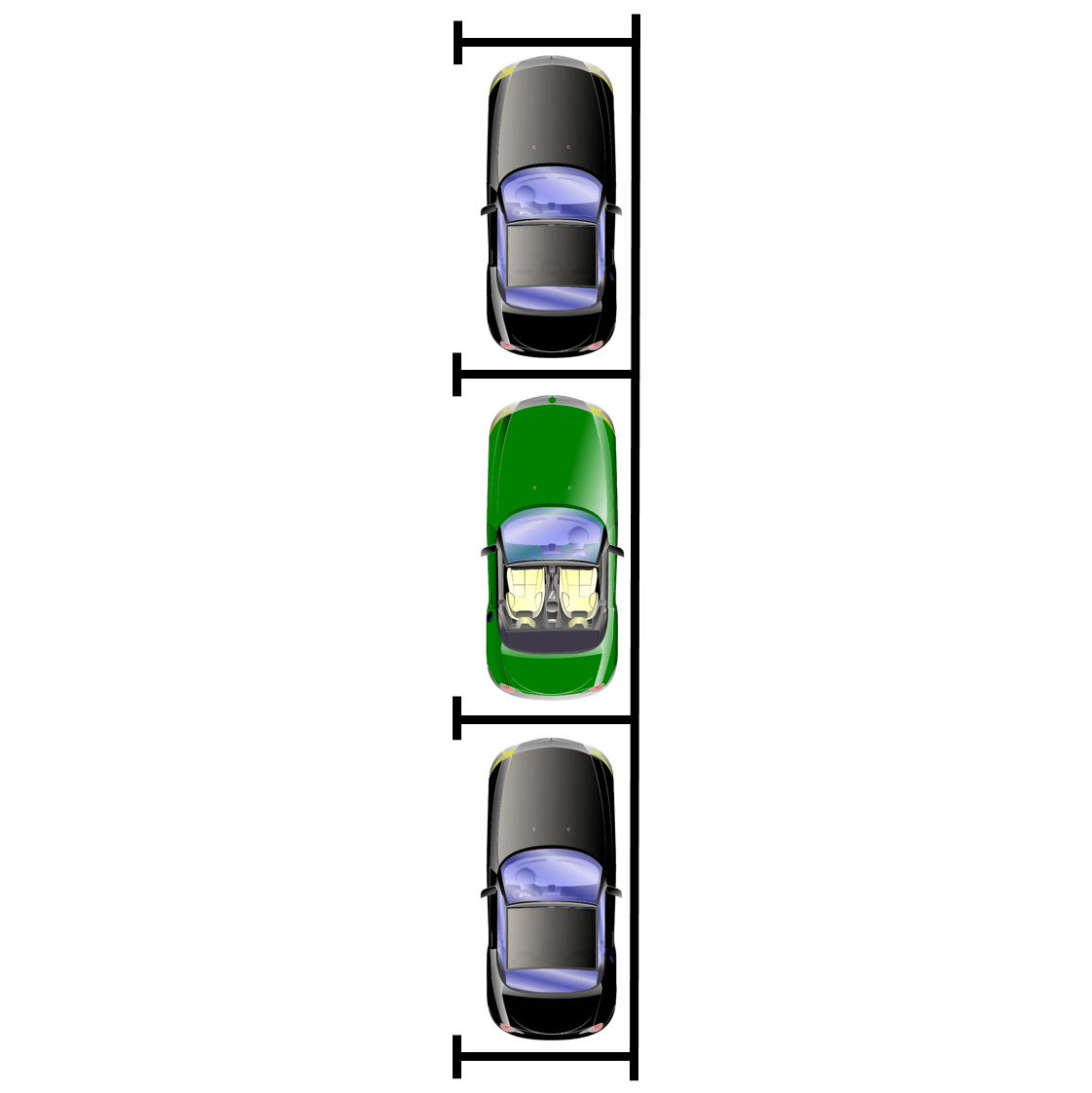
A. Transversal
In this position the long axis of the probe is situted perpendiculary (transversal position) to the long axis of the extremity.
Examples: carpal tunnel, subscapularis muscle, transvesal view at the elbow joint, etc.
B. Longitudinal
In this position the long axis of the probe is situated parallel (longitudinal position) to the long axis of the extremity.
Examples: long head of biceps brachii, patellar ligament, tibialis anterior tendon, etc.
C. Oblique
There are also some specific cases when we want to scan a structure with an oblique course, so the probe has to be angled.
Examples: distal biceps tendon, ligamentum cruciatum anterius, femoral neck, etc.