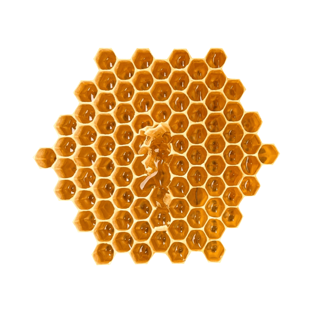
Peripheral nerves are made up of multiple nerve fibers that are organized into bundles called fascicles. When a nerve is examined using ultrasound, the scan shows several small, hypoechoic “bubbles” within the larger, hyperechoic epineurium. These bubbles represent the fascicles and have a honeycomb-like structure. It is important to be able to recognize this organized structure in order to distinguish it from a tendon, which has a fibrillar structure rather than fascicles.
Transverse – Honeycomb
Longitudinal –
Pathologies
Neuropathy
Definition: A term that describes various nerve conditions.
US appearence: Variable.
Compression neuropathy
Definition:Disorder characterised by nerve dysfunction as a result of nerve entrapment or extrinsic impingement.
US appearence: Hypoechoic appearance of nerve from epineural oedema with possible fascicular enlargement typically proximal and sometimes distal to the compression site.
Transection
Definition:Partial or complete discontinuity of a nerve due to disruption of some or all the nerve fascicles.
US appearence: Discontinuity of some or all nerve fascicles with possible retraction of the discontinuous nerve and focal or mass-like thickening at the retracted end.
Neuroma
Definition: The focal enlargement of an injured nerve or fascicle, which may be associated with nerve or fascicular retraction if due to transection
US appearence: Hypoechoic focal nerve or fascicle enlargement at the site of injury with possible retraction.
Neuritis
Definition:Nerve inflammation as seen with inflammatory, infectious or autoimmune conditions.
US appearence:Abnormally hypoechoic nerve with possible increased flow on Doppler imaging.