 MSK Academy
1. Ventrale Ansicht
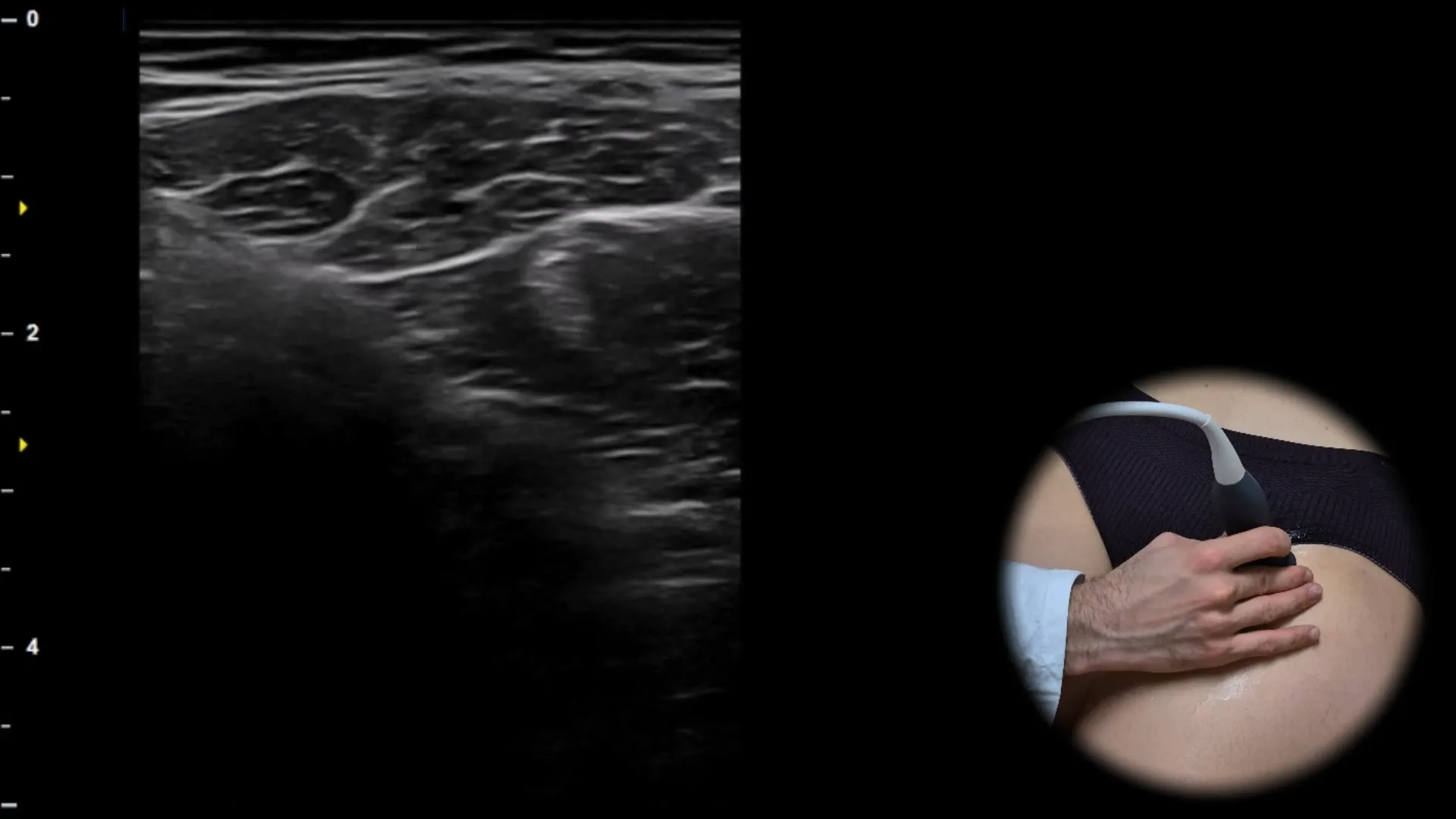
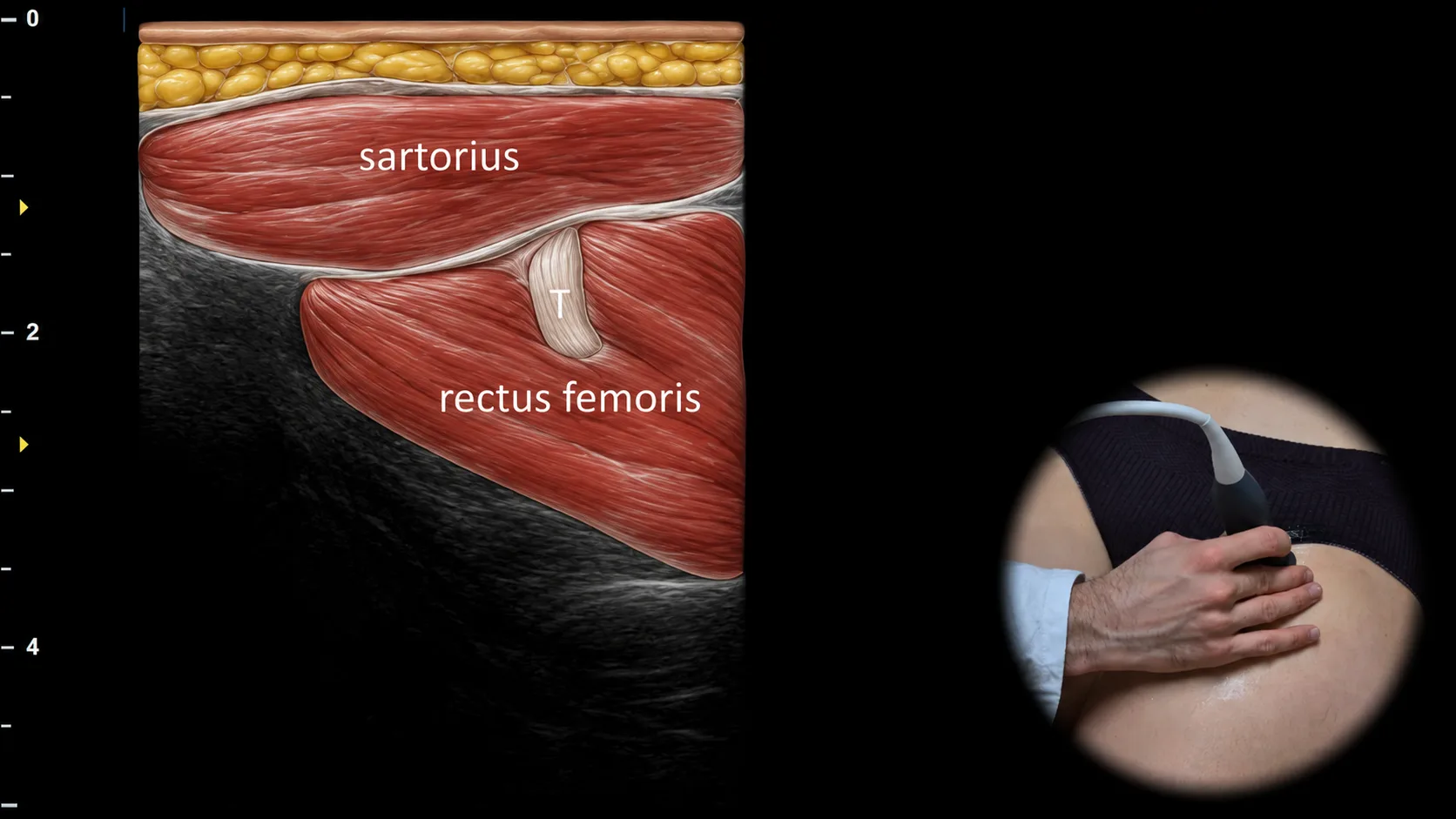
Abbildung 1. Ventrale Ansicht, Transversalebene. T: Sehne des M. rectus femoris
Transversaler sonografischer Schnitt durch den vorderen Oberschenkel mit Darstellung des oberflächlich gelegenen M. sartorius, der schräg von lateral nach medial verläuft, und des tiefer gelegenen M. rectus femoris. Innerhalb seines Muskelbauchs ist die zentrale Sehne (T) sichtbar, die sich als hyperechogene lineare Struktur darstellt und in ihrem tieferen Verlauf zum Ansatz hin deutlicher abgrenzbar wird. Diese Projektion ist für die Beurteilung des myotendinösen Übergangs des M. rectus femoris und für die Erkennung von muskulärer Überlastung, Zerrung oder Tendinopathie wichtig.
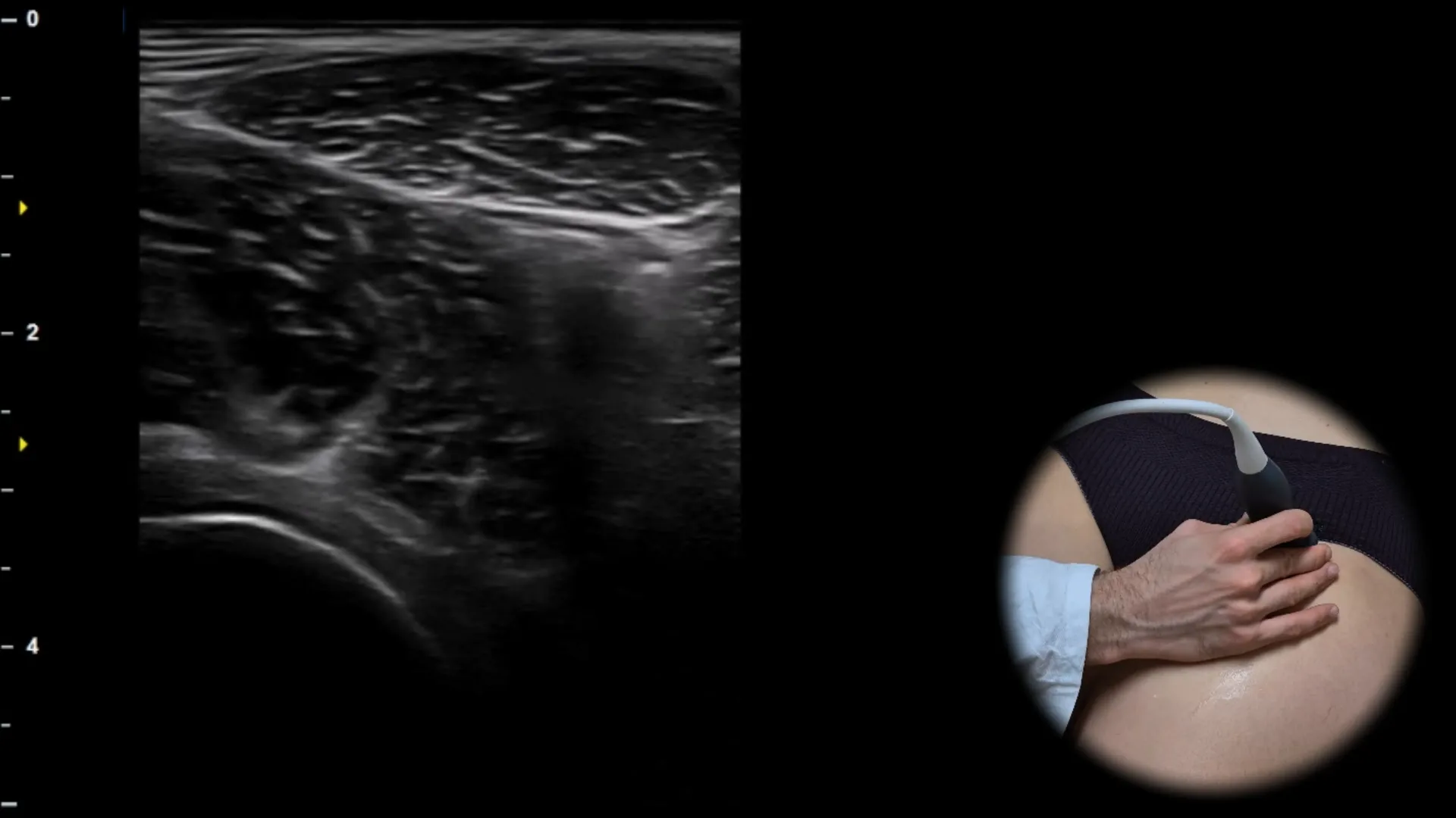
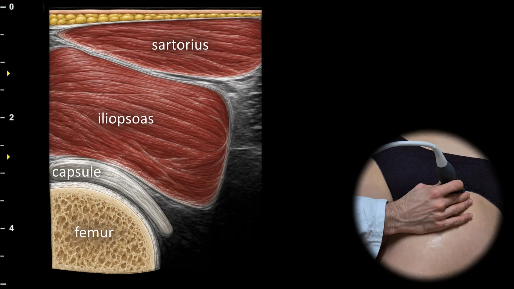
Abbildung 2. Ventrale Ansicht, Transversalebene.
Transversaler sonografischer Schnitt durch den anterioren Anteil des Hüftgelenks. Oberflächlich ist der m. sartorius sichtbar, darunter liegt in der Tiefe der m. iliopsoas, unter dem die Gelenkkapsel angrenzend an die anteriore Kontur des Femurs dargestellt ist, wobei der Femur die wichtigste knöcherne Landmarke dieser Projektion bildet. Diese Projektion ist für die Beurteilung des anterioren Rezessus des Hüftgelenks essenziell, insbesondere zum Nachweis eines Gelenkergusses, einer Synovitis und von Pathologien im Bereich des m. iliopsoas.
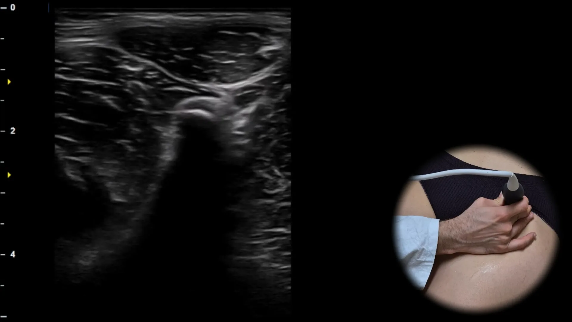
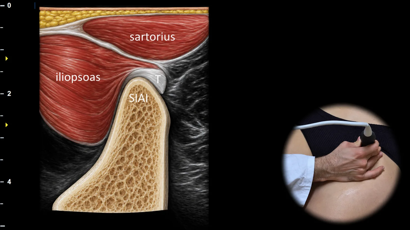
Abbildung 3. Ventrale Ansicht, Transversalebene. T: Sehne des Musculus rectus femoris, SIAI: spina iliaca anterior inferior
Transversaler sonografischer Schnitt durch die vordere Hüftregion mit Darstellung der direkten Sehne des Musculus rectus femoris (T) an ihrer Anheftungsstelle an der spina iliaca anterior inferior (SIAI). Die SIAI ist als knöcherne Prominenz mit der angrenzenden hyperechogenen fibrillären Sehne sichtbar, während in der Umgebung gleichzeitig auch der Musculus sartorius und der Musculus iliopsoas als wichtige Weichteillandmarken dargestellt werden können. Diese Projektion ist wesentlich für die Beurteilung der Anheftung des Musculus rectus femoris und für die Erkennung von Apophysenverletzungen, Avulsionsfrakturen und Tendinopathien in dieser Region, insbesondere bei sportlich aktiven Personen.
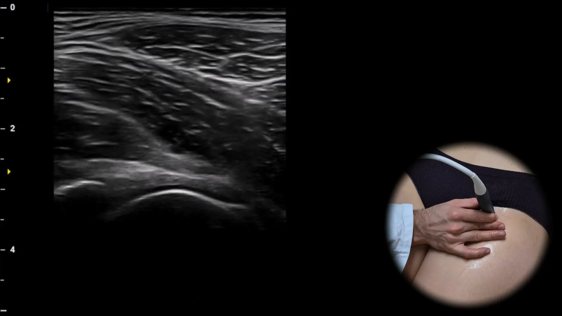
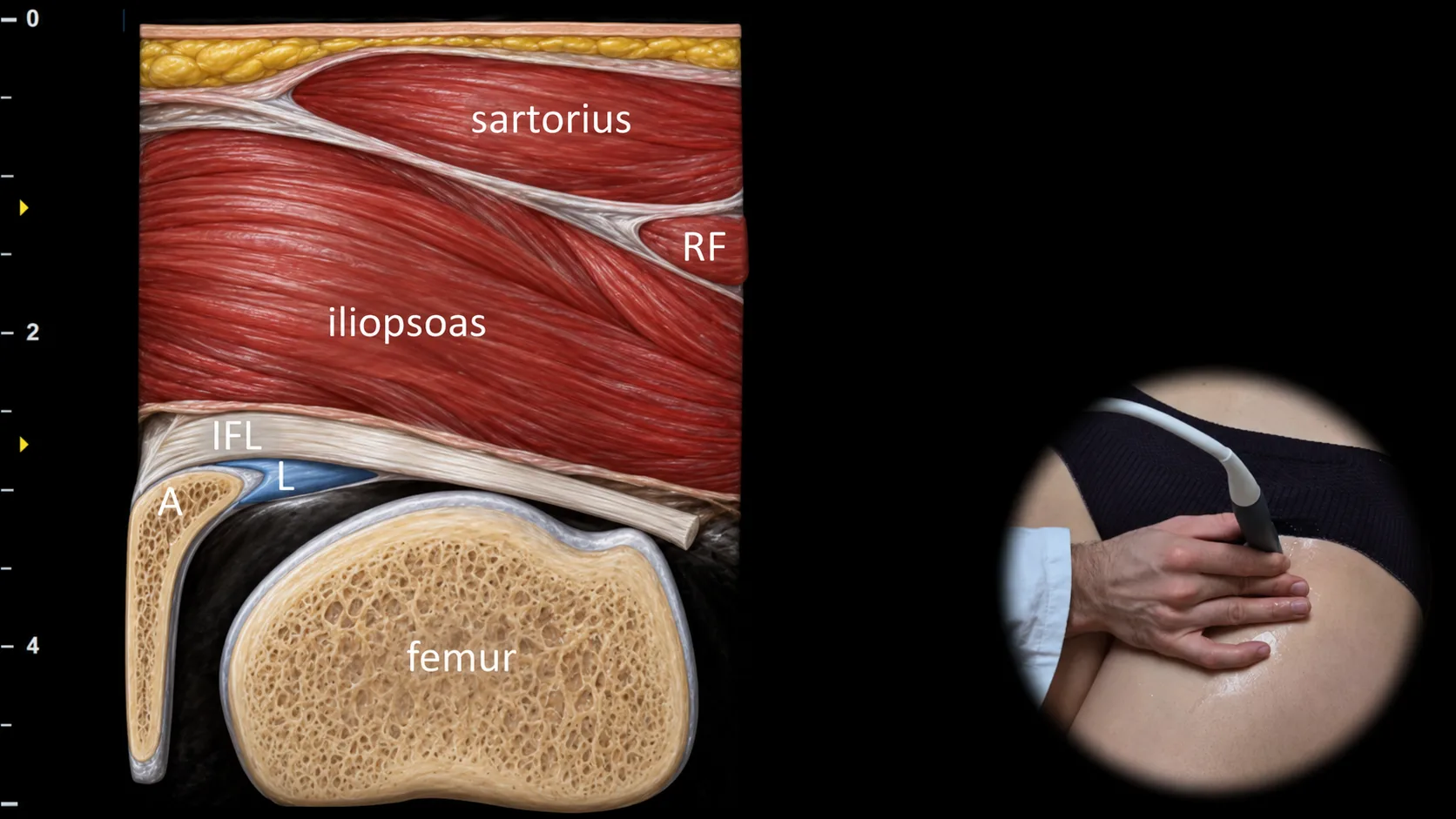
Abbildung 4. Ventralansicht, schräge Ebene. IFL: Lig. iliofemorale, A: Acetabulum, L: Labrum, RF: M. rectus femoris
Schräger Ultraschallschnitt durch den anterioren Anteil des Hüftgelenks mit Darstellung der femoroacetabulären Artikulation. Der Femurkopf ist in Artikulation mit dem Acetabulum (A) sichtbar, an dessen anteriorem Rand das Labrum acetabulare (L) als dreieckige, der knöchernen Kontur aufliegende Struktur dargestellt werden kann; oberflächlich sind außerdem das Lig. iliofemorale (IFL) sowie umgebende Weichteilleitstrukturen sichtbar, insbesondere der M. rectus femoris (RF), der M. sartorius und der M. iliopsoas. Diese Projektion ist für die Beurteilung der Labrumintegrität, der anterioren Gelenkkongruenz und von Zeichen eines femoroacetabulären Impingements unerlässlich.
Klinischer Hinweis
In dieser Projektion lässt sich Flüssigkeit im vorderen Abschnitt des Hüftgelenks gut nachweisen, insbesondere bei einer Distension des anterioren Gelenkrezessus.
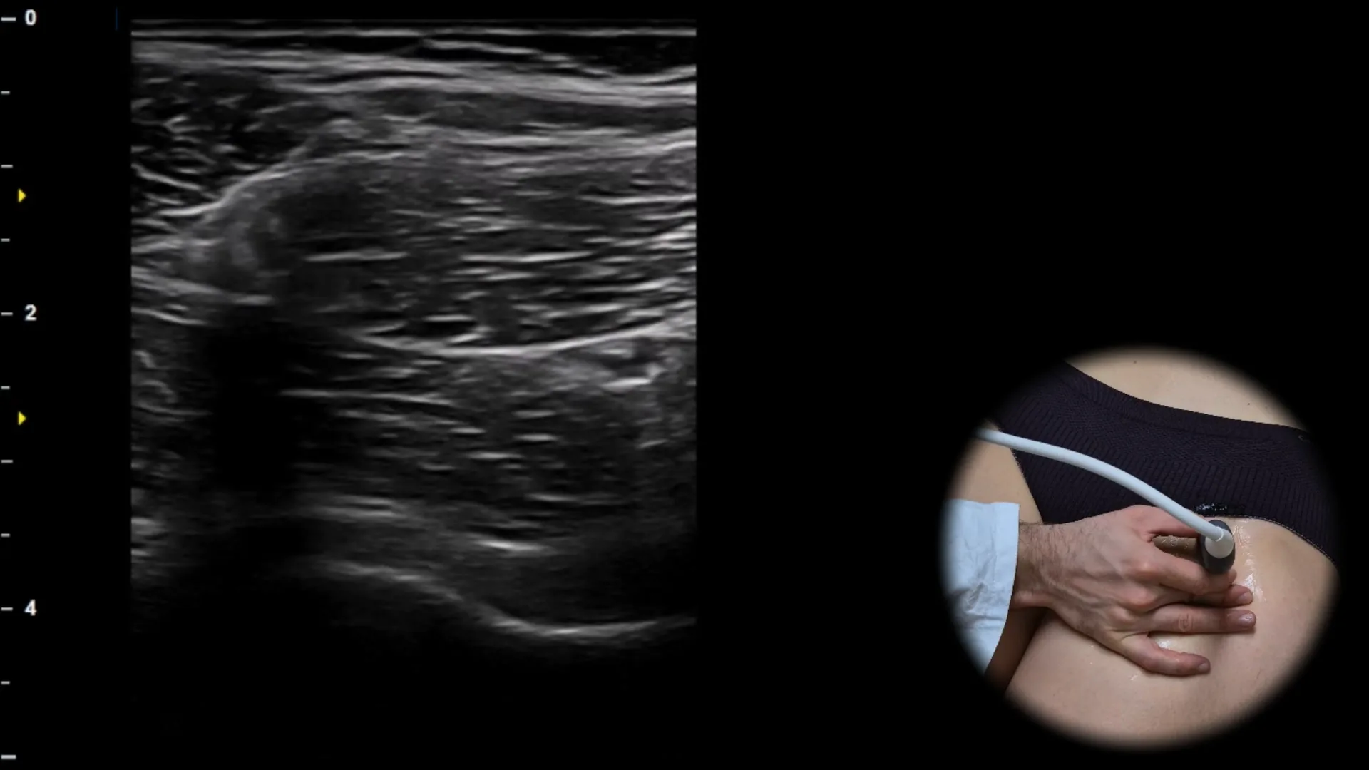
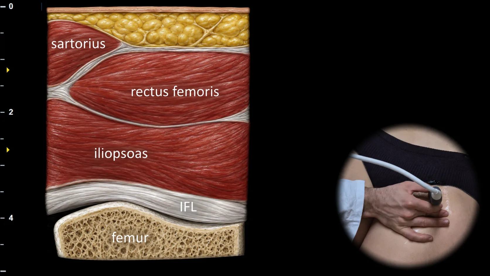
Abbildung 5. Ventrale Ansicht, schräge Ebene. IFL: Ligamentum iliofemorale
Weiter distal geführter schräger sonografischer Schnitt durch den anterioren Anteil des Hüftgelenks, der den anterioren Gelenkrezessus im Bereich des Femurkopfes zeigt. Unter den oberflächlich gelegenen Muskeln, insbesondere dem Musculus rectus femoris, Musculus sartorius und Musculus iliopsoas, ist die Gelenkkapsel sichtbar; darunter verläuft das Ligamentum iliofemorale (IFL), das als hyperechogenes Band angrenzend an die anteriore Kontur des Femurs erscheint. Diese Projektion ist entscheidend für die Beurteilung von Gelenkerguss, Synovitis und Verdickung der anterioren Kapsel oder des Ligamentum iliofemorale bei entzündlichen und degenerativen Erkrankungen des Hüftgelenks.
Klinischer Hinweis
Bei Vorliegen einer intraartikulären Flüssigkeitsansammlung kann es zu einer Vorwölbung des anterioren Rezessus und zu einer ventralen Verlagerung des Ligamentum iliofemorale kommen, wodurch der Erguss in dieser Projektion deutlicher erkennbar wird.
Zugang zur gesamten Bibliothek
Voller Zugriff auf Scan-Protokolle, Anatomie und klinische Referenzen. Jederzeit kündbar.
- Alle Protokolle und Anatomiereferenzen
- Originale Ultraschall-Illustrationen und Video-Demonstrationen
- Synchronisierung zwischen Mobilgerät und Web