 MSK Academy
4. Dorsale Ansicht
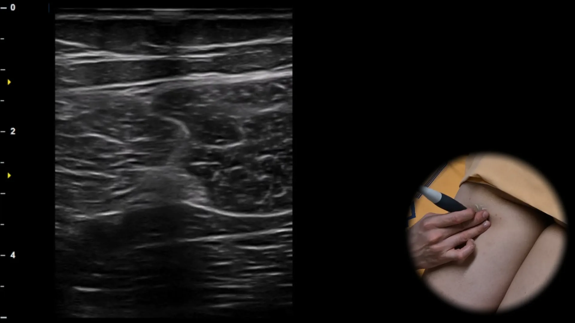
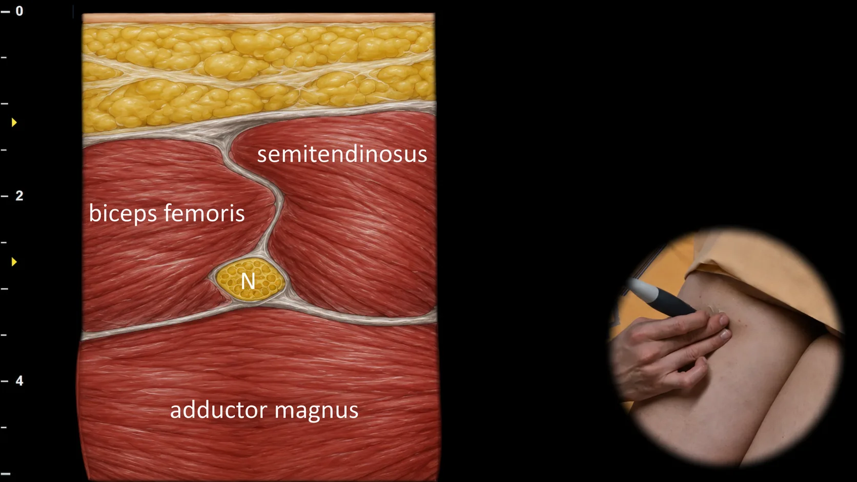
Abbildung 1. Dorsale Ansicht, Transversalebene. N: N. ischiadicus
Transversaler sonografischer Schnitt durch den dorsalen Oberschenkel mit Darstellung des N. ischiadicus als ovale Struktur mit typischem faszikulärem Erscheinungsbild, gelegen zwischen dem M. semitendinosus medial und dem Caput longum des M. biceps femoris lateral. In der Tiefe ist der M. adductor magnus sichtbar, der den Boden dieses anatomischen Fensters bildet. Diese Projektion ist für die Lokalisation des N. ischiadicus und die Beurteilung seiner Pathologie im Bereich des dorsalen Oberschenkels von wesentlicher Bedeutung. Klinischer Hinweis: Die typische Anordnung der umgebenden Muskeln erzeugt das sogenannte "windmill sign", das die schnelle Orientierung und Identifikation des Nervs bei der sonografischen Untersuchung erleichtert.
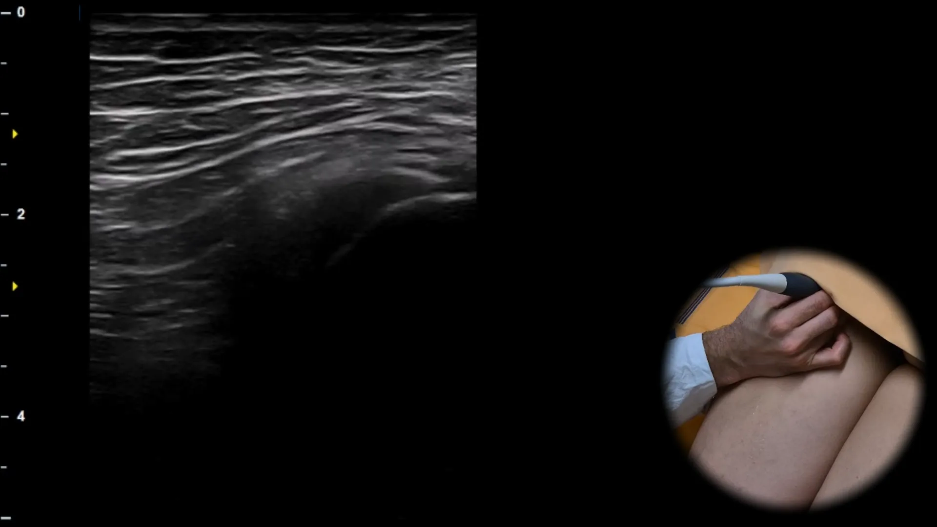
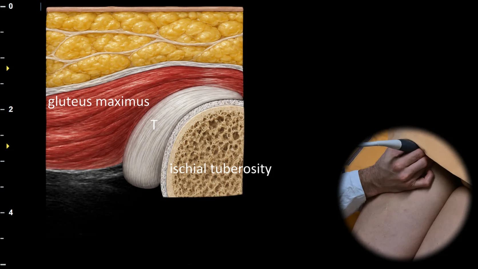
Abbildung 2. Dorsale Ansicht, Transversalebene. T: Hamstringsehne
Transversaler Ultraschallschnitt des proximalen dorsalen Oberschenkels mit Darstellung der Region des Tuber ischiadicum. Das Tuber ischiadicum erscheint als bogenförmige hyperechogene knöcherne Kontur, an der der gemeinsame Sehnenansatz der Hamstrings (T) ansetzt, einschließlich der Sehnen des M. semitendinosus, des M. semimembranosus und des langen Kopfes des M. biceps femoris; oberflächlich ist auch der M. gluteus maximus sichtbar. Diese Projektion ist für die Beurteilung des proximalen Hamstringansatzes und für den Nachweis von Tendinopathien, Teilrupturen oder Avulsionsverletzungen in der Region des Tuber ischiadicum von wesentlicher Bedeutung, insbesondere bei Sportlern.
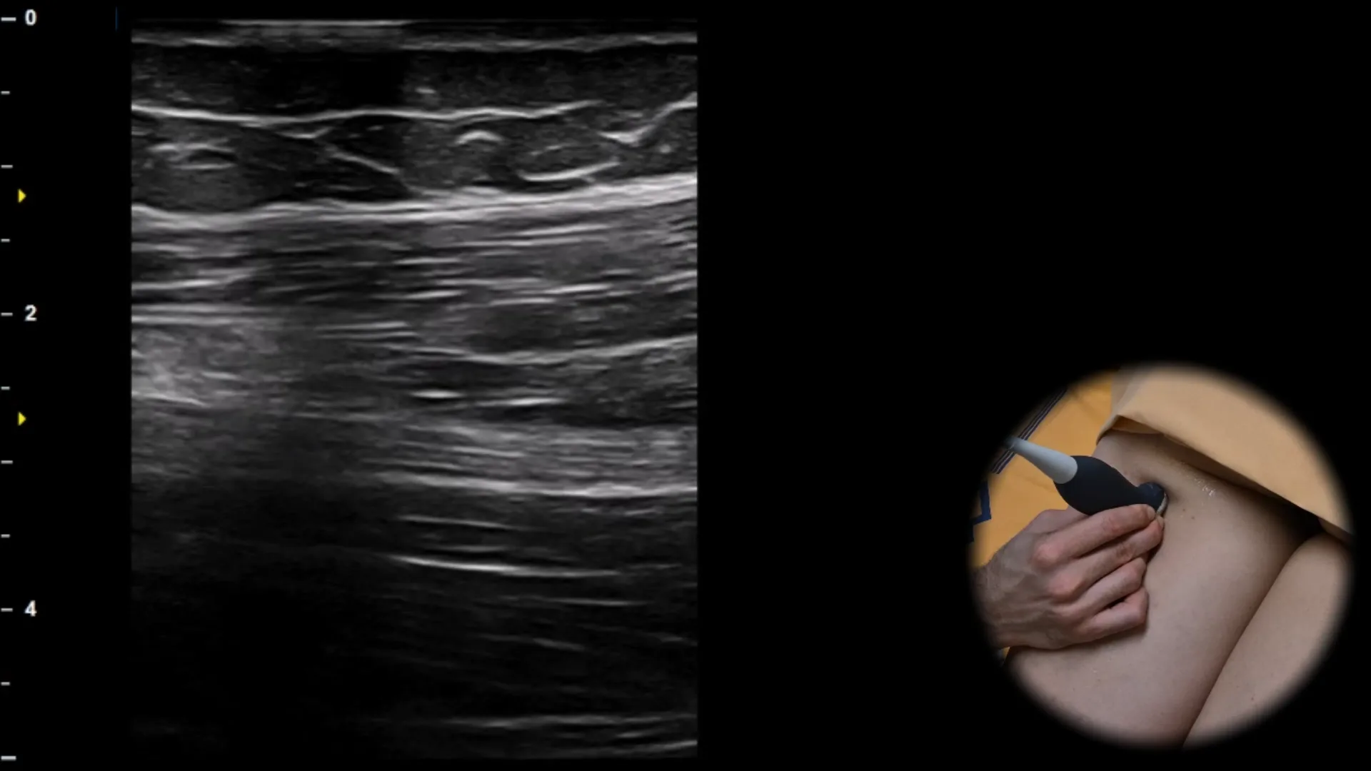
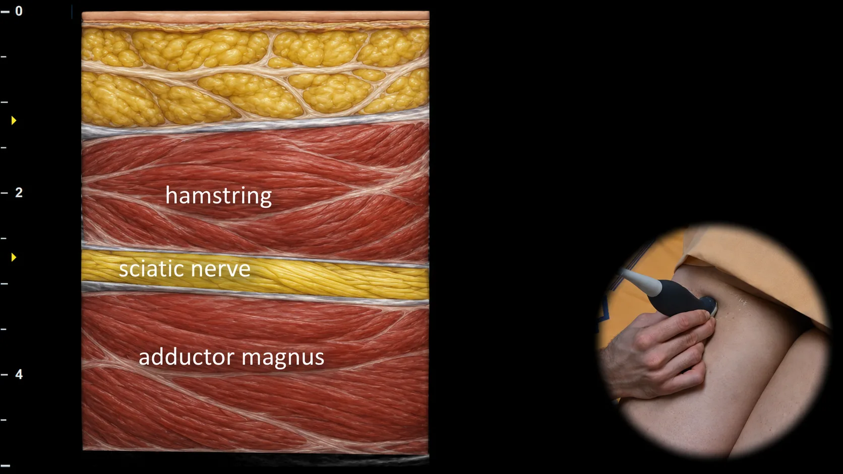
Abbildung 3. Dorsalansicht, Sagittalebene.
Longitudinaler Ultraschallschnitt durch den dorsalen Oberschenkel mit Darstellung des n. ischiadicus in der Längsachse. Der Nerv ist in dieser Projektion als gestreifte Struktur mit typischer faszikulärer Anordnung sichtbar und verläuft zwischen den oberflächlich gelegenen ischiokruralen Muskeln und dem tiefer gelegenen m. adductor magnus. Diese Projektion ist für die Beurteilung der Kontinuität des n. ischiadicus, seines Verlaufs und für die Erkennung pathologischer Veränderungen im Bereich des dorsalen Oberschenkels unerlässlich.
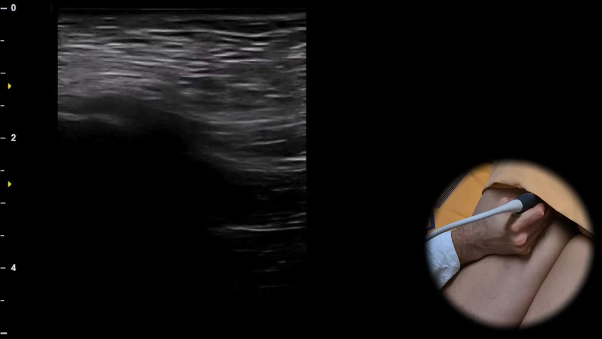
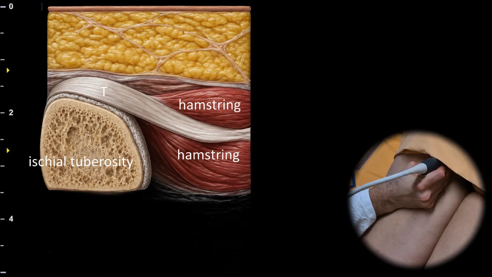
Abbildung 4. Dorsale Ansicht, Sagittalebene. T: Hamstringsehne
Longitudinaler Ultraschallschnitt im Bereich des Tuber ischiadicum mit Darstellung des gemeinsamen Hamstringansatzes. Das Tuber ischiadicum ist als gekrümmte hyperechogene knöcherne Kontur sichtbar, an der der gemeinsame sehnige Ursprung der Hamstrings (T) ansetzt, einschließlich der Sehnen des m. semitendinosus, des m. semimembranosus und des langen Kopfes des m. biceps femoris; distal des Ansatzes geht die Sehne in die Muskelbäuche der posterioren Oberschenkelmuskelgruppe über. Diese Projektion ist für die Beurteilung des proximalen Hamstringansatzes und für den Nachweis von Tendinopathien, Partialrupturen und vollständigen Avulsionen von wesentlicher Bedeutung, insbesondere bei Sportlern mit Schmerzen in der Glutealregion oder am posterioren Oberschenkel.
Zugang zur gesamten Bibliothek
Voller Zugriff auf Scan-Protokolle, Anatomie und klinische Referenzen. Jederzeit kündbar.
- Alle Protokolle und Anatomiereferenzen
- Originale Ultraschall-Illustrationen und Video-Demonstrationen
- Synchronisierung zwischen Mobilgerät und Web