 MSK Academy
Mittlerer Abschnitt
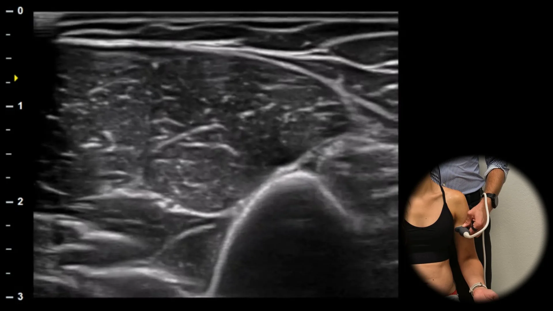
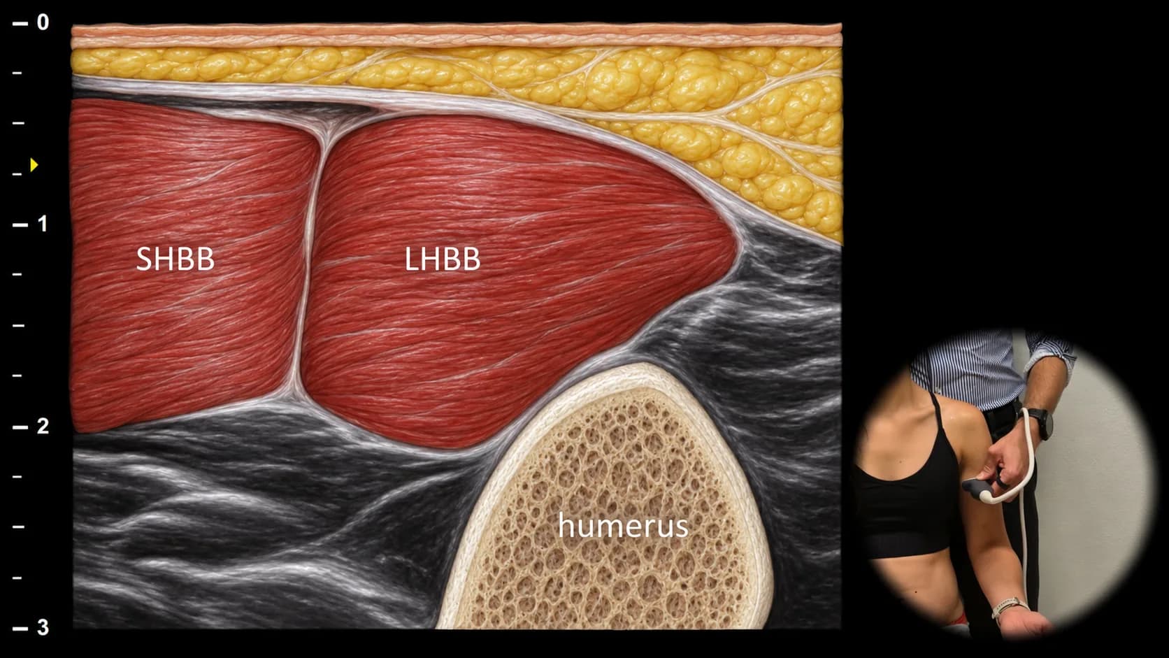
Abbildung 1. Ventrale Ansicht, Transversalebene. LHBB: Muskelbauch des langen Kopfes des M. biceps brachii, SHBB: Muskelbauch des kurzen Kopfes des M. biceps brachii
Der transversale Ultraschallschnitt durch den mittleren Abschnitt des M. biceps brachii zeigt die Muskelbäuche des langen und kurzen Bizepskopfes. Das Muskelgewebe weist das typische Erscheinungsbild eines "Sternenhimmels" auf, bei dem hypoechogene Muskelfasern von feinen hyperechogenen Septen aus Bindegewebe und Perimysium durchzogen sind, wodurch das feinkörnige Muster eines gesunden Muskels entsteht. Der lange Kopf (LHBB) liegt lateral, der kurze Kopf (SHBB) medial. In der Tiefe ist der Humerus sichtbar. Diese Ansicht eignet sich zur Beurteilung von Symmetrie, Volumen und Echotextur des Muskelgewebes sowie zum Nachweis von Ödemen, Atrophie oder fokalen Läsionen.
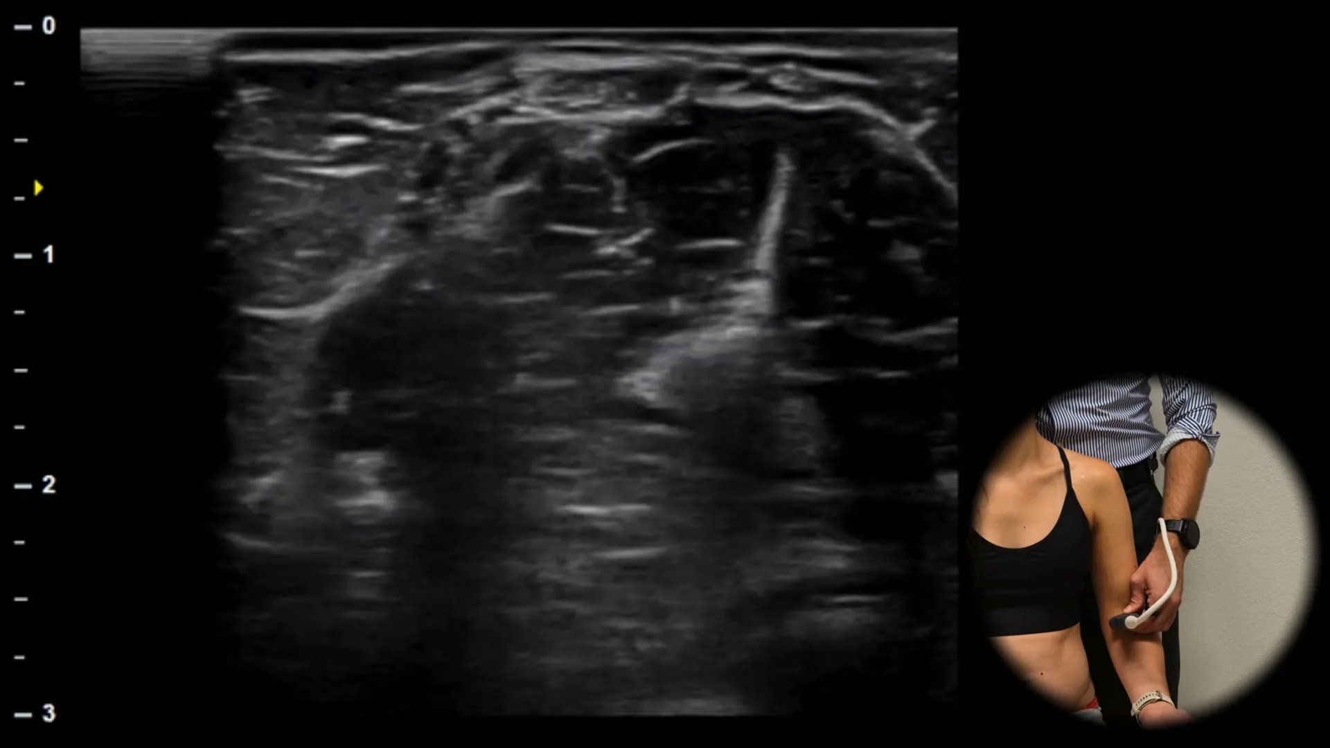
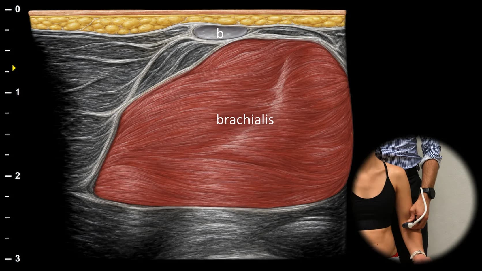
Abbildung 2. Ventralansicht, Transversalebene. b: Sehne des M. biceps brachii, brachialis: m. brachialis
Ein weiter distal gelegener transversaler sonografischer Schnitt durch den ventralen Oberarm zeigt die oberflächlich gelegene Sehne des M. biceps brachii (b) als hyperechogene fibrilläre Struktur. Darunter ist der m. brachialis sichtbar, der sich als breiterer, weniger echogener Muskelbauch in enger Beziehung zum Humerus darstellt. Die klare Abgrenzung der Bizepssehne vom tiefer gelegenen m. brachialis ermöglicht eine präzise Orientierung in diesem Bereich und ist wichtig für die Beurteilung von Sehnenverletzungen, myotendinösen Läsionen und Flüssigkeitsansammlungen zwischen den Muskelschichten.
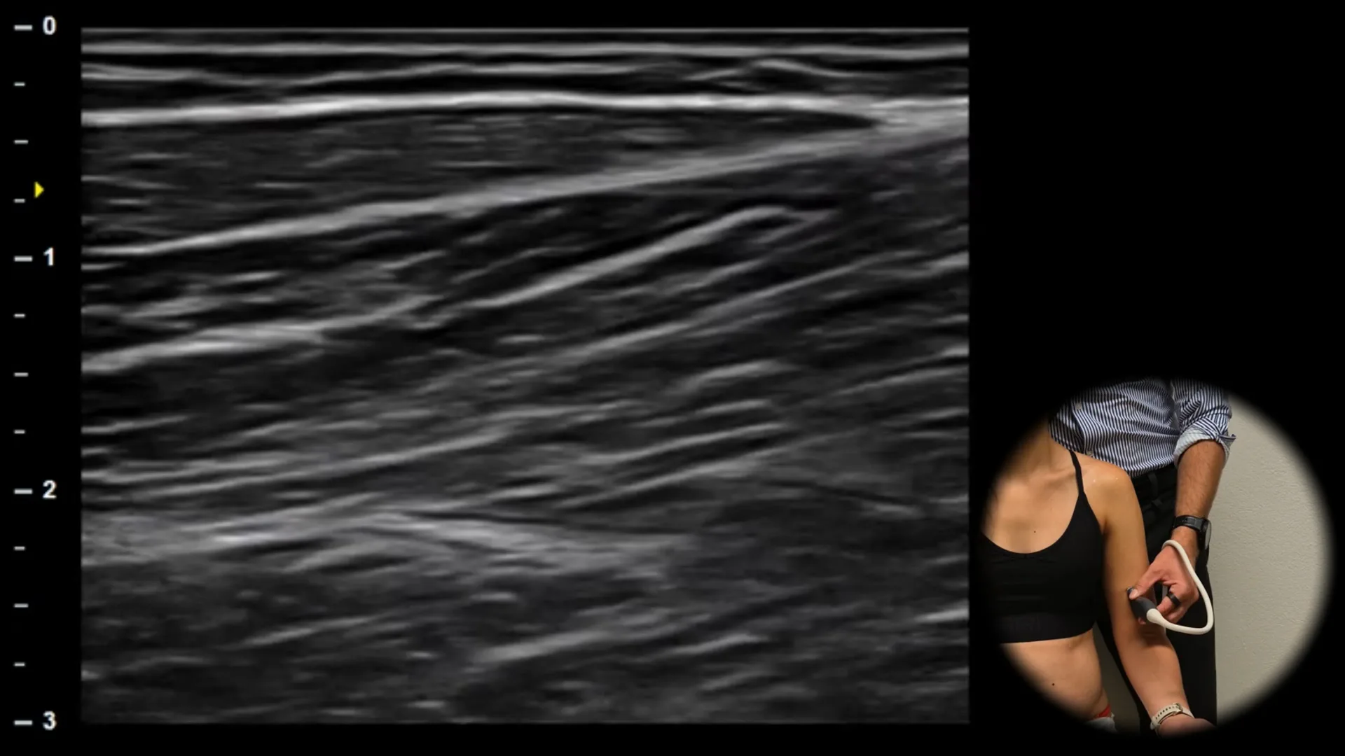
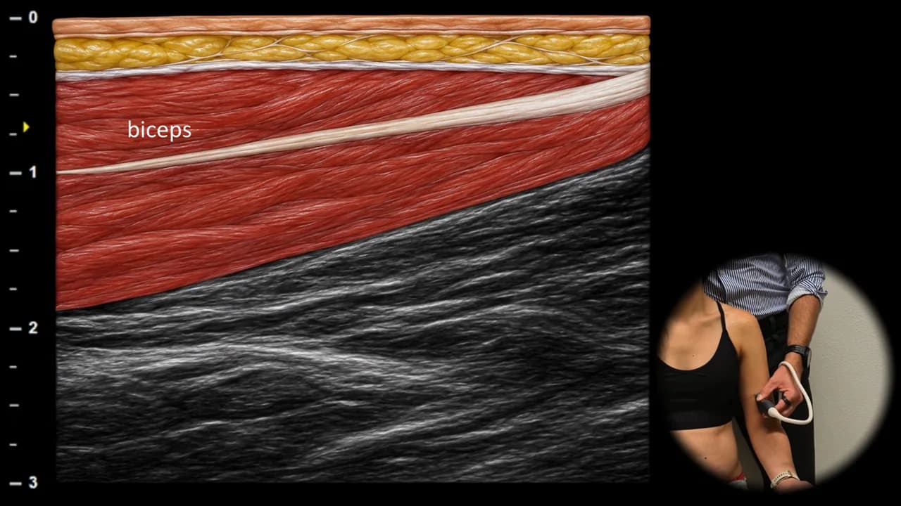
Abbildung 3. Ventrale Ansicht, Longitudinalebene. biceps: m. biceps brachii
Der longitudinale sonografische Schnitt durch den distalen Anteil des m. biceps brachii zeigt den distalen myotendinösen Übergang, an dem sich die Muskelfasern allmählich verjüngen und in die distale Sehne übergehen. Die Sehne stellt sich als gut abgrenzbare hyperechogene fibrilläre Struktur dar, die sich nach distal bis zu ihrem Ansatz erstreckt. Proximal ist der Übergang des Muskelgewebes in diese dichte, regelmäßig organisierte fibrilläre Architektur erkennbar. Tiefer liegt der m. brachialis, der im Vergleich zur Sehne eine geringere Echogenität aufweist. Diese Projektion ist wichtig für die Beurteilung partieller und vollständiger Läsionen des myotendinösen Übergangs, die Verlaufskontrolle der Heilung und die Planung der weiteren Behandlung oder Rehabilitation.
Zugang zur gesamten Bibliothek
Voller Zugriff auf Scan-Protokolle, Anatomie und klinische Referenzen. Jederzeit kündbar.
- Alle Protokolle und Anatomiereferenzen
- Originale Ultraschall-Illustrationen und Video-Demonstrationen
- Synchronisierung zwischen Mobilgerät und Web