Probe types
In musculoskeletal sonography, three basic types of probes are used: linear, convex, and high-frequency. The choice of probe directly affects image quality and proper interpretation of findings.
High-frequency probe
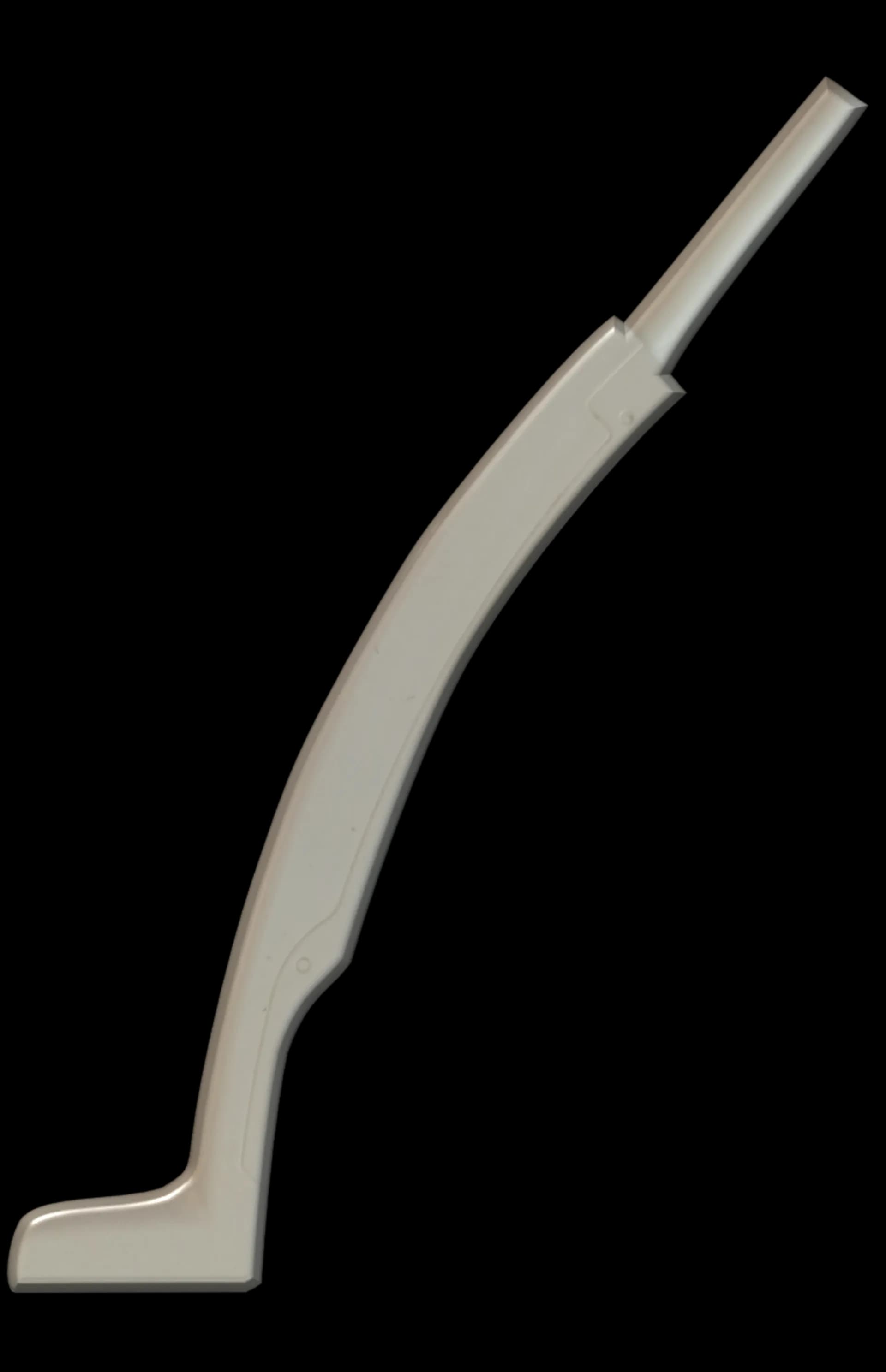
It is used to examine superficially located structures and provides very high spatial resolution. This allows detailed visualization of tendons, ligaments, muscles, nerves, and small joints. Its disadvantage is limited depth penetration, making it unsuitable for deeply located tissues.
Linear Probe
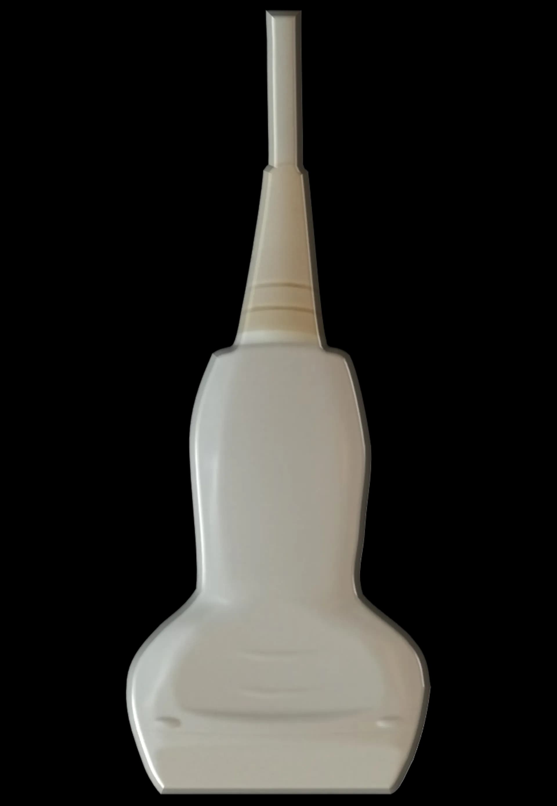
This is the most commonly used probe in musculoskeletal ultrasonography. It has a straight contact surface and produces a rectangular image that is clear and precise. It is best suited for examination of superficial soft tissues, where it enables very high-quality visualization of anatomical details.
Convex probe
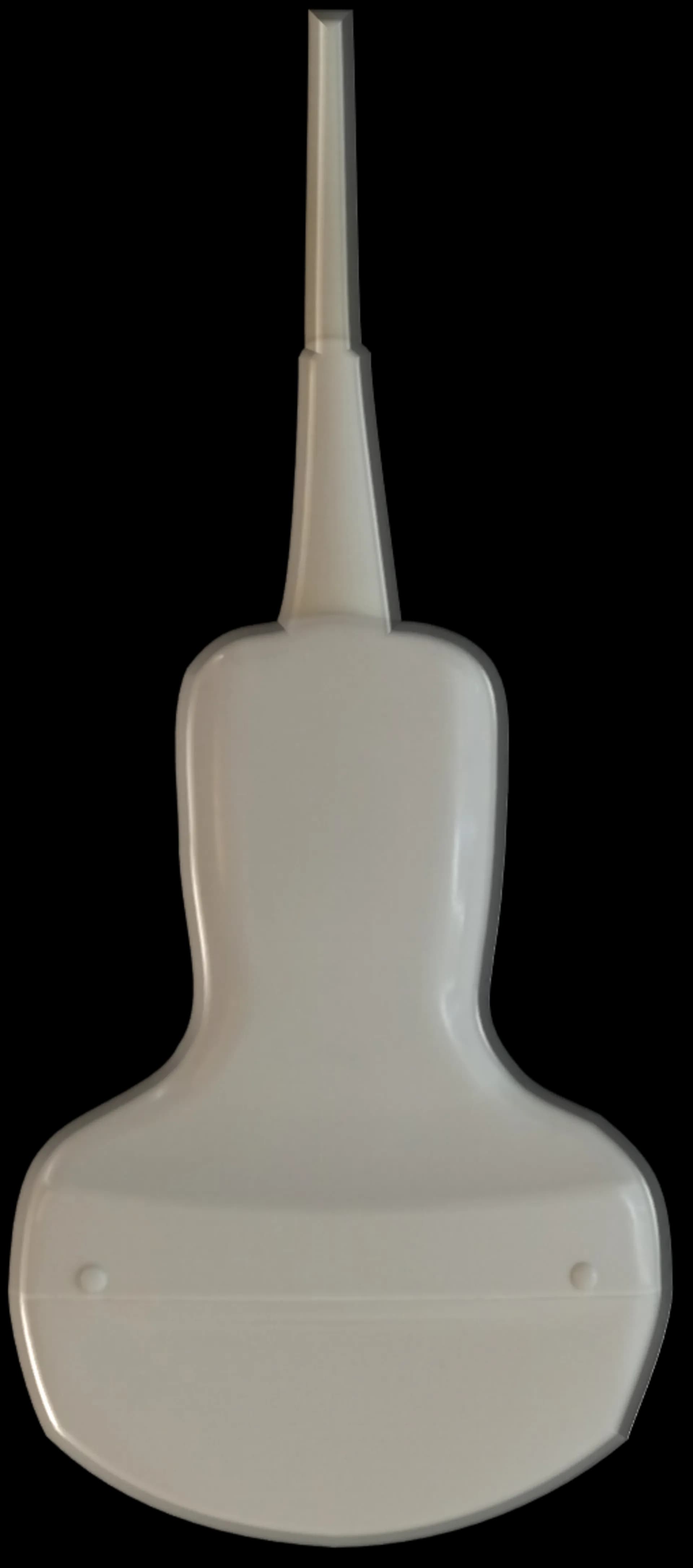
This probe is used for examining deeper structures. It has a wider field of view and better penetration depth, but compared to a linear probe provides lower resolution. In musculoskeletal ultrasonography, it is useful for example when imaging the hip joint, deeper muscle groups, or in patients with a thicker subcutaneous layer.
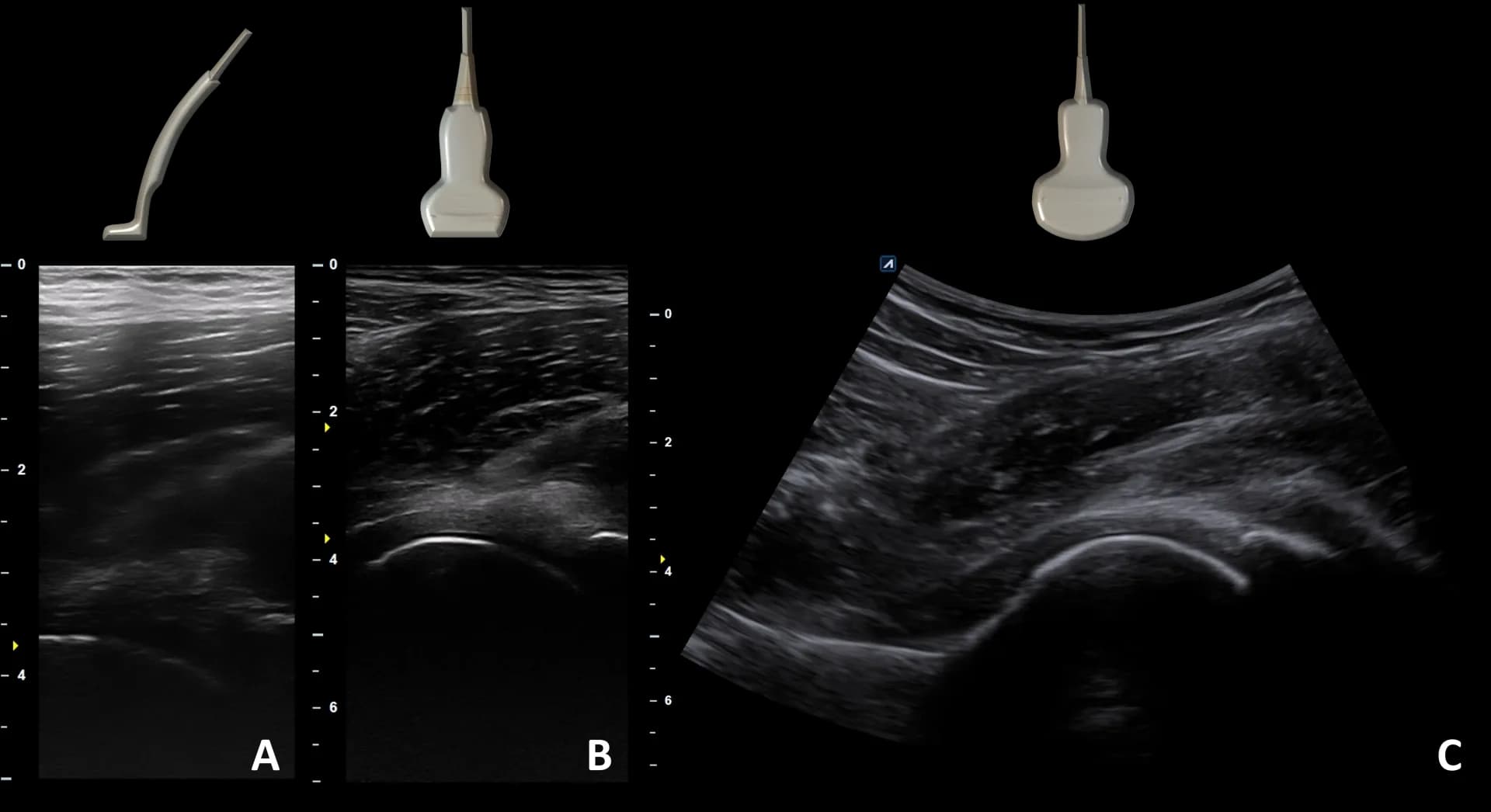
Sonographic imaging of the hip joint using different types of ultrasound probes. A: High-frequency probe (images superficial structures such as subcutaneous tissue and superficial muscles well), B: Linear probe (images both superficial and deep structures well), C: Convex probe (images deep structures well, over a wider range).