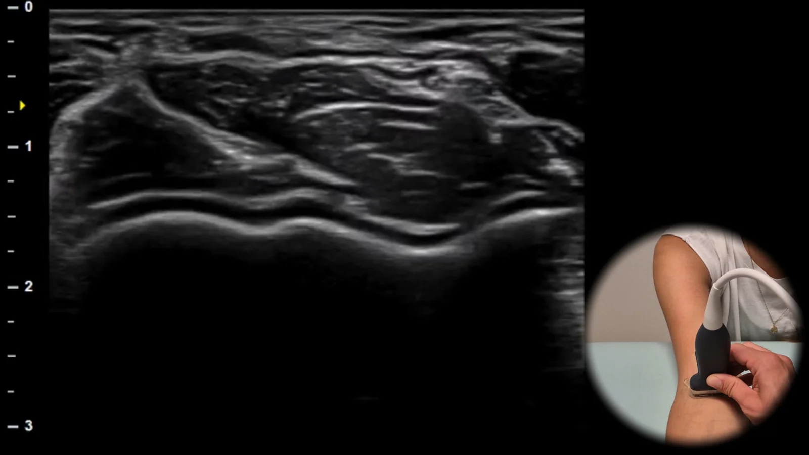
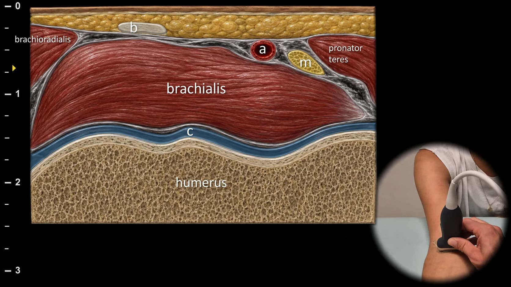
Figure 1. Ventral view, transverse plane. b: distal tendon of m. biceps brachii, a: a. brachialis, m: n. medianus, c: cartilage
Transverse ultrasound section of the anterior part of the elbow joint. The distal humerus is displayed as a hyperechoic cortical line covered by a hypoechoic cartilage layer. In the surrounding area, m. brachialis, m. pronator teres, m. brachioradialis and the distal tendon of m. biceps brachii (b) are visible. In this projection, neurovascular structures are also clearly visible, particularly a. brachialis (a) and n. medianus (m).
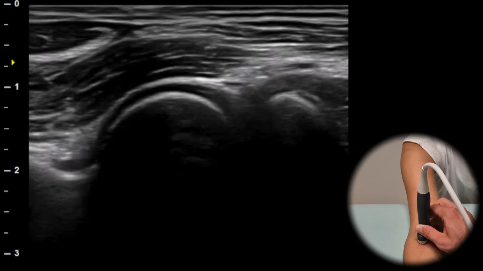
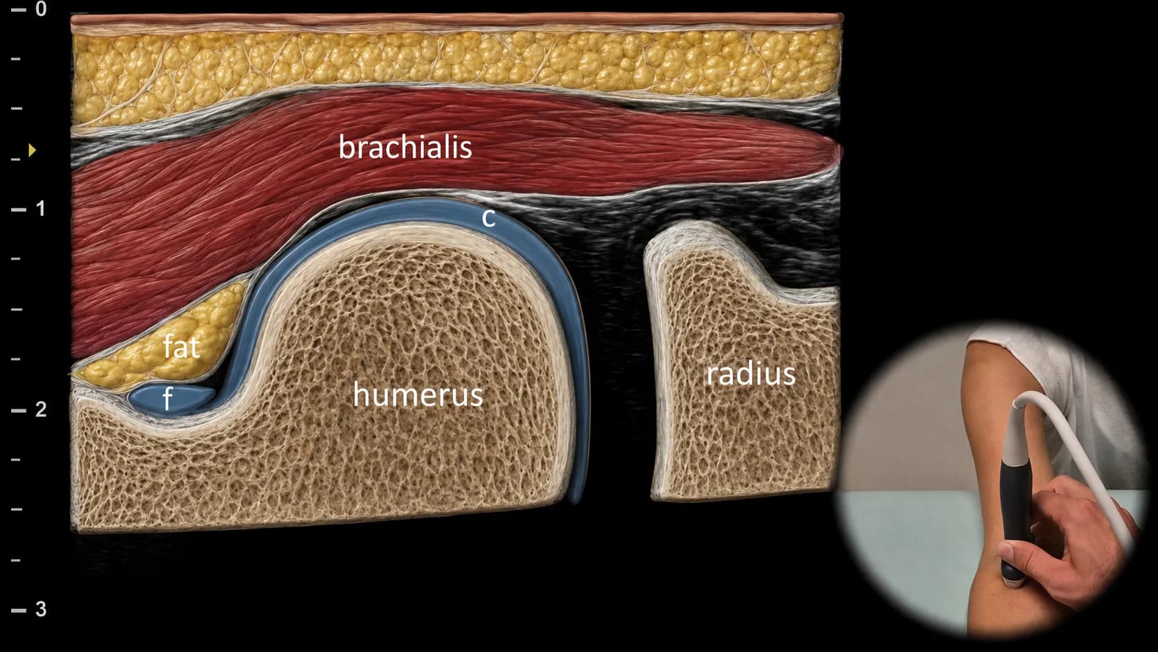
Figure 2. Ventral view, sagittal plane, radial side. c: cartilage, f: fluid in the elbow joint
Longitudinal ultrasound section of the anterior radial part of the elbow joint focusing on the radiohumeral articulation. The radial head, capitulum humeri and radial fossa are visible, which may contain a small amount of fat and fluid (f). A layer of hyaline cartilage (c) is shown on the surface of the humerus. Among the surrounding soft tissues, the brachialis muscle is most clearly visible.
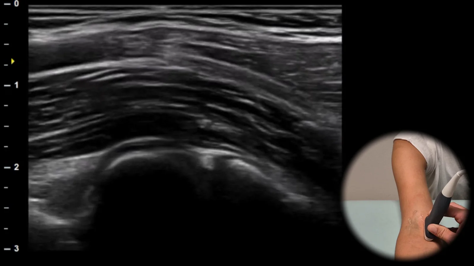
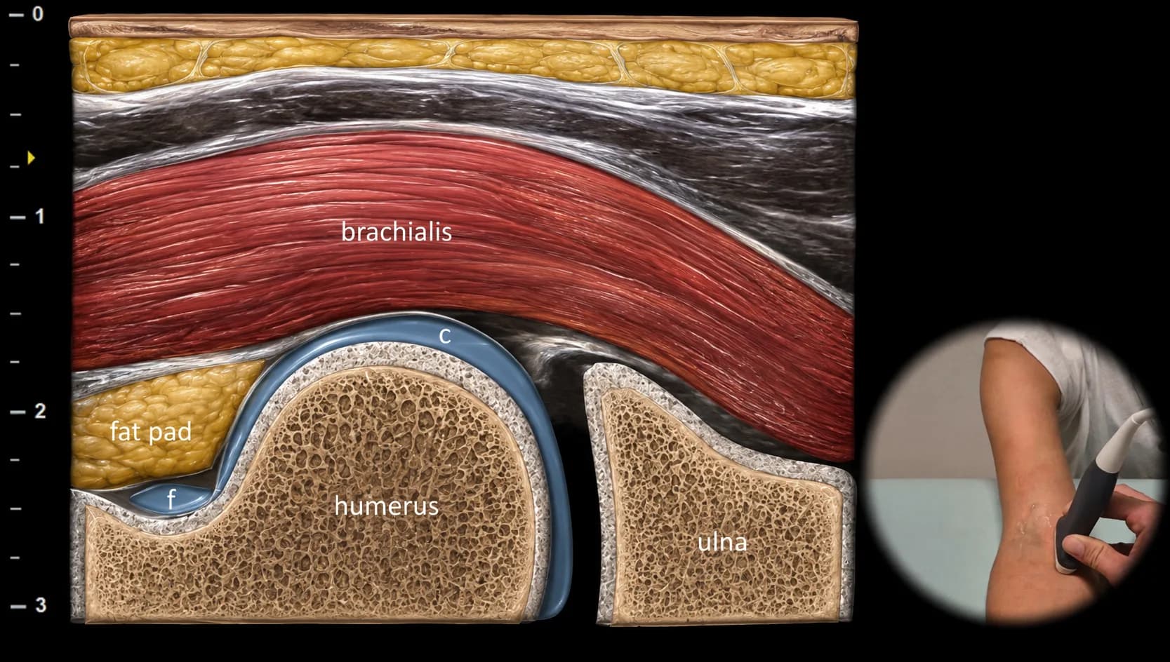
Figure 3. Ventral view, sagittal plane, ulnar side. c: cartilage, f: fluid in the elbow joint
Longitudinal ultrasound section of the anterior ulnar part of the elbow joint. Shows the humeroulnar articulation, particularly the trochlea humeri and processus coronoideus ulnae. We evaluate the joint space, fossa coronoidea and anterior synovial recess, where fluid (f) may accumulate. Among the surrounding soft tissues, primarily m. brachialis and m. pronator teres are visible in this projection.
 MSK Academy