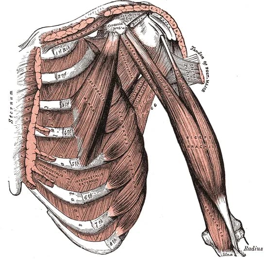
Anatomy of the m. biceps brachii is an important component of musculoskeletal ultrasonography in rehabilitation, orthopedics, and sports medicine, because this muscle significantly affects shoulder and elbow function and is a frequent source of pain and movement disorders. Knowledge of its two-headed arrangement, the course of the muscle belly, and the distal tendon is essential for proper evaluation of ultrasound findings.
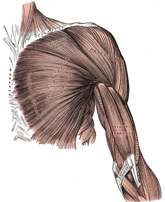
Proximally, the biceps is formed by the long and short heads. The long head runs in the sulcus intertubercularis humeri and on ultrasound is well visualized as an oval fibrillar structure. The short head originates from the processus coracoideus and runs more medially and superficially.
In the middle portion of the arm, both heads merge into a unified muscle belly, which lies superficially over the m. brachialis. In ultrasound imaging, it has a typical spindle shape and homogeneous muscle architecture. This area is important when evaluating muscle lesions, hematomas, or edema.
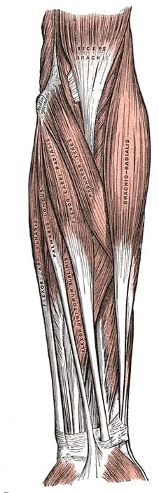
Distally, the biceps transitions into a distal tendon, which attaches to the tuberositas radii. This part is functionally significant for elbow flexion and forearm supination and is essential in diagnosing partial and complete ruptures. The examination may also include imaging of the bicipitoradial bursa.