 MSK Academy
Proximaler Abschnitt
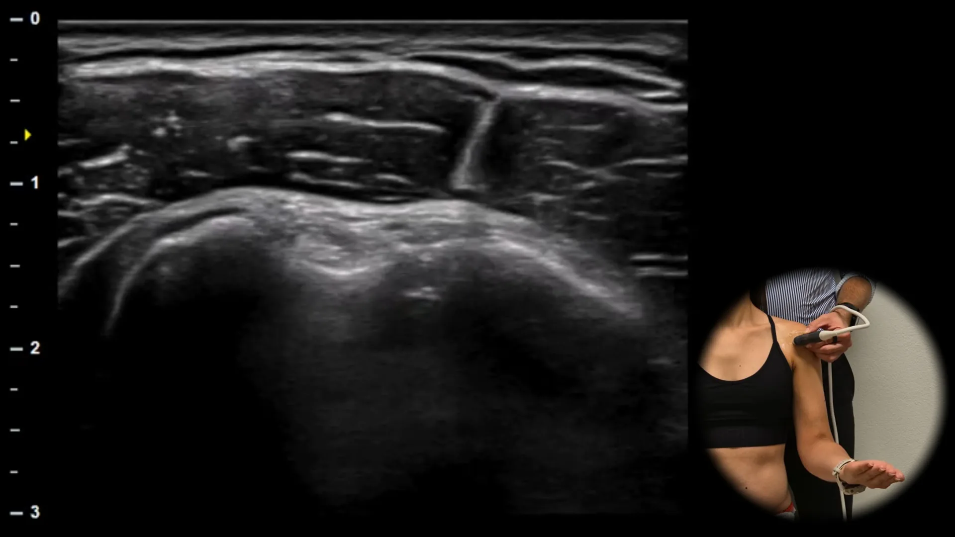
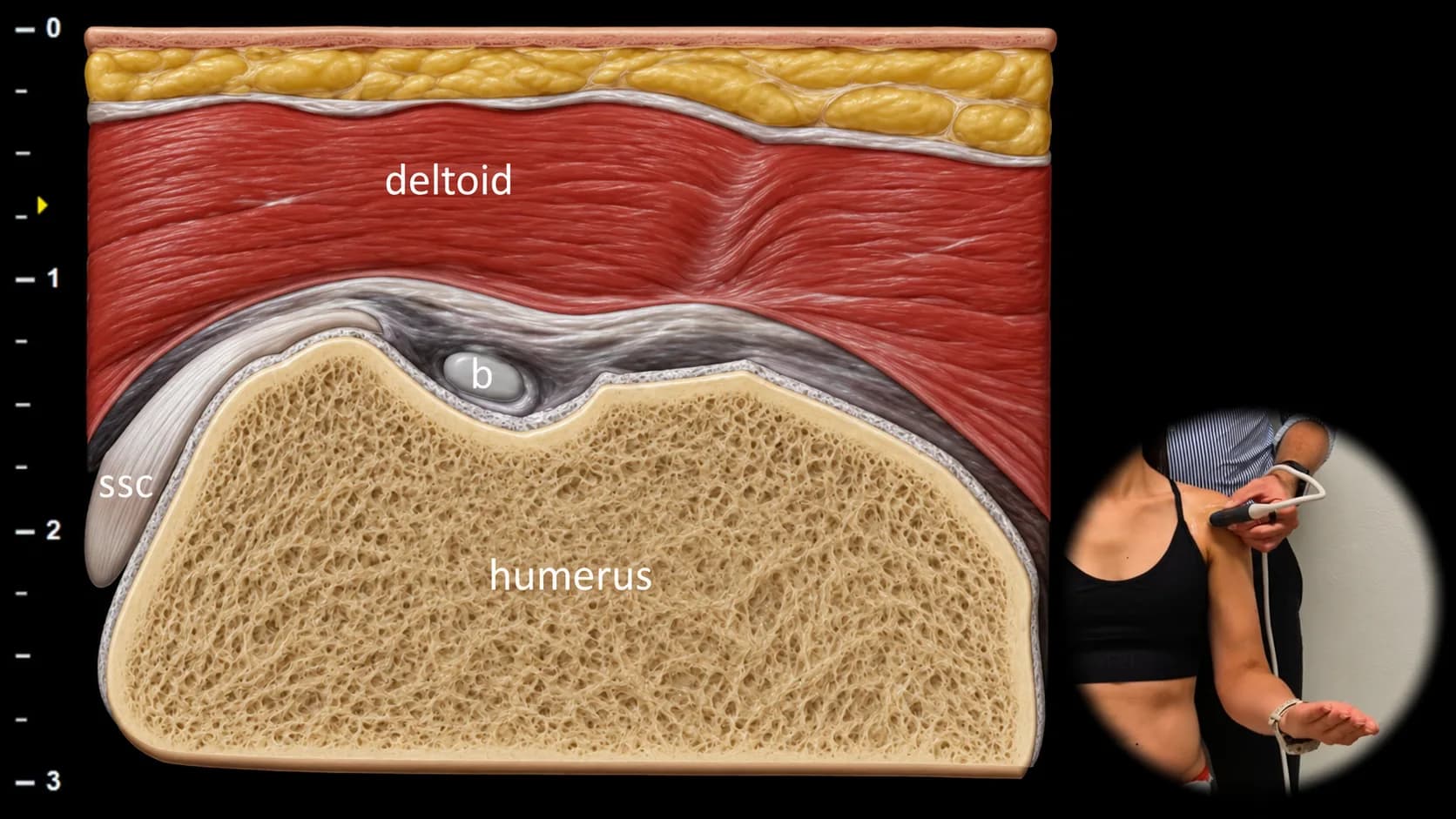
Abbildung 1. Ventralansicht, Transversalebene. b: Sehne des langen Kopfes des m. biceps brachii, ssc: Sehne des m. subscapularis.
Im Sulcus intertubercularis des Humerus liegt die Sehne des langen Kopfes des m. biceps brachii (b), die als gut abgegrenzte ovale hyperechogene fibrilläre Struktur erscheint. Um die Sehne ist ein dünner hypoechogener Saum der Synovialscheide sichtbar. Das Tuberculum majus ist lateral, das Tuberculum minus medial dargestellt. Am medialen Rand des Sulcus ist ein Teil der Sehne des m. subscapularis (ssc) sichtbar, die am Tuberculum minus des Humerus ansetzt. Oberflächlich liegt der m. deltoideus.
Klinischer Hinweis
Flüssigkeit um die Sehne des langen Bizepskopfes kann Ausdruck einer Beteiligung der Sehnenscheide selbst sein, aufgrund der Verbindung der Synovialscheide mit der Gelenkhöhle jedoch auch aus dem Glenohumeralgelenk stammen.
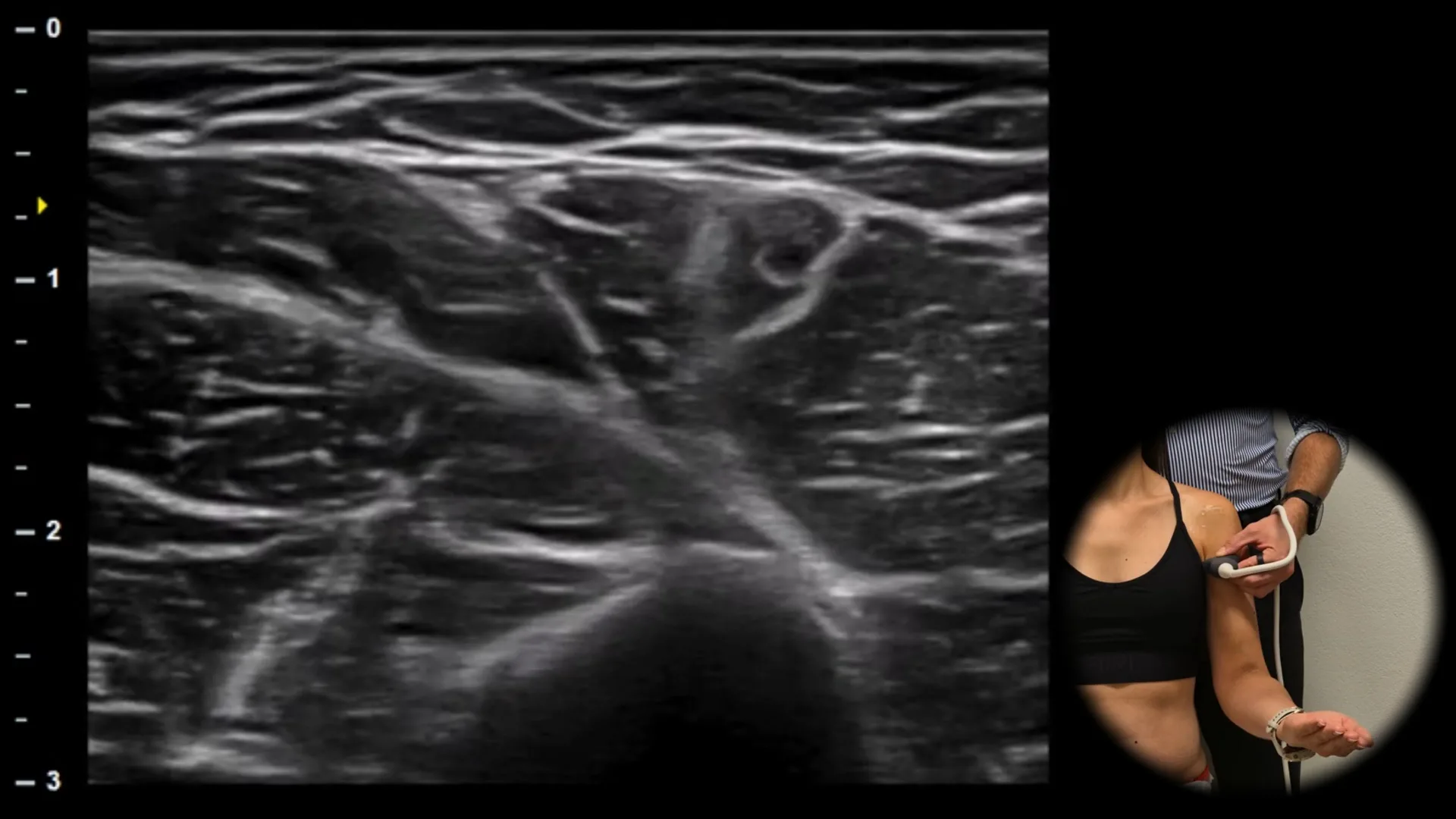
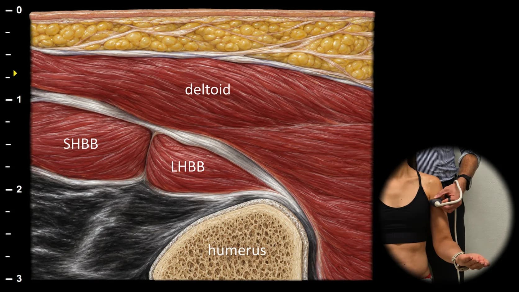
Abbildung 2. Ventrale Ansicht, Transversalebene. LHBB: Muskelbauch des langen Kopfes des Biceps brachii, SHBB: Muskelbauch des kurzen Kopfes des Biceps brachii
Ein weiter distal gelegener transversaler Ultraschallschnitt des ventralen Oberarms zeigt die Muskelbäuche beider Köpfe des Biceps brachii. Der lange Kopf (LHBB) liegt lateral, der kurze Kopf (SHBB) medial. Der Musculus deltoideus liegt oberflächlich, und der proximale Humerus ist in der Tiefe sichtbar. Diese Projektion eignet sich zur Beurteilung der Integrität des Muskelgewebes, der wechselseitigen Symmetrie beider Bizepsköpfe und der dynamischen Muskelkontraktion.
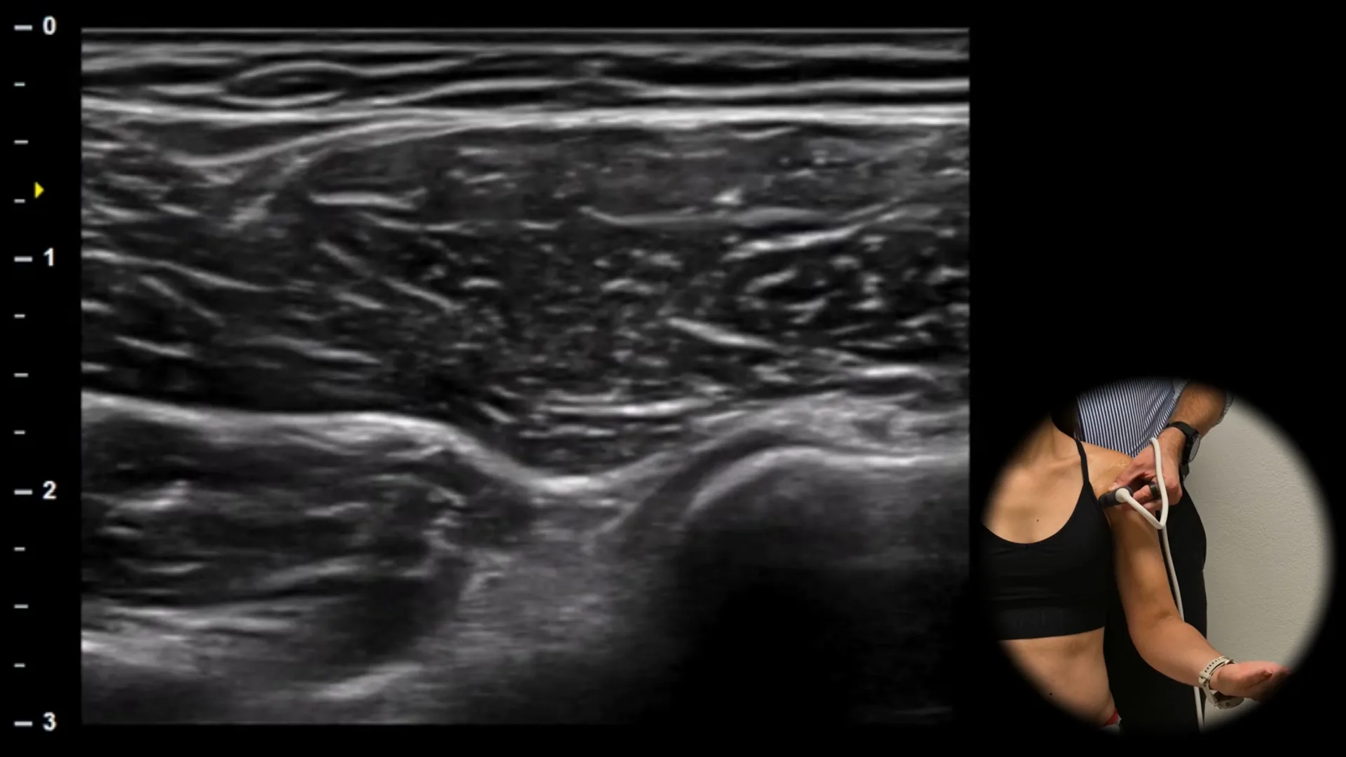
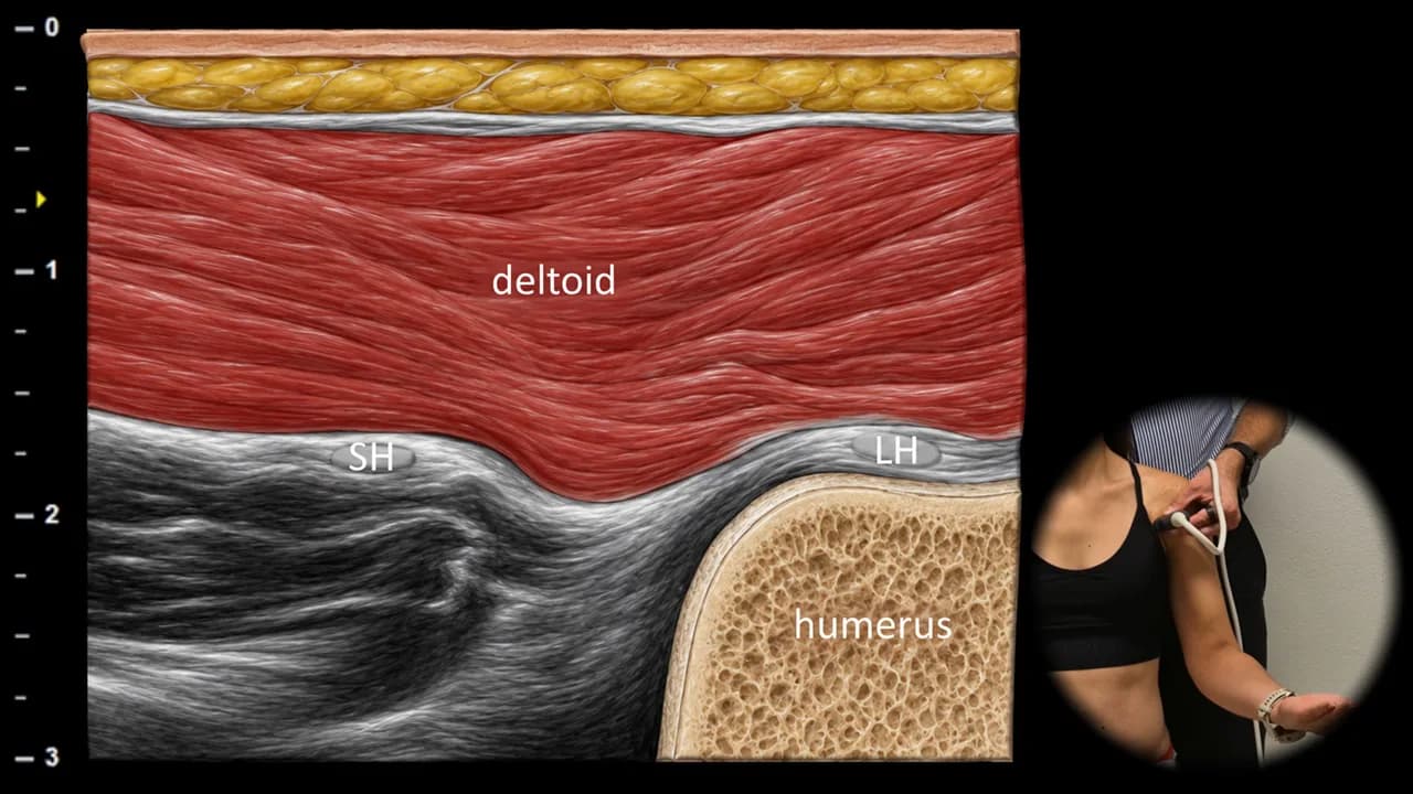
Abbildung 3. Ansicht von ventral, Transversalebene. LH: langer Kopf des M. biceps brachii, SH: kurzer Kopf des M. biceps brachii
Ein weiter medial und proximal gelegener transversaler sonografischer Schnitt der anterioren Schulter zeigt den kurzen Kopf des M. biceps brachii (SH) als runde echoarme Struktur, die unter dem M. deltoideus und medial des langen Kopfes (LH) liegt. Der kurze Kopf erstreckt sich hier in Richtung seines Ursprungs im Bereich des Processus coracoideus. Der M. deltoideus liegt oberflächlich, während der proximale Humerus in der Tiefe sichtbar ist. Diese Projektion ist wichtig für die vollständige Beurteilung der proximalen Anatomie des Bizeps und zur Unterscheidung einer isolierten Beteiligung des kurzen und des langen Kopfes.
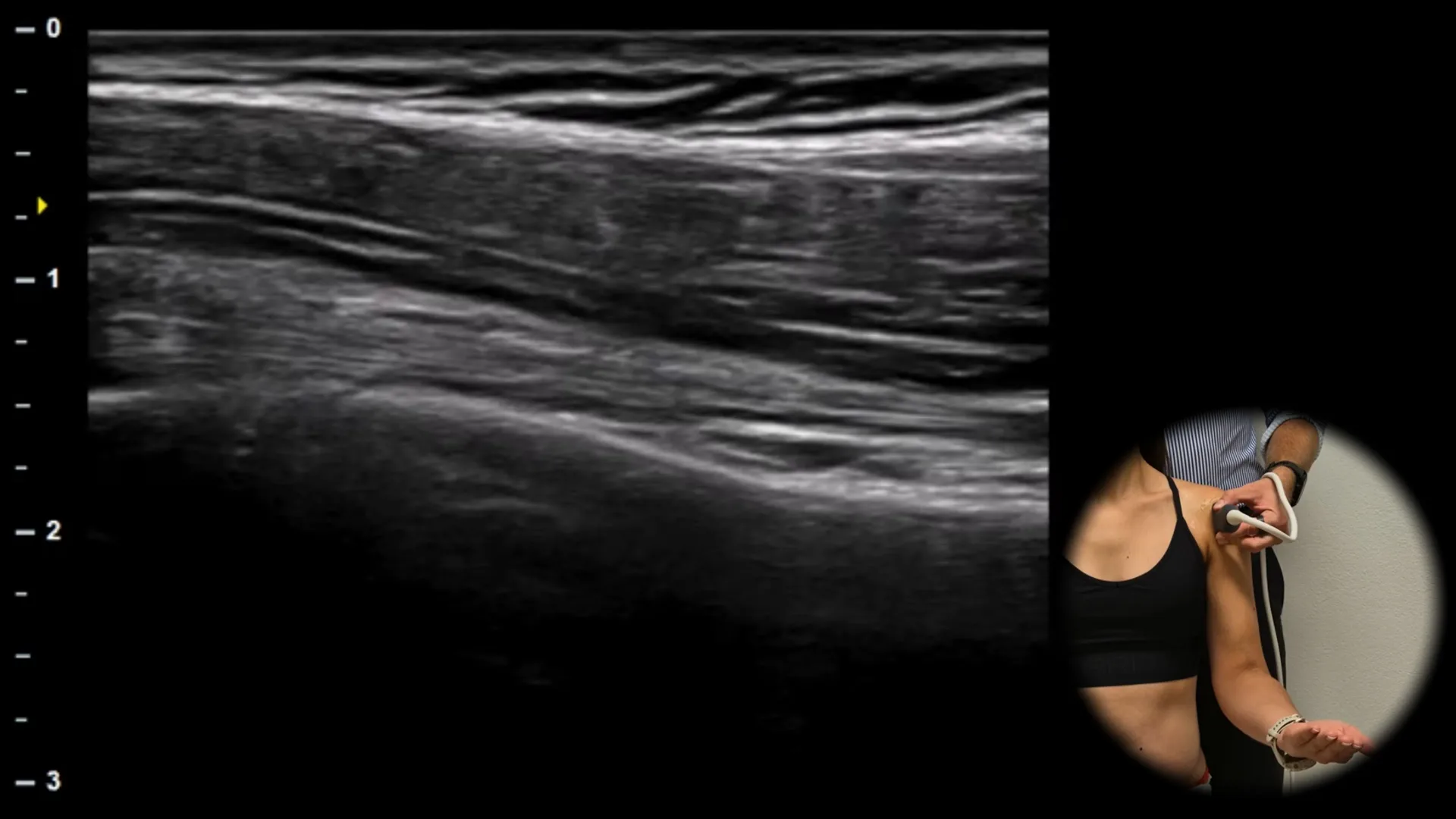
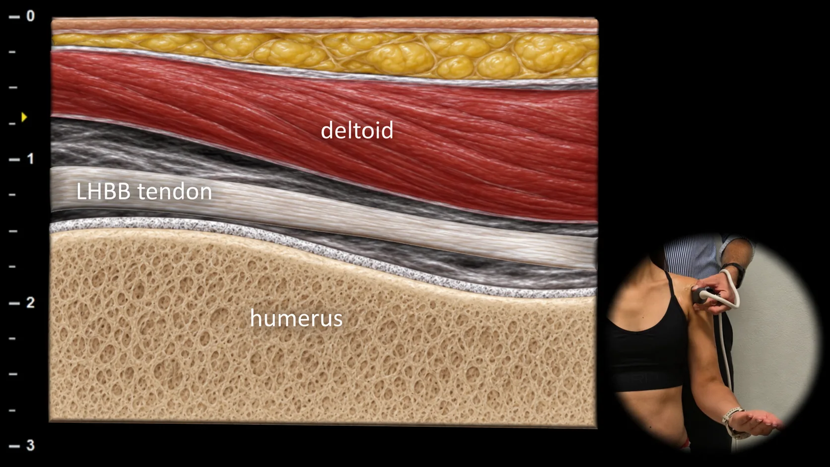
Abbildung 4. Ventralansicht, Longitudinalebene. LHBB-Sehne: Sehne des langen Kopfes des M. biceps brachii
Der sonografische Längsschnitt der anterioren Schulterregion zeigt die Sehne des langen Kopfes des M. biceps brachii als hyperechogene fibrilläre Struktur, die im Sulcus intertubercularis des Humerus verläuft. Die knöchernen Wände des Sulcus werden durch die kortikalen Ränder von tuberculum majus und tuberculum minus gebildet, die eine zuverlässige anatomische Orientierung ermöglichen. Oberflächlich liegt der M. deltoideus. In dieser Projektion beurteilen wir primär die Kontinuität, Kontur und Dicke der Sehne sowie das mögliche Vorliegen von Flüssigkeit in der Sehnenscheide.
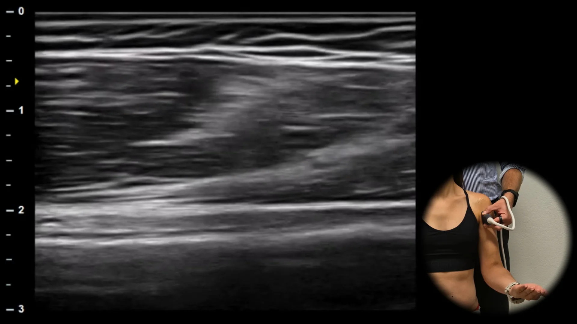
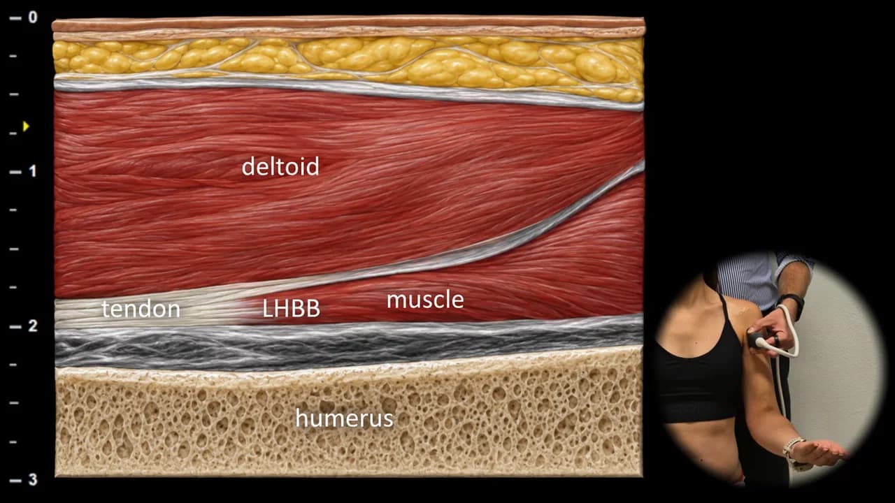
Abbildung 5. Ventralansicht, Longitudinalebene. LHBB: langer Kopf des M. biceps brachii
Longitudinaler sonografischer Schnitt durch den vorderen Teil der Schulter im Bereich des myotendinösen Übergangs des langen Kopfes des M. biceps brachii. Ein gleichmäßiger Übergang von der Sehne zum Muskelbauch ist erkennbar, der einen Wechsel von der hyperechogenen fibrillären Struktur der Sehne zu hypoechogenem Muskelgewebe mit feiner streifiger Architektur darstellt. Der M. deltoideus liegt oberflächlich, und der Humerus ist in der Tiefe sichtbar.
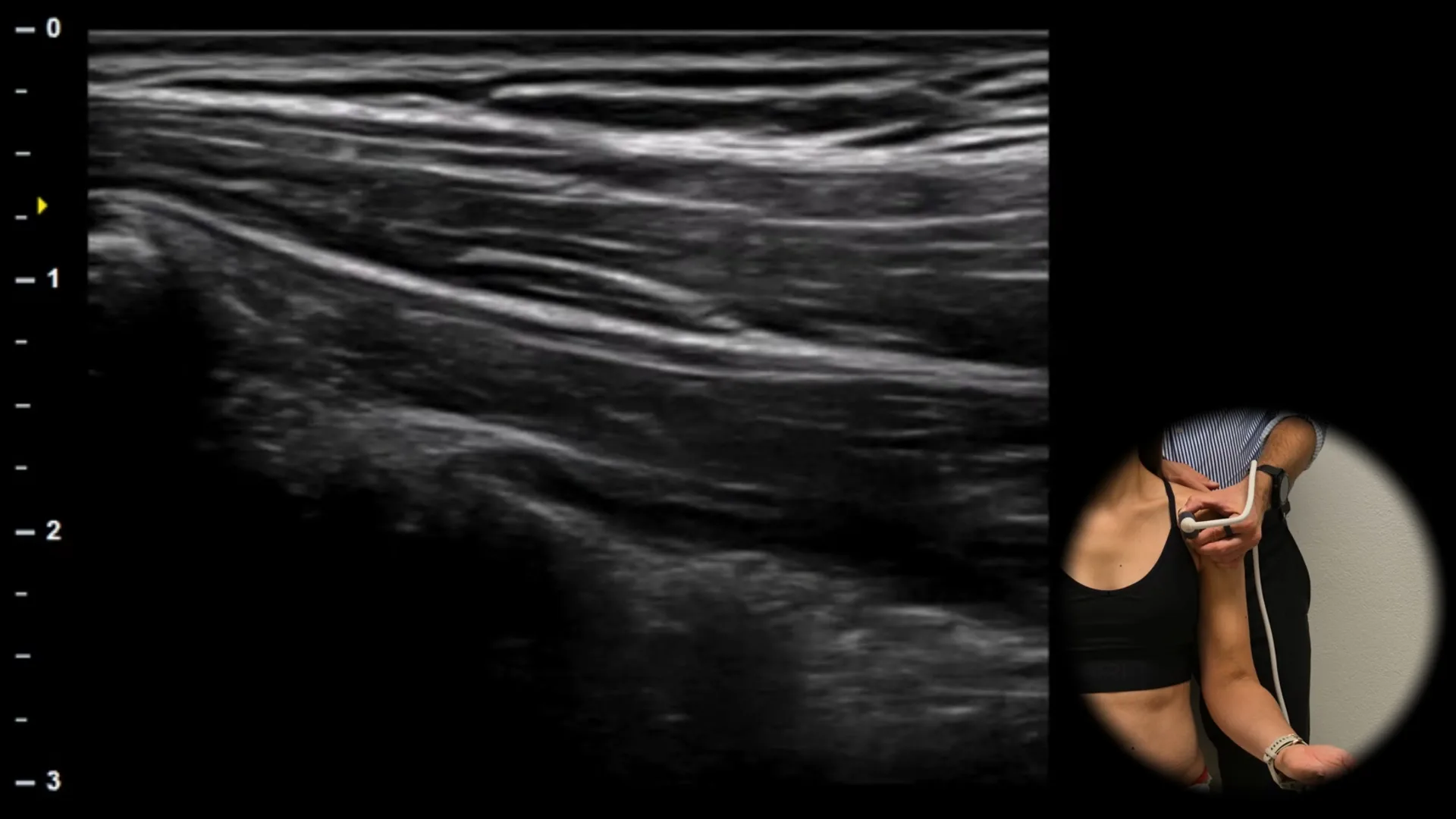
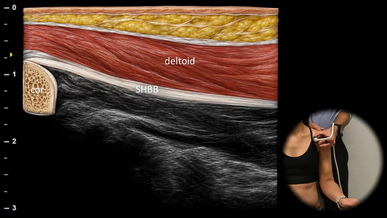
Abbildung 6. Ventralansicht, Longitudinalebene. SHBB: kurzer Kopf des m. biceps brachii, cor: processus coracoideus
Der sonografische Längsschnitt durch den anterioren Abschnitt der Schulter zeigt den kurzen Kopf des m. biceps brachii (SHBB), der nach proximal zu seinem Ursprung am processus coracoideus der Scapula (cor) verläuft. Der Muskel erscheint als hypoechogene Struktur mit feiner streifiger Architektur angrenzend an die kortikale Oberfläche des Korakoids. Der m. deltoideus liegt oberflächlich. Diese Projektion ist zur Differenzierung des kurzen und langen Bizepskopfes sowie zur Beurteilung des Bereichs bei proximalen Bizepsschmerzen oder bei Verdacht auf eine traumatische Avulsionsverletzung hilfreich.
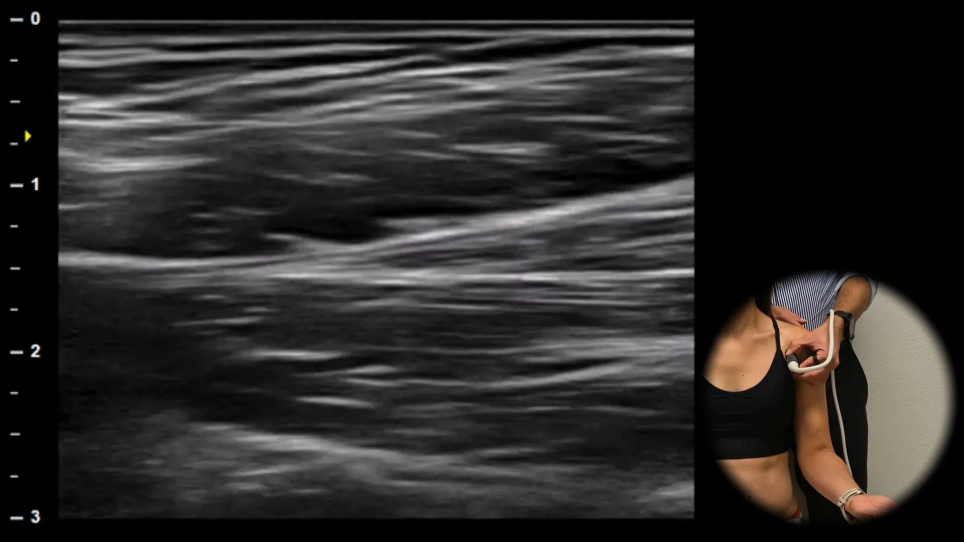
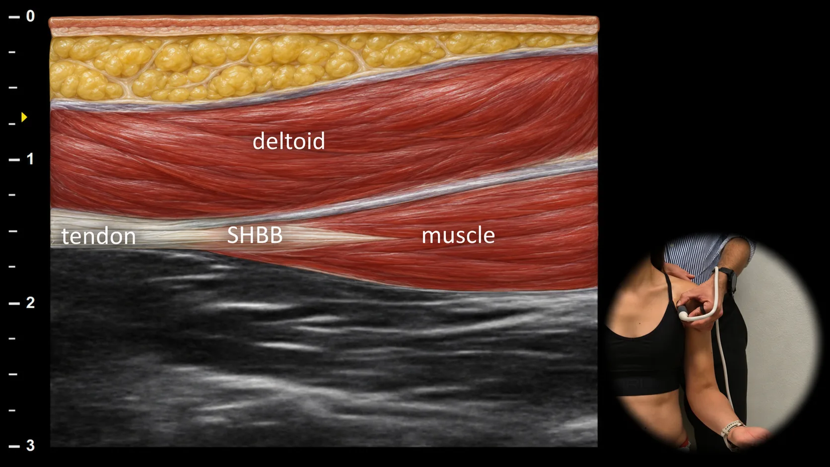
Abbildung 7. Ventralansicht, Longitudinalebene. SHBB: kurzer Kopf des M. biceps brachii
Longitudinaler Ultraschallschnitt durch den anterioren Teil des Oberarms im Bereich des myotendinösen Übergangs des kurzen Kopfes des M. biceps brachii (SHBB). Sichtbar ist ein gleichmäßiger Übergang der Sehnenfasern in den proximalen Muskelbauch, der eine homogene Echogenität und eine feine streifige Architektur aufweist. Der M. deltoideus liegt oberflächlich. In dieser Projektion beurteilen wir primär die Kontinuität und Struktur des myotendinösen Übergangs.
Zugang zur gesamten Bibliothek
Voller Zugriff auf Scan-Protokolle, Anatomie und klinische Referenzen. Jederzeit kündbar.
- Alle Protokolle und Anatomiereferenzen
- Originale Ultraschall-Illustrationen und Video-Demonstrationen
- Synchronisierung zwischen Mobilgerät und Web