 MSK Academy
Distaler Abschnitt
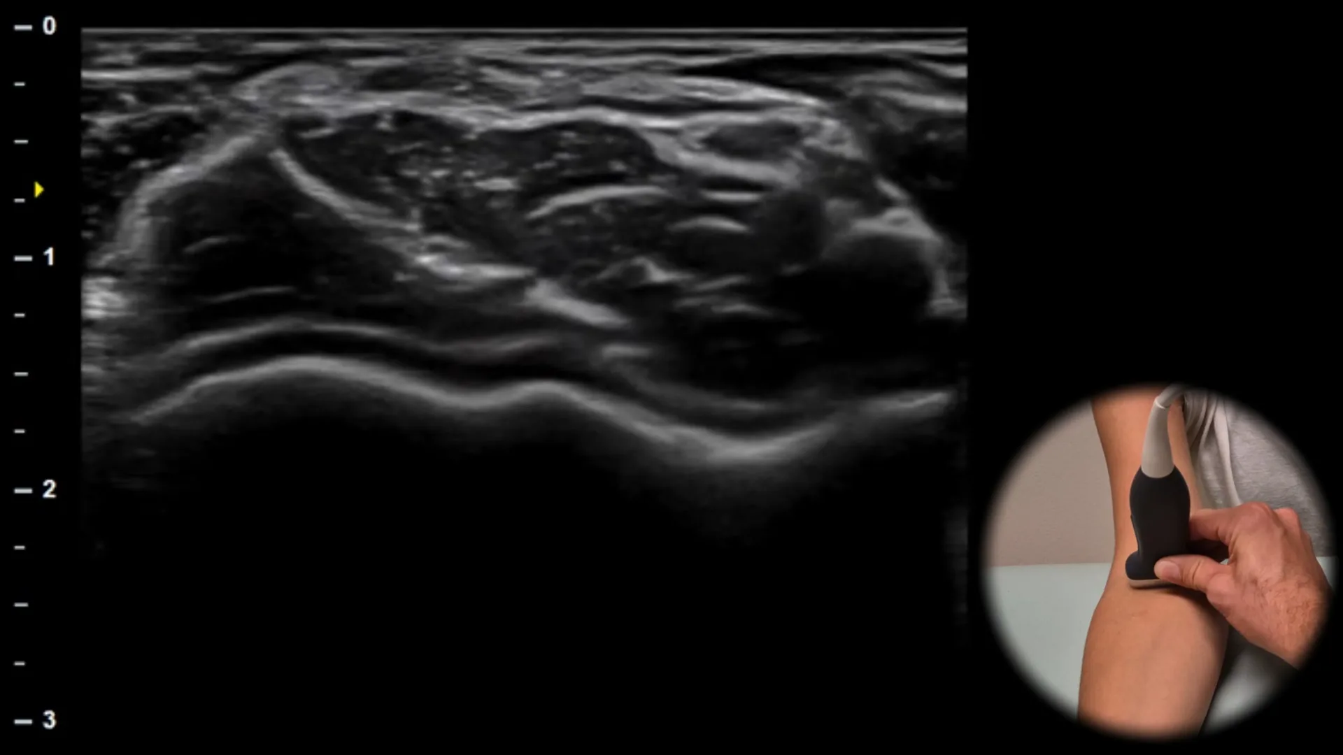
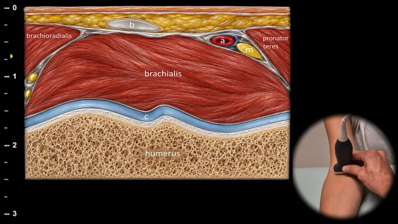
Abbildung 1. Ventrale Ansicht, Transversalebene. b: distale Sehne des m. biceps brachii, m: n. medianus, r: n. radialis, a: a. brachialis, c: Humerusknorpel
Der transversale Ultraschallschnitt durch den anterioren Anteil des distalen Oberarms zeigt die distale Sehne des m. biceps brachii (b) im Querschnitt als oberflächlich gelegene hyperechogene ovale Struktur. Unmittelbar darunter ist der m. brachialis sichtbar, der ein homogeneres und weniger echogenes Erscheinungsbild aufweist. Lateral befindet sich der m. brachioradialis, medial der m. pronator teres. In diesem Bereich sind auch der medial der Sehne gelegene n. medianus (m) und der lateral entlang des m. brachioradialis verlaufende n. radialis (r) gut identifizierbar; in unmittelbarer Nähe ist außerdem die a. brachialis (a) sichtbar. Diese Projektion ist für die anatomische Orientierung, das Nerven-Mapping und die sichere Planung interventioneller Eingriffe im Bereich der Fossa cubitalis von Bedeutung.
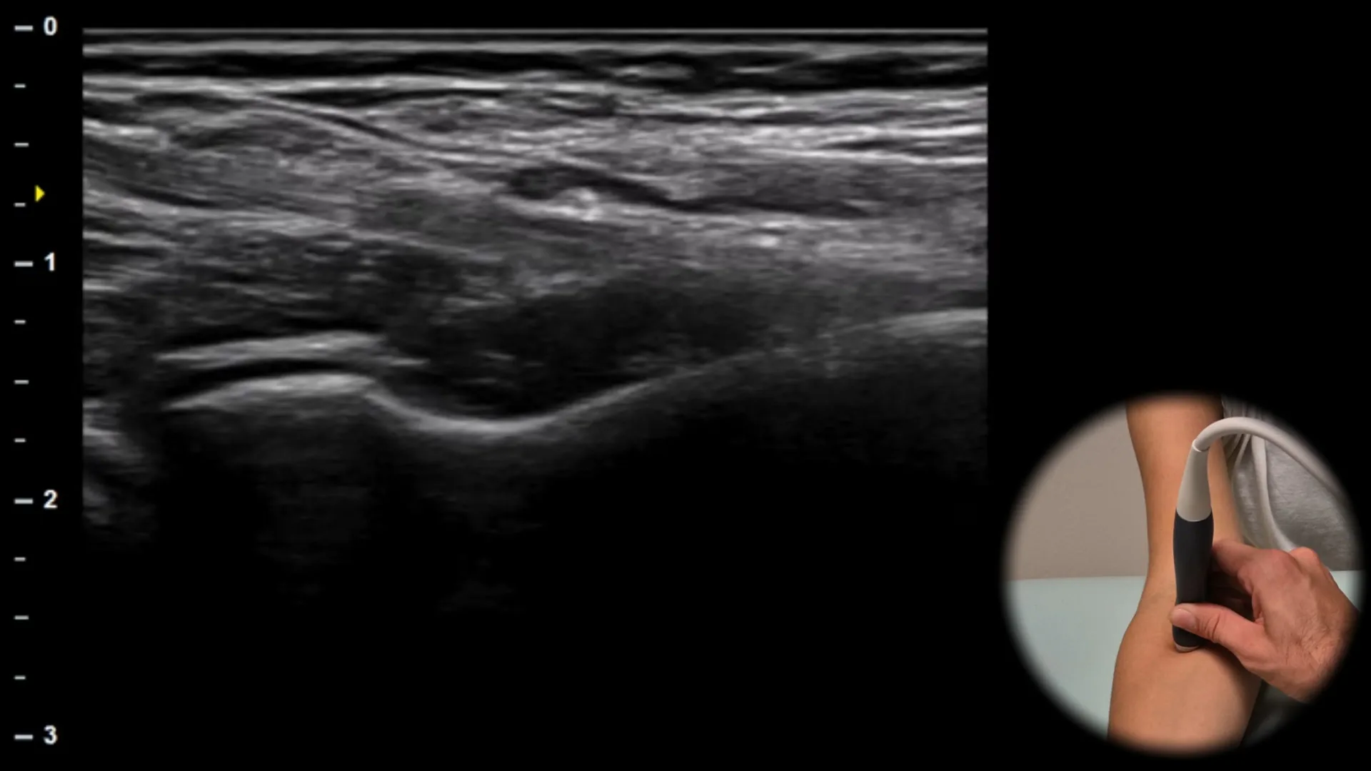
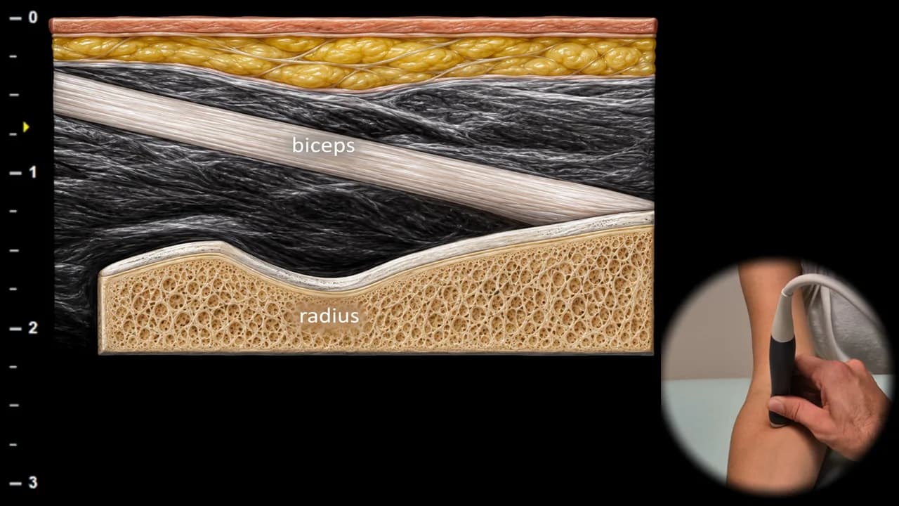
Figure 12. Ventral view, longitudinal oblique plane. biceps: distal tendon of m. biceps brachii
The longitudinal oblique ultrasound section follows the natural course of the distal tendon of m. biceps brachii to its insertion on the tuberositas radii. The tendon appears as a hyperechoic fibrillar structure with parallel linear architecture that narrows distally. The probe orientation corresponds to the oblique course of the tendon. Deep to the tendon, the m. brachialis is visible, with the cortex of the radius forming the bottom of the image. This projection is essential for assessing the continuity of the distal biceps tendon and for demonstrating partial or complete ruptures.
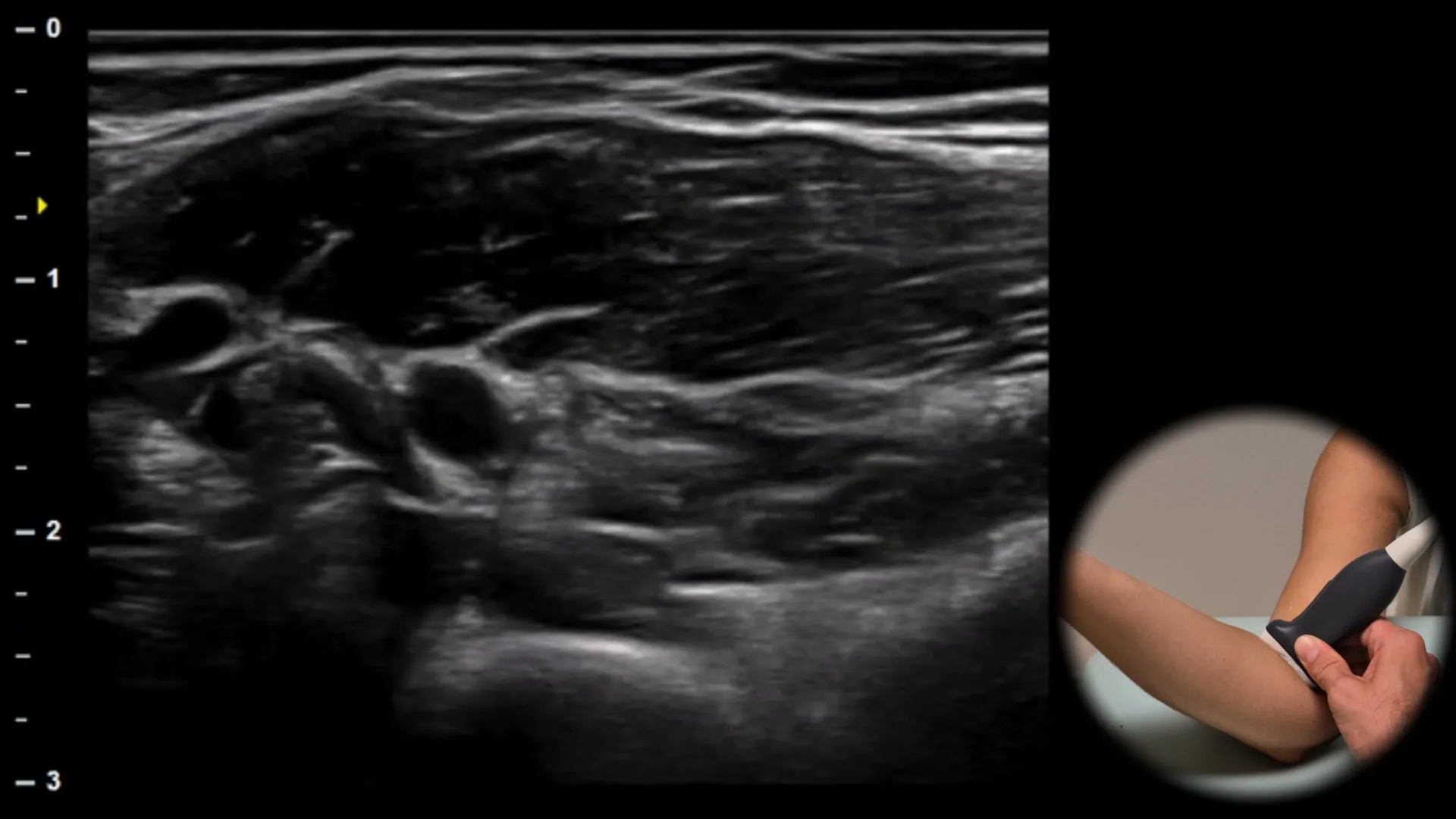
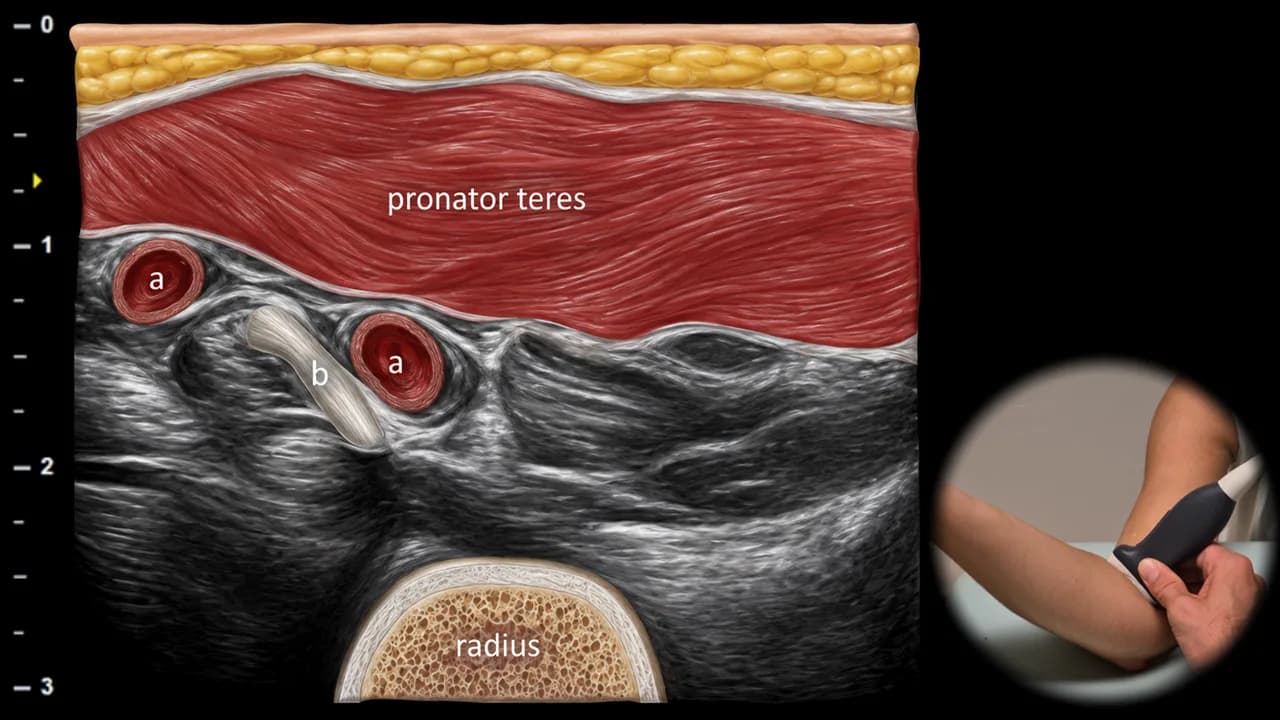
Abbildung 3. Ventrale Ansicht, Transversalebene – Pronatorfenster. b: distale Sehne des M. biceps brachii, a: A. brachialis
Der transversale Ultraschallschnitt im Bereich des Pronatorfensters zeigt die distale Sehne des M. biceps brachii (b) im Querschnitt in ihrem Verlauf zum Ansatz an der tuberositas radii. Die Sehne erscheint als ovale hyperechogene fibrilläre Struktur. Tief unterhalb der Sehne ist der Radius als hyperechogene Kortikalislinie mit dorsaler Schallauslöschung sichtbar. Oberflächlich liegt der M. pronator teres, der ein geeignetes akustisches Fenster für die Darstellung dieses Bereichs bildet. Diese Einstellung ist besonders nützlich für die Beurteilung von Pathologien der distalen Bizepssehne, insbesondere von Partialrupturen und Enthesopathien.
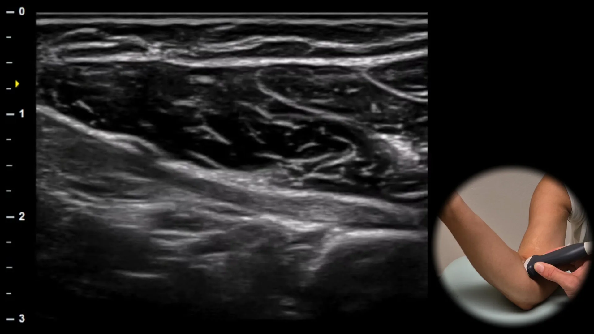
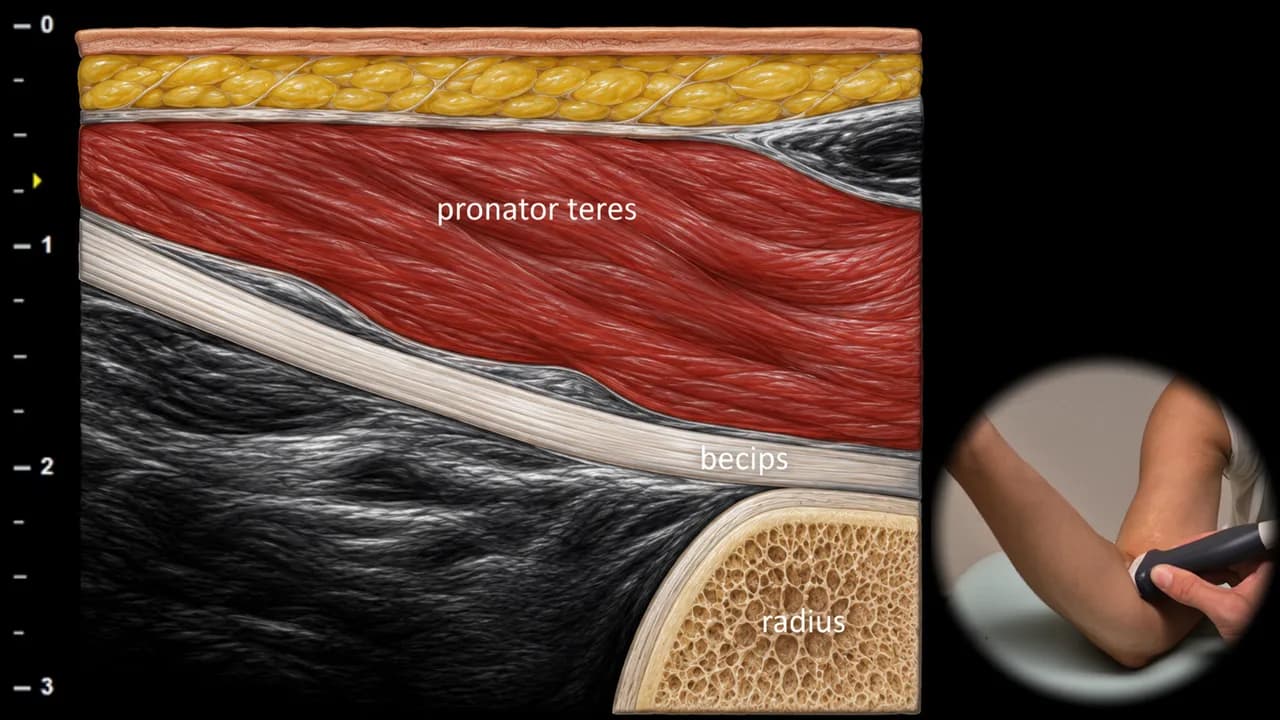
Abbildung 4. Ventrale Ansicht, Longitudinalebene. biceps: distale Sehne des M. biceps brachii
Der longitudinale Ultraschallschnitt zeigt die distale Sehne des M. biceps brachii in ihrem schrägen Verlauf vom distalen myotendinösen Übergang bis zu ihrem Ansatz an der Tuberositas radii. Die Sehne stellt sich als lineare hyperechogene fibrilläre Struktur dar, die unter dem M. pronator teres verläuft. Die Tuberositas radii ist als helle, durchgehende Kortikalislinie sichtbar, an der die Sehne ansetzt. Diese Projektion ermöglicht die Beurteilung der Sehnenkontinuität, die dynamische Untersuchung und die Beurteilung von Tendinopathien oder partiellen und vollständigen Rupturen.
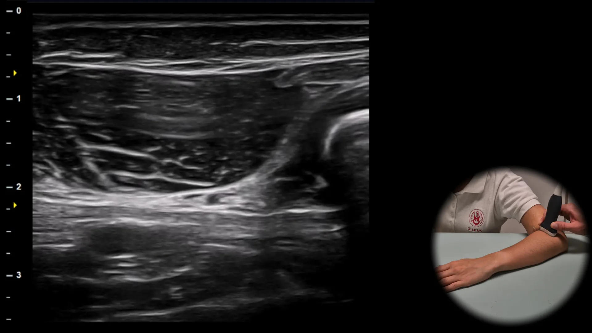
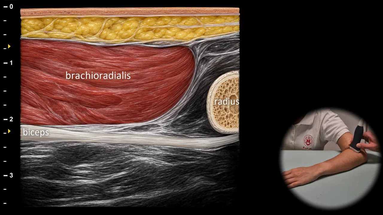
Abbildung 5. Lateral-roll-Position, Longitudinalebene. biceps: distale Sehne des m. biceps brachii
Der sonografische Längsschnitt in Lateral-roll-Position zeigt die distale Sehne des m. biceps brachii als hyperechogene fibrilläre Struktur, die schräg vom myotendinösen Übergang bis zu ihrem Ansatz an der tuberositas radii verläuft. In dieser Projektion liegt die Sehne unterhalb des m. brachioradialis. Diese Ansicht eignet sich zur dynamischen Untersuchung, zur Beurteilung der Kontinuität der distalen Sehne und zur Beurteilung des Bereichs des myotendinösen Übergangs.
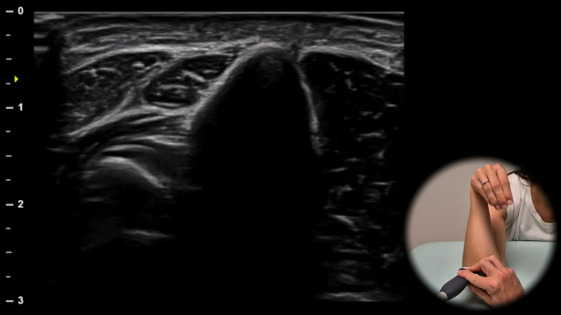
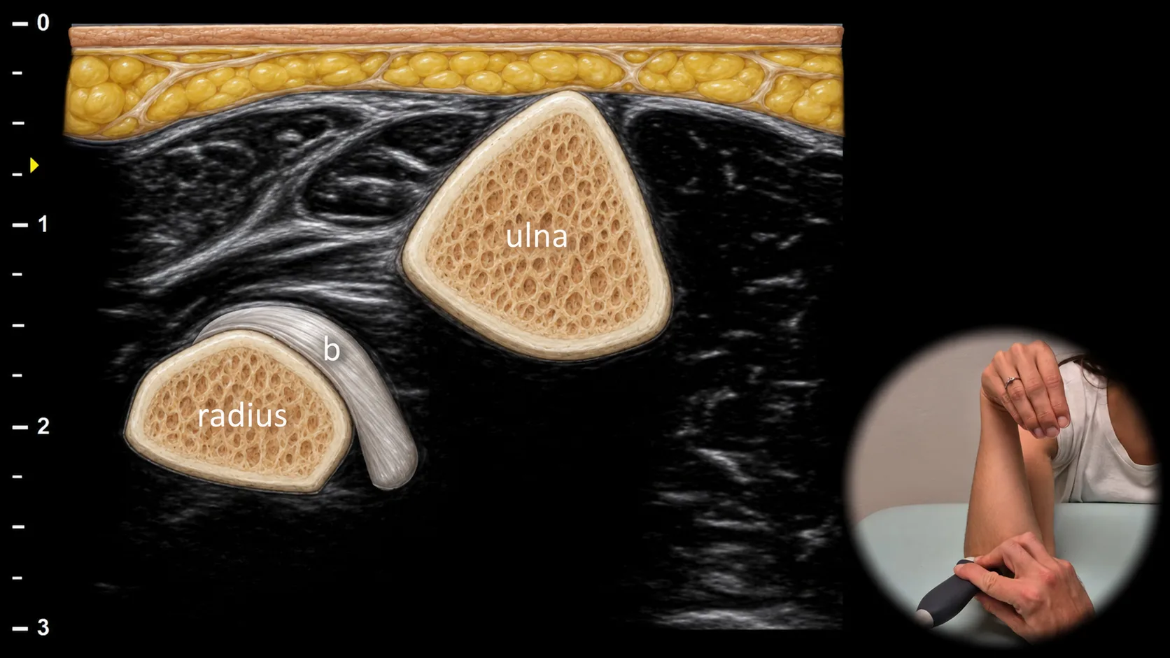
Abbildung 6. Cobra-Position, Transversalebene. b: distale Sehne des Musculus biceps brachii
Der transversale Ultraschallschnitt in Cobra-Position (Ellenbogenflexion und Pronation des Unterarms, wobei die Hand mit der Handfläche nach unten an der Schulter platziert wird) zeigt die distale Sehne des Musculus biceps brachii (b) in ihrem gekrümmten Verlauf zum Ansatz an der tuberositas radii. Die Sehne ist zwischen Radius und Ulna sichtbar, was eine klarere Darstellung ihres distalen Anteils und ihrer Beziehung zur Ansatzstelle ermöglicht. Diese Projektion ist besonders nützlich zur Beurteilung der Kontinuität der distalen Bizepssehne und bei der Diagnostik partieller oder kompletter Rupturen.
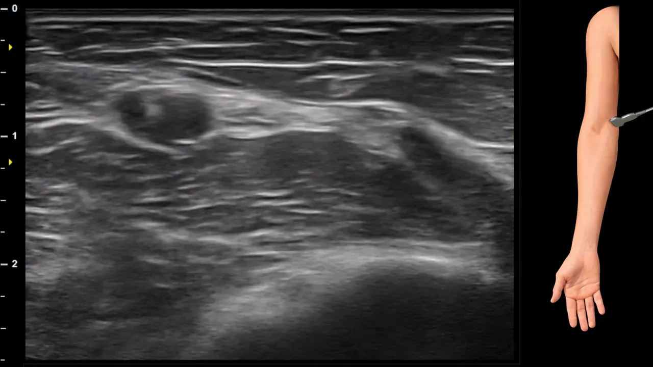
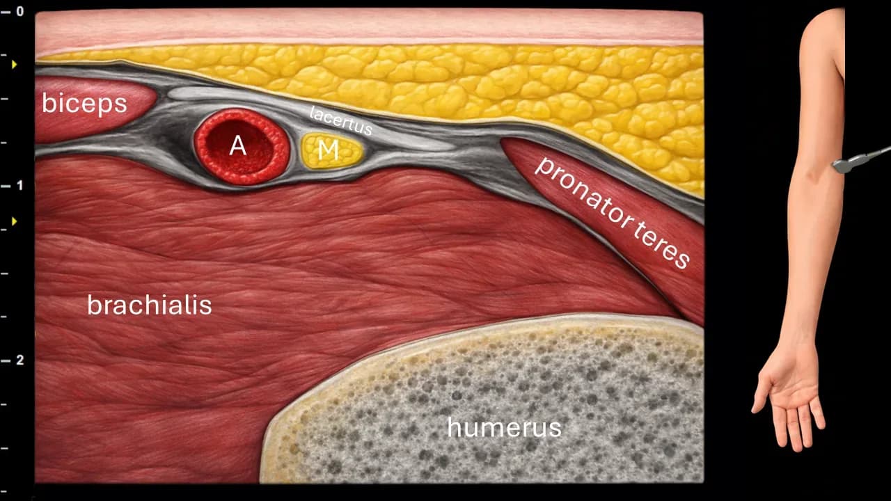
Abbildung 7. Cubitum, Transversalebene. A: a. brachialis, M: n. medianus.
Transversaler Ultraschallschnitt im Bereich der Fossa cubitalis. Das Bild zeigt die a. brachialis (A) und den n. medianus (M), der in dieser Projektion medial der a. brachialis liegt. Der n. medianus verläuft weiter unter dem Lacertus fibrosus und befindet sich im Raum zwischen dem m. brachialis und dem m. pronator teres. Das Bild erfasst außerdem den m. biceps brachii, den m. brachialis, den m. pronator teres und die Kontur des Humerus.
Zugang zur gesamten Bibliothek
Voller Zugriff auf Scan-Protokolle, Anatomie und klinische Referenzen. Jederzeit kündbar.
- Alle Protokolle und Anatomiereferenzen
- Originale Ultraschall-Illustrationen und Video-Demonstrationen
- Synchronisierung zwischen Mobilgerät und Web