Anatomy
The ankle joint is a very dynamic joint complex. It plays a crucial role in weight bearing, balance maintenance, and locomotion, and is therefore essential for daily activities as well as athletic performance. Compared to the shoulder, it has a smaller range of motion, but must withstand considerable mechanical stress and rapid directional changes. This is precisely why the ankle is a frequent site of sprains, tendon injuries, and overuse conditions. For ultrasound examination, orientation to bony landmarks, ligaments, tendons, bursae, and joint recesses is essential.
Bone Landmarks
Bone landmarks are fundamental reference points during scanning. They help guide the probe correctly and quickly distinguish normal anatomy from pathology.
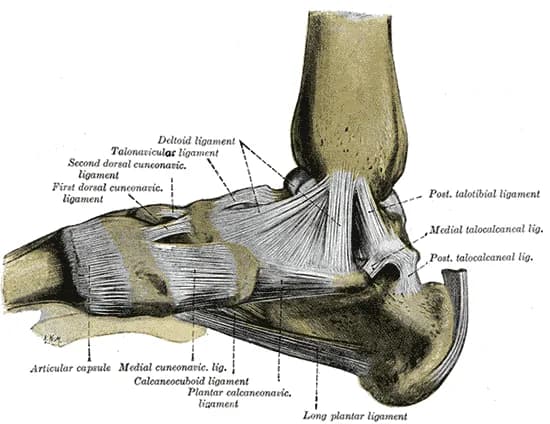
- Malleolus medialis – distal process of the tibia on the medial side, an important landmark for the deltoid ligament, tendon of m. tibialis posterior, and flexor retinaculum.
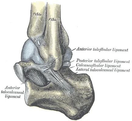
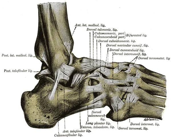
- Malleolus lateralis – distal end of the fibula, a key point for examination of the lateral ligamentous complex and peroneal tendons.
- Trochlea tali – the superior articular surface of the talus, visible in the anterior recess of the ankle, important when evaluating effusion and osteochondral lesions.
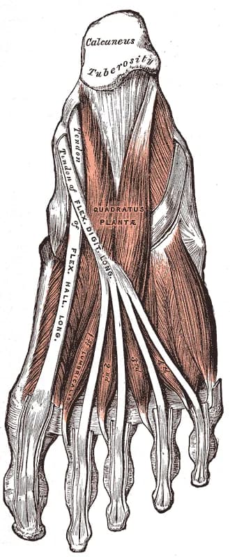
- Calcaneus – heel bone, insertion site of the Achilles tendon and reference point for surrounding bursae and soft tissues.
- Tuberositas ossis navicularis – bony prominence on the medial side of the foot, important for orientation when examining the m. tibialis posterior tendon and spring ligament.
- Base of the 5th metatarsal – lateral bony landmark, insertion site of the m. peroneus brevis tendon.
- Anterior margin of the distal tibia – landmark for the anterior joint recess and assessment of synovitis or effusion.
Muscles
Muscles – anterior group
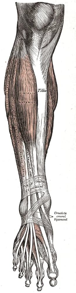
- Tibialis anterior – dorsiflexion and inversion of the foot.
- Extensor hallucis longus – extension of the thumb and dorsiflexion.
- Extensor digitorum longus – extension of toes 2–5 and dorsiflexion.
Muscles – lateral group
- Peroneus longus – eversion and plantar flexion.
- Peroneus brevis – eversion and plantar flexion.
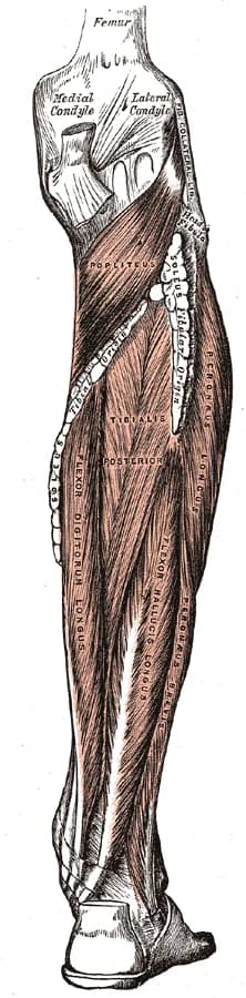
Muscles – posterior deep group
- Tibialis posterior – inversion and plantar flexion.
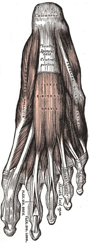
- Flexor digitorum longus – flexion of toes 2–5 and plantar flexion.
- Flexor hallucis longus – flexion of the big toe and plantar flexion.
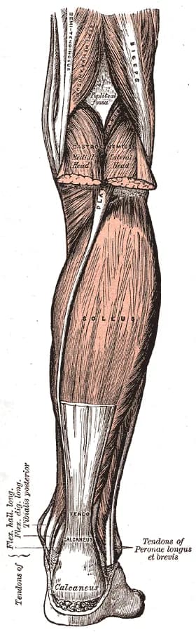
Muscles – posterior superficial group
- Gastrocnemius – plantar flexion of the foot, also assists with knee flexion.
- Soleus – plantar flexion of the foot.
- Achilles tendon – common insertion of m. gastrocnemius and m. soleus on the posterior surface of the calcaneus.
Unlock the full Health Library
Full access to scanning protocols, anatomy, and clinical references. Cancel anytime.
- Every protocol and anatomy reference
- Original ultrasound illustrations and video demonstrations
- Sync across mobile and web