Anatomy
The knee joint is one of the largest and most complex joints in the human body. It enables basic movements such as walking, running, squatting, or jumping, while simultaneously combining a wide range of motion with stability when bearing high loads. Due to these functional demands, the knee is a frequent site of injury, especially in active individuals. For ultrasound examination, orientation regarding bony landmarks, tendon insertions, ligaments, bursae, and joint recesses is essential.
Bone Landmarks
Bone landmarks are fundamental reference points during scanning. They help guide the probe correctly and quickly distinguish normal anatomy from pathology.
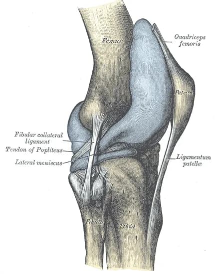
- Patella – a sesamoid bone located within the tendon of m. quadriceps femoris, easily palpable and readily visualized with ultrasound, key for orientation when examining the anterior part of the knee.
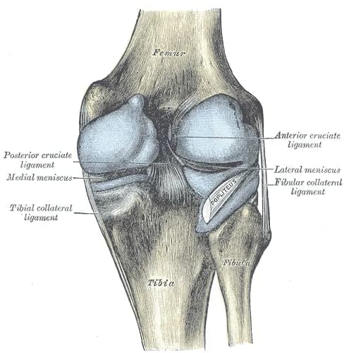
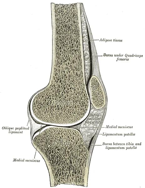
- Femoral condyles – medial and lateral rounded distal parts of the femur, important for evaluation of the femorotibial joint space, menisci and collateral ligaments.
- Tibial plateau – proximal articular surface of the tibia, significant in evaluating meniscal attachments, effusion, and ligamentous structures.
- Tuberositas tibiae – prominent bony prominence below the patella, insertion site of the patellar tendon.
- Fibular head – palpable laterally, important landmark for the lateral knee, insertion of m. biceps femoris and area of the lateral collateral ligament.
- Femoral epicondyles – medial and lateral bony prominences above the condyles, landmark points for the origins of collateral ligaments.
Muscles
Muscles – anterior group
- Quadriceps femoris – main extensor of the knee, attaches to the tuberositas tibiae via the quadriceps tendon and ligamentum patellae.
- Rectus femoris – knee extension, also assists with hip flexion.
- Vastus medialis – knee extension, participates in medial stabilization of the patella.
- Vastus lateralis – knee extension, participates in lateral stabilization of the patella.
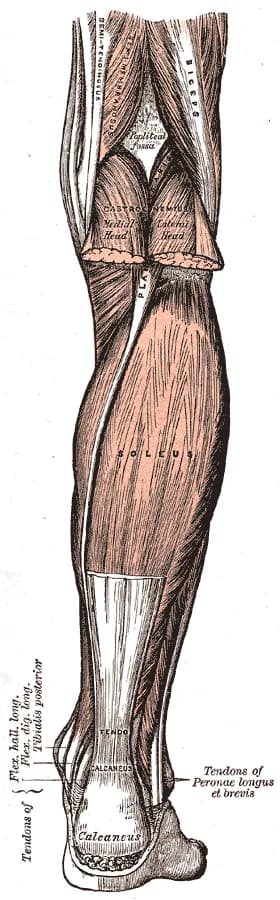
Muscles – posterior group
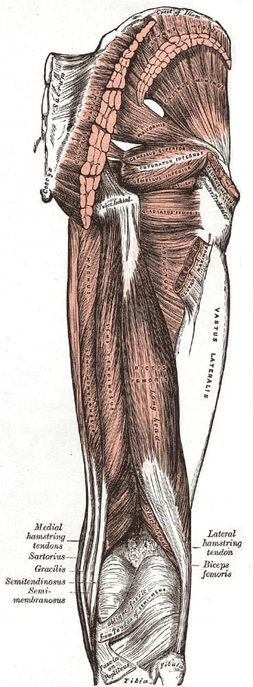
- Biceps femoris – knee flexion, external rotation of the leg, the long head also participates in hip extension.
- Semitendinosus – knee flexion, internal rotation of the leg, hip extension.
- Semimembranosus – knee flexion, internal rotation of the leg, hip extension.
- Gastrocnemius – plantar flexion of the ankle, while also assisting with knee flexion.
- Popliteus – unlocks the knee at the beginning of flexion by internal rotation of the tibia.


Unlock the full Health Library
Full access to scanning protocols, anatomy, and clinical references. Cancel anytime.
- Every protocol and anatomy reference
- Original ultrasound illustrations and video demonstrations
- Sync across mobile and web