 MSK Academy
3. Dorsal view
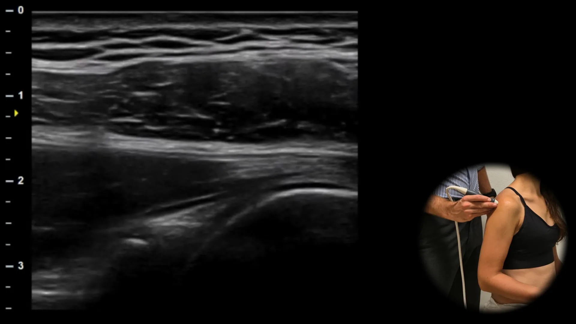
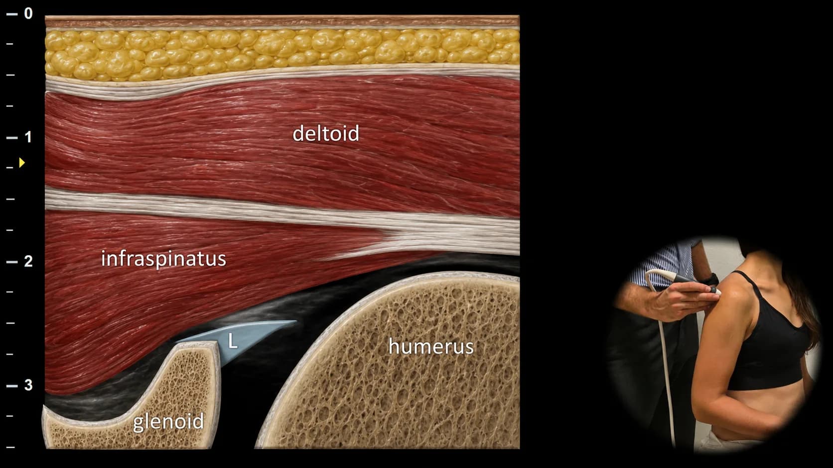
Figure 1. Dorsal view, transverse plane. L: glenoid labrum
Transverse ultrasound section of the posterior part of the shoulder joint with the probe positioned below the scapular spine. The humeral head and glenoid form the main bony landmarks, with a triangular hyperechoic structure visible in the upper part of the glenoid corresponding to the glenoid labrum (L), which contributes to shoulder joint stability.
Clinical Note
This projection is significant for detecting intraarticular fluid in the posterior part of the shoulder joint, whose presence around the glenoid labrum may be better visible during external rotation of the shoulder.
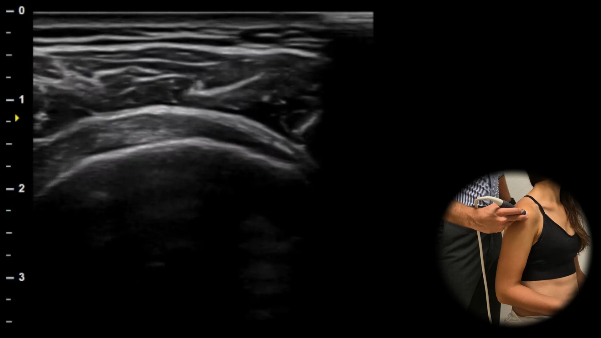
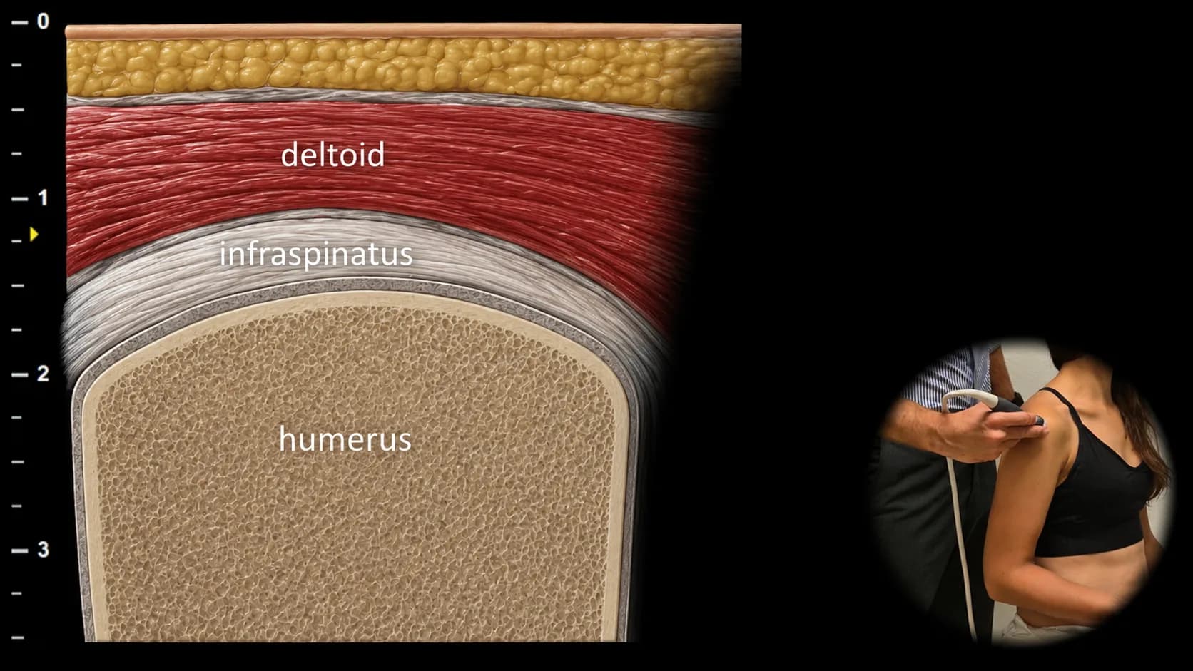
Figure 2. Dorsal view, transverse plane.
Transverse ultrasound section of the posterior part of the shoulder with lateral displacement of the probe, showing the infraspinatus tendon as a fibrillar structure overlying the posterior aspect of the humeral head. With caudal displacement of the probe, the teres minor tendon also comes into the field of view, located directly below the infraspinatus tendon. This projection is important for assessing the integrity of the posterior part of the rotator cuff and for distinguishing isolated infraspinatus tendon involvement from combined pathology that also includes the teres minor.