Anatomy
The wrist is a very complex and mobile joint complex. It enables fine motor skills and a wide range of hand movements, which is essential for daily activities as well as athletic performance. However, this high mobility comes at the cost of greater susceptibility to overuse and injury. For ultrasound examination, orientation to bony landmarks, joint spaces, tendon courses, and localization of neurovascular structures is crucial.
Bone Landmarks
Bone landmarks are fundamental reference points during scanning. They help guide the probe correctly and quickly distinguish normal anatomy from pathology.
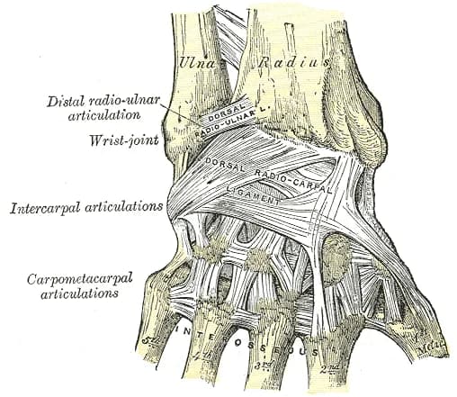
- Distal radius – wide, easily palpable anatomical landmark for the radiocarpal joint and flexor and extensor tendons.
- Lister's tubercle – dorsal bony prominence on the distal radius, important for orientation of the extensor pollicis longus tendon.
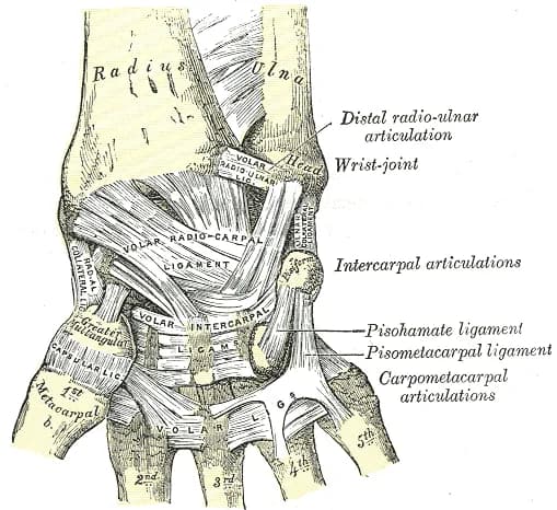
- Distal ulna / processus styloideus ulnae – orientation point on the ulnar side for the DRUJ and TFCC region.
- Os scaphoideum – important anatomical landmark in the anatomical snuffbox region, significant when evaluating the dorsal aspect of the wrist.
- Os pisiforme – easily palpable on the volar ulnar side, landmark for Guyon's canal and the ulnar nerve with artery.
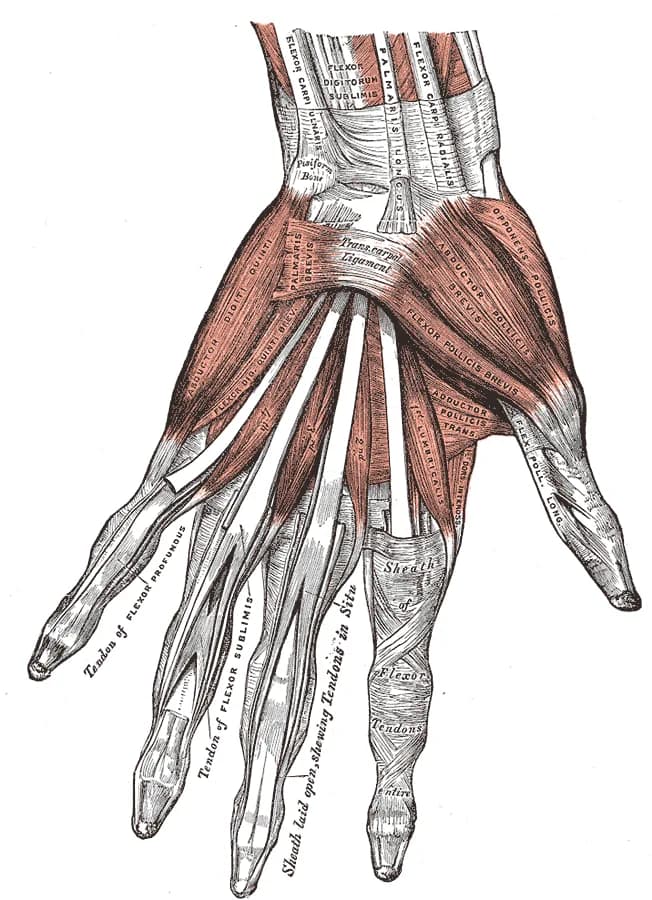
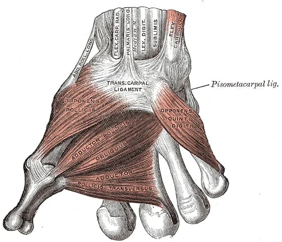
Muscles
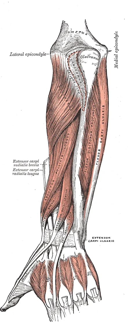
Muscles – dorsal side
- Abductor pollicis longus – abduction and extension of the thumb.
- Extensor pollicis brevis – thumb extension at the MCP joint.
- Extensor carpi radialis longus – wrist extension and radial deviation.
- Extensor carpi radialis brevis – wrist extension.
- Extensor pollicis longus – extends the thumb, wraps around Lister's tubercle.
- Extensor digitorum – extension of fingers 2–5.
- Extensor indicis – extension of the index finger.
- Extensor digiti minimi – extension of the little finger.
- Extensor carpi ulnaris – wrist extension and ulnar deviation.
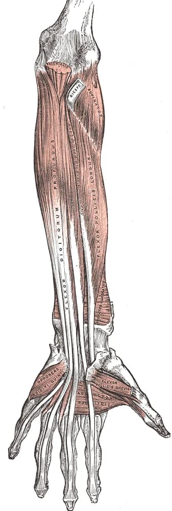
Muscles – volar side
- Flexor carpi radialis – wrist flexion and radial deviation.
- Palmaris longus – assists with wrist flexion, muscle may be absent.
- Flexor carpi ulnaris – wrist flexion and ulnar deviation.
- Flexor digitorum superficialis – flexes PIP joints and wrist.
- Flexor digitorum profundus – flexes DIP joints and wrist.
- Flexor pollicis longus – thumb flexion.
- Pronator quadratus – pronation of the forearm, deeply located muscle visible on distal transverse scan.
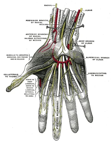
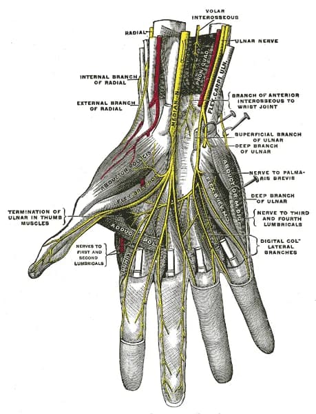
Other Important Structures
- Nervus medianus – runs through the carpal tunnel, crucial in evaluating carpal tunnel syndrome.
- Nervus ulnaris and arteria ulnaris – run through Guyon's canal on the ulnar side of the wrist.
- Extensor compartments – six dorsal compartments beneath the extensor retinaculum, important for orientation during tendon examination.
- TFCC – important stabilizing structure of the ulnar side of the wrist.
Premium
Unlock the full Health Library
Full access to scanning protocols, anatomy, and clinical references. Cancel anytime.
- Every protocol and anatomy reference
- Original ultrasound illustrations and video demonstrations
- Sync across mobile and web