Ultrasound examination
Examination Protocol
Radial nerve in the course of the upper extremity
- Armpit
- Brachium
- Elbow
Superficial branch of the radial nerve
- Elbow
- Forearm
- Carpus
Deep branch of the radial nerve
- Elbow
- Forearm
- Carpus
Interactive feature, available in the app
1. Radial nerve in the course of the upper extremity
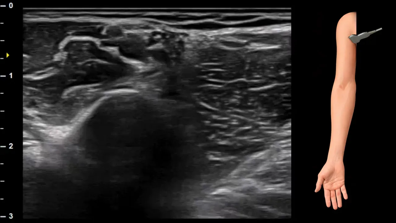
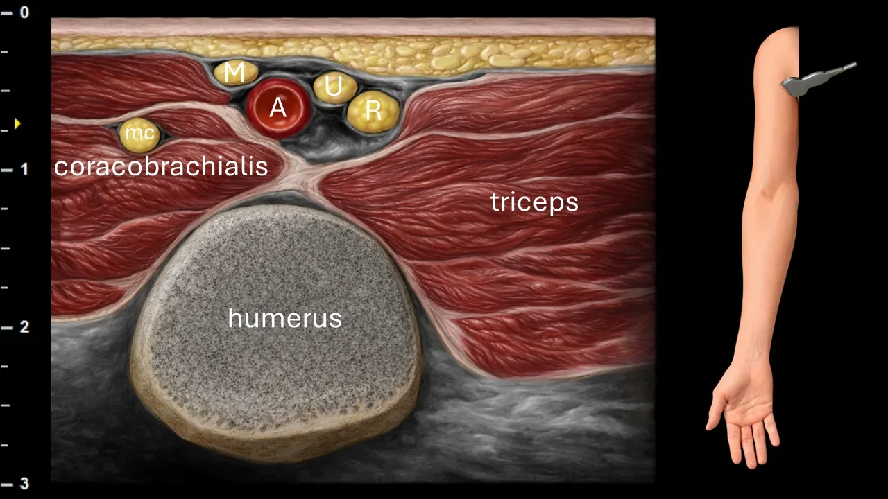
Figure 1. Axilla, transverse plane. A: axillary artery, M: median nerve, U: ulnar nerve, R: radial nerve, mc: musculocutaneus nerve.
Transverse ultrasound section of the axilla. The image shows the axillary artery (A), which represents the main landmark structure for identifying nerve elements of the brachial plexus. The radial nerve (R) is located dorsal to the axillary artery in this projection. Also visible near the vessel are the median nerve (M), ulnar nerve (U), and musculocutaneus nerve (mc). The image also captures surrounding muscle structures, particularly the coracobrachialis muscle and triceps brachii muscle, as well as the contour of the humerus.
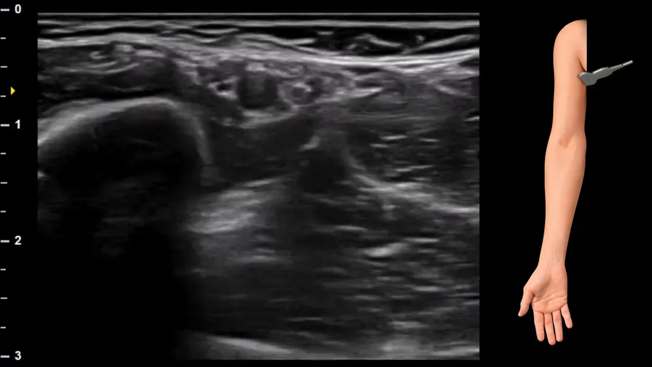
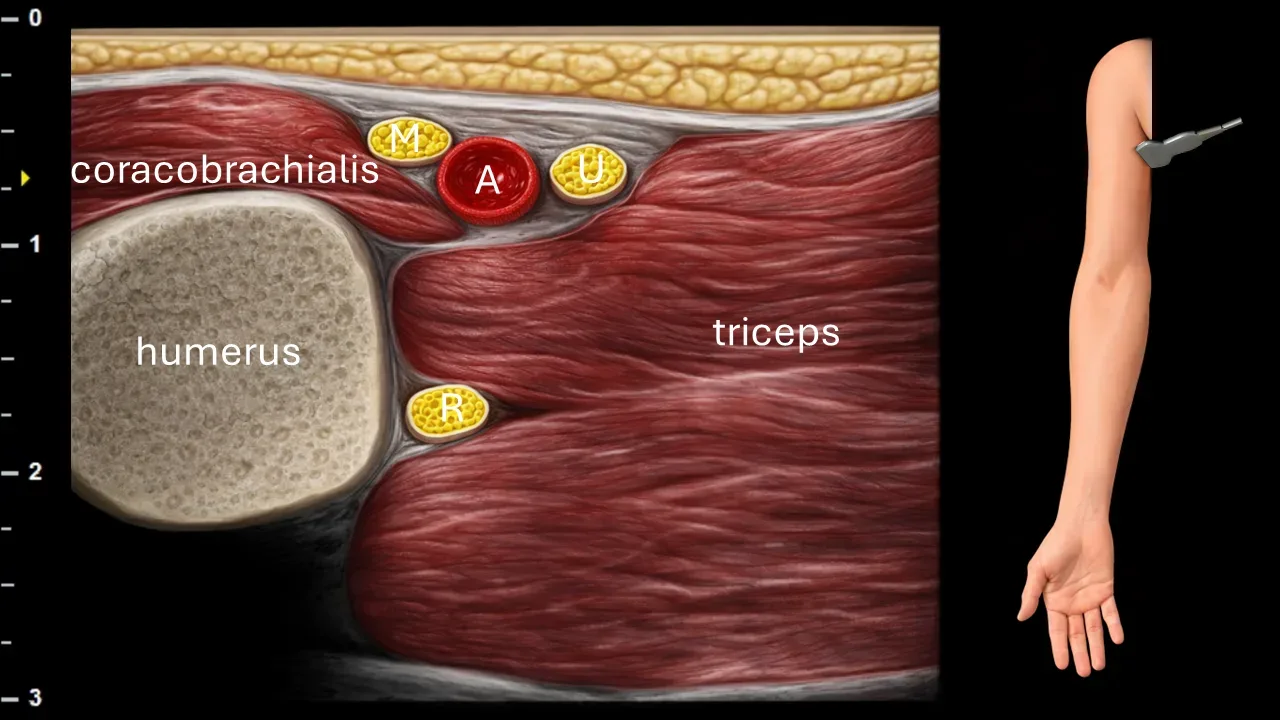
Figure 2. Brachium, transverse plane. A: a. brachialis/axillaris, M: n. medianus, U: n. ulnaris, R: n. radialis.
Transverse ultrasound section of the arm. N. radialis (R) runs in this projection between the long and medial heads of m. triceps brachii at the posterior aspect of the humerus. The image also shows the artery (A) with adjacent nerve structures, particularly n. medianus (M) and n. ulnaris (U). Among the surrounding anatomical structures captured are m. coracobrachialis, m. triceps brachii, and the bony contour of the humerus.
Clinical Note
In the proximal part of the arm and axilla, the n. radialis can be affected by compression from improper use of axillary crutches, leading to the development of so-called crutch palsy. Ultrasound can help localize the site of compression and assess potential changes in nerve caliber or echostructure.
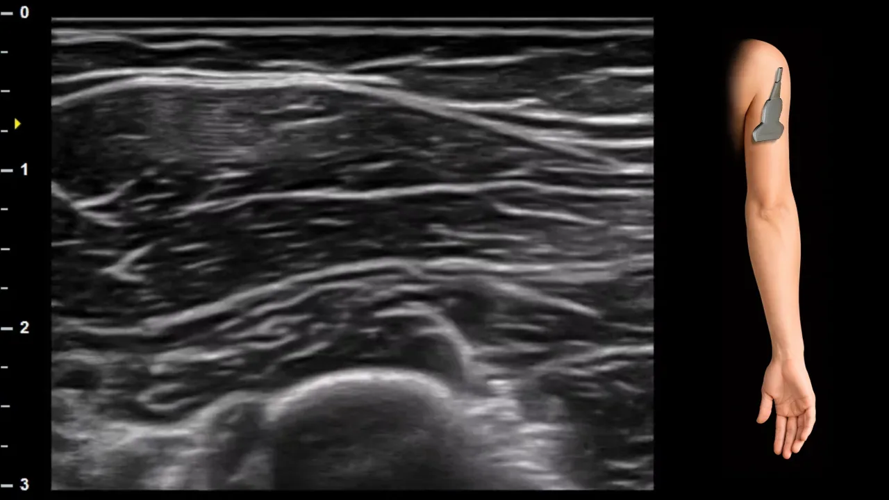
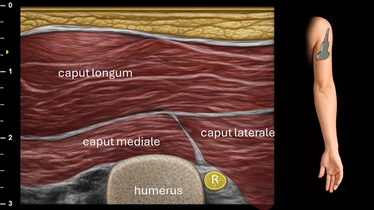
Figure 3. Brachium, transverse plane. R: n. radialis.
Transverse ultrasound section of the arm at the level of the sulcus nervi radialis. N. radialis (R) runs in this projection along the posterior surface of the humerus in the sulcus nervi radialis, between the medial and lateral heads of m. triceps brachii. The image shows the individual heads of m. triceps brachii – caput longum, caput mediale and caput laterale – and the bony contour of the humerus.
Clinical Note
In the area of the spiral groove, the n. radialis may be compressed during prolonged external pressure on the arm, typically in so-called Saturday night palsy. Ultrasound can help identify the site of compression and assess potential nerve thickening or changes in its echostructure.
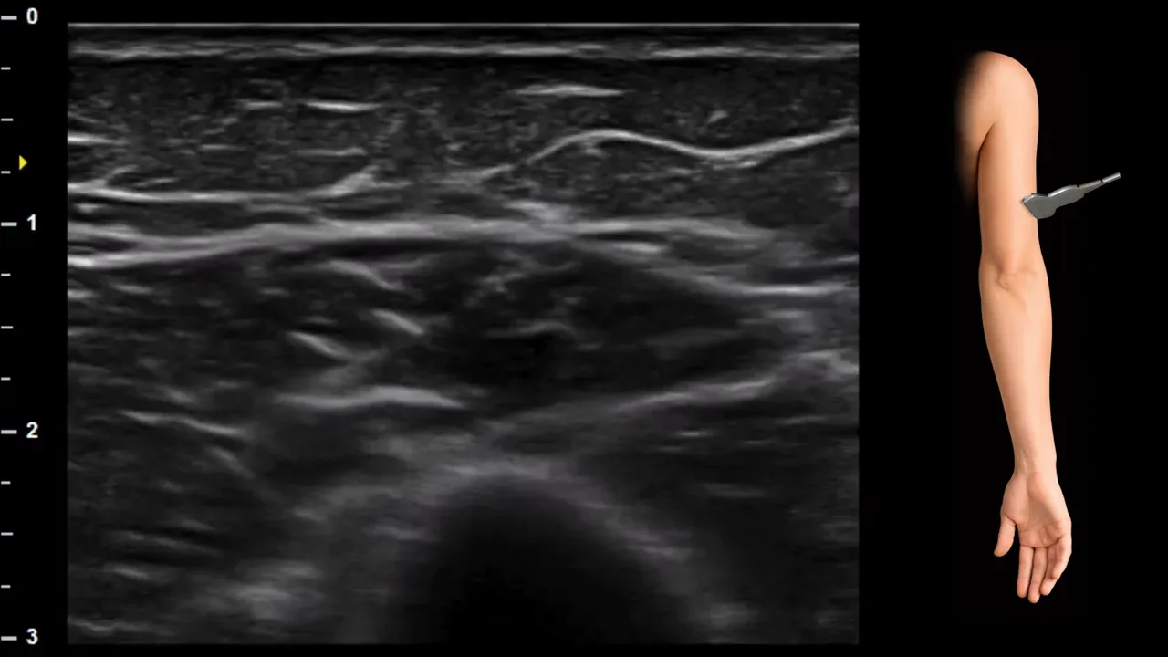
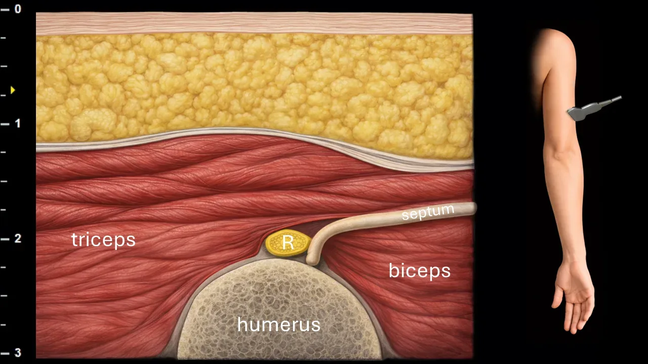
Figure 4. Brachium, transverse plane. R: n. radialis.
Transverse ultrasound section of the arm at the level where the n. radialis (R) passes through the lateral intermuscular septum and transitions from the posterior compartment ventrally. In this projection, the nerve is located at the lateral surface of the humerus in close relation to the lateral intermuscular septum, between the muscle structures m. triceps brachii and m. biceps brachii. The bony contour of the humerus is also visible in the image.
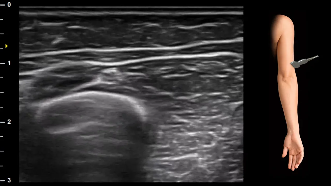
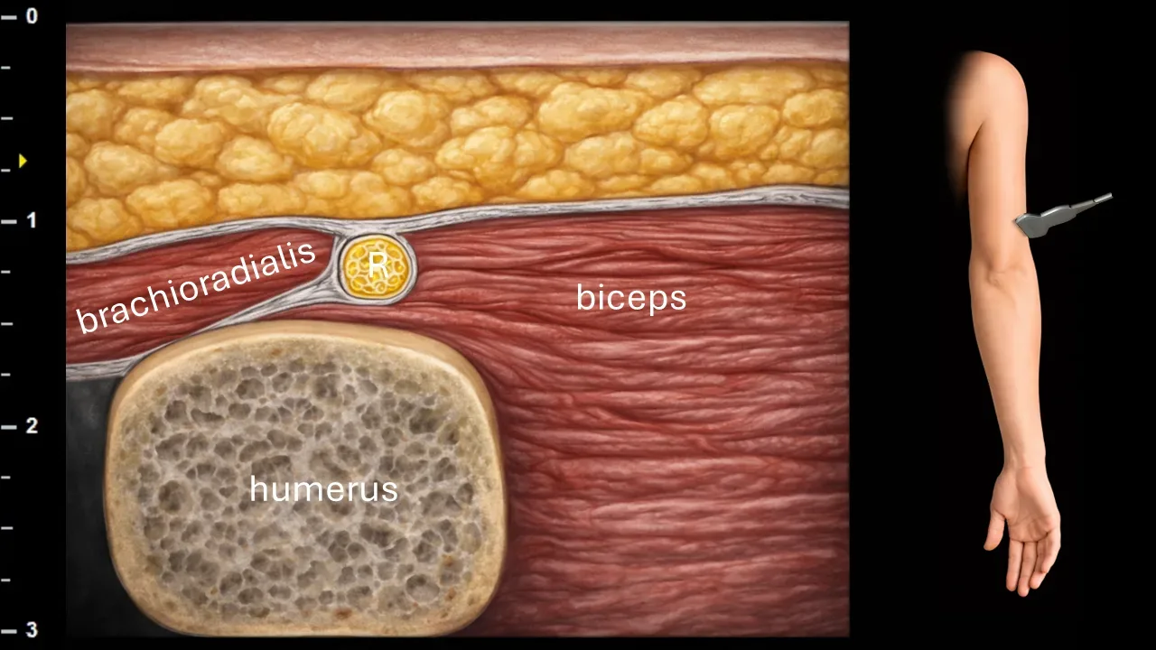
Figure 5. Brachium, transverse plane. R: n. radialis.
Transverse ultrasound section of the distal arm. N. radialis (R) is located in this projection between m. brachioradialis and m. biceps brachii on the ventrolateral aspect of the humerus. The image also shows surrounding muscle structures, particularly m. brachioradialis and m. biceps brachii, and the bony contour of the humerus.
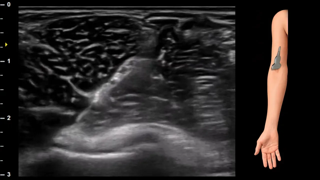
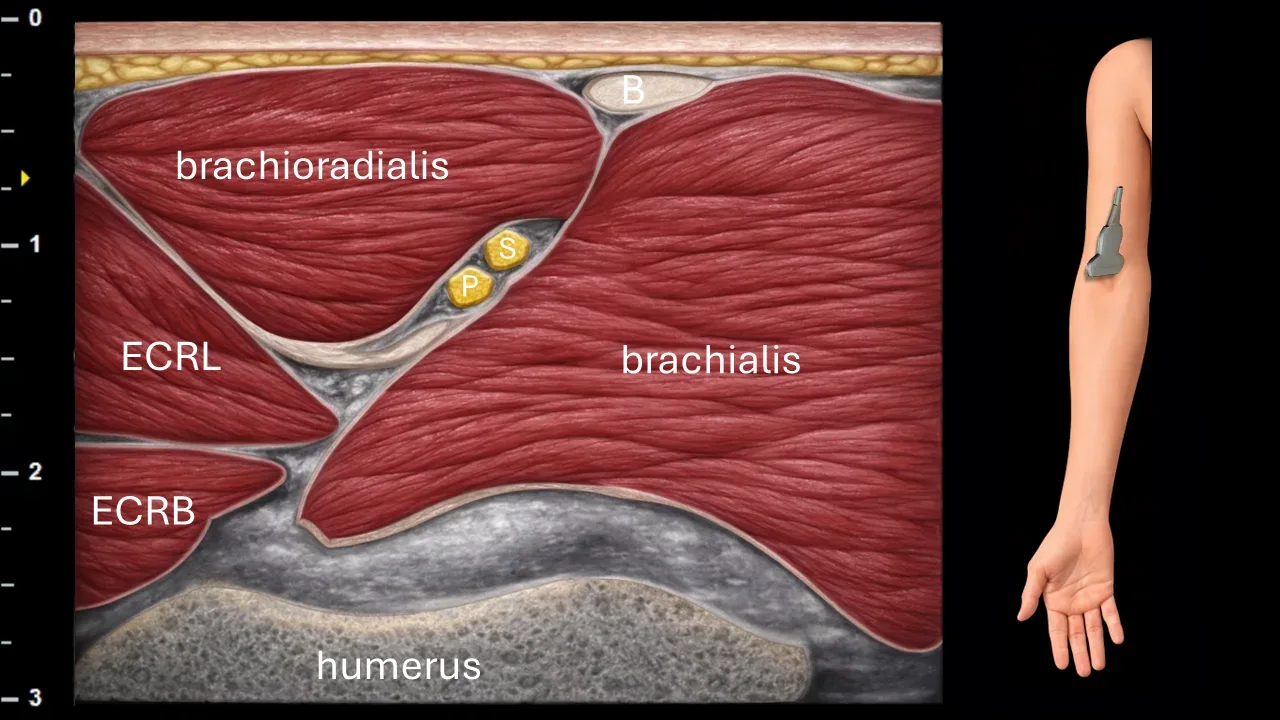
Figure 6. Elbow, transverse plane. P: superficial branch of radial nerve, S: deep branch of radial nerve, B: biceps brachii tendon, ECRL: extensor carpi radialis longus muscle, ECRB: extensor carpi radialis brevis muscle.
Transverse ultrasound section in the elbow region. The radial nerve runs in this projection between the brachioradialis and brachialis muscles, where it subsequently divides into superficial (P) and deep (S) branches. The superficial branch continues more superficially, while the deep branch extends deeper toward the supinator canal. The image also shows surrounding anatomical structures, particularly the brachioradialis muscle, brachialis muscle, extensor carpi radialis longus muscle (ECRL), extensor carpi radialis brevis muscle (ECRB), biceps brachii tendon (B), and the bony contour of the humerus.
2. Superficial branch of the radial nerve
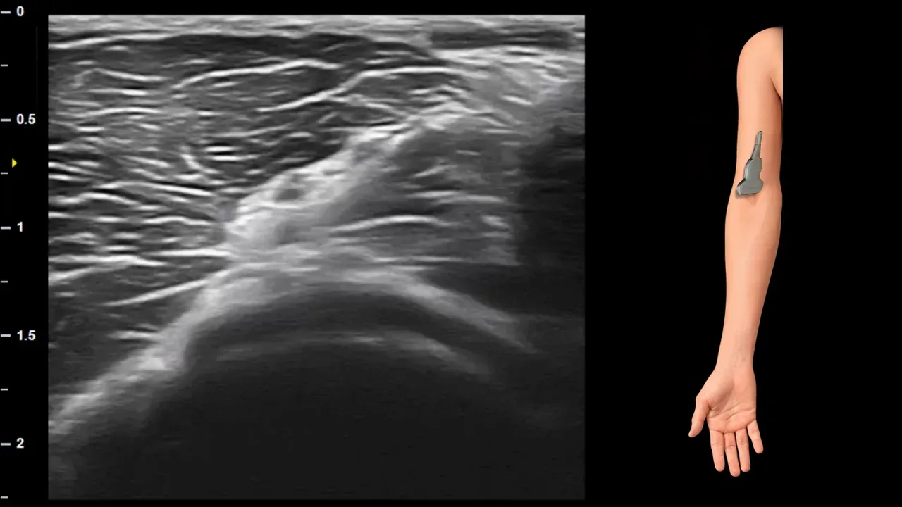
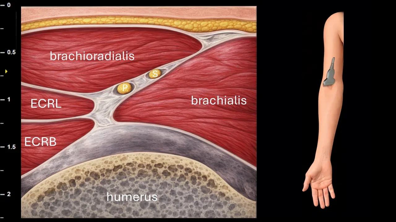
Figure 7. Cubitum, transverse plane. P: superficial branch of n. radialis, S: deep branch of n. radialis, ECRL: m. extensor carpi radialis longus, ECRB: m. extensor carpi radialis brevis.
Transverse ultrasound section in the elbow region. At this level, the n. radialis divides into the superficial branch (P) and the deep branch (S). Both branches are captured in the space between the m. brachioradialis and m. brachialis, in close proximity to the muscles of the radial extensor compartment, particularly the m. extensor carpi radialis longus (ECRL) and m. extensor carpi radialis brevis (ECRB). The image also shows the bony contour of the distal humerus.
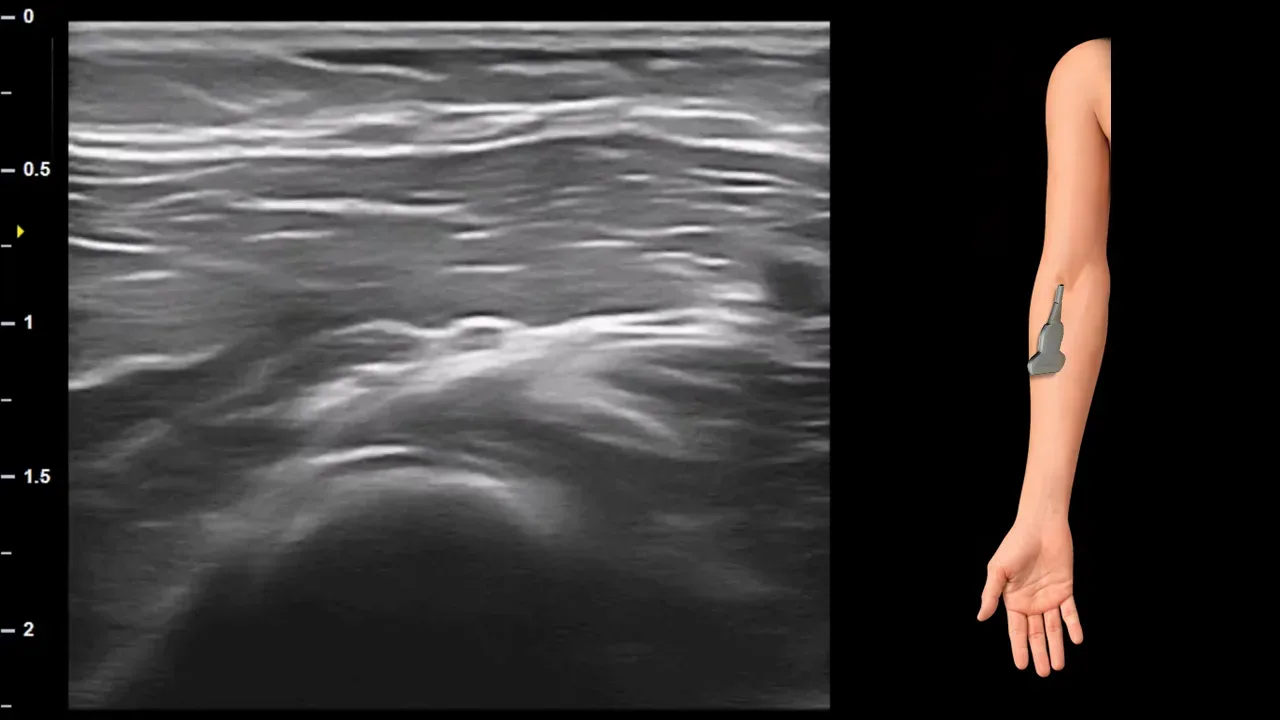
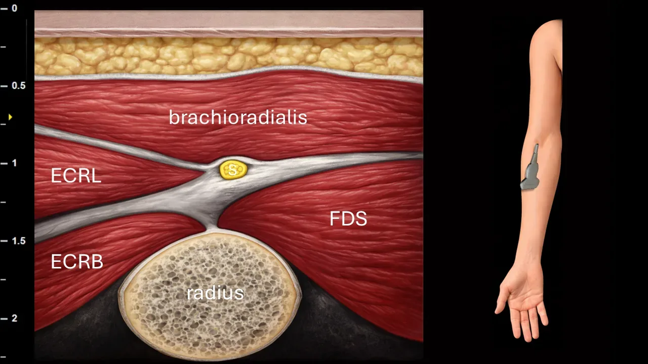
Figure 8. Antebrachium, transverse plane. S: superficial branch of n. radialis, ECRL: m. extensor carpi radialis longus, ECRB: m. extensor carpi radialis brevis, FDS: m. flexor digitorum superficialis.
Transverse ultrasound section of the proximal forearm. The superficial branch of n. radialis (S) runs in this projection beneath m. brachioradialis, in the space between the muscles of the radial compartment of the forearm. Also visible in the image are m. extensor carpi radialis longus (ECRL), m. extensor carpi radialis brevis (ECRB), m. flexor digitorum superficialis (FDS) and the bony contour of the radius.
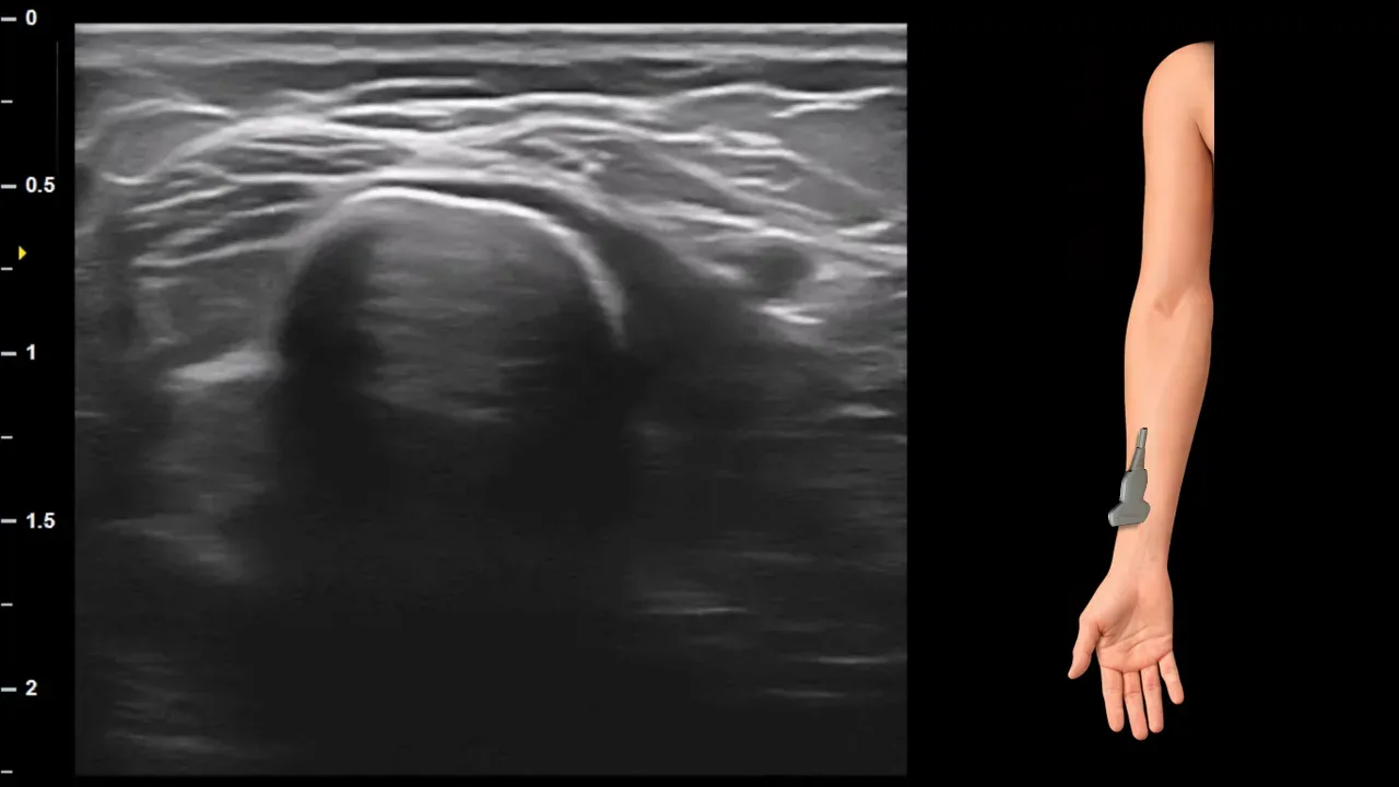
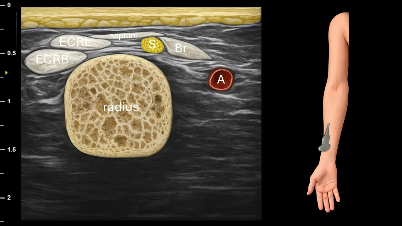
Figure 9. Antebrachium, transverse plane. S: superficial branch of radial nerve, Br: brachioradialis muscle, ECRL: extensor carpi radialis longus muscle, ECRB: extensor carpi radialis brevis muscle, A: radial artery.
Transverse ultrasound section of the distal forearm. The superficial branch of the radial nerve (S) at this level perforates the fascia and becomes positioned more superficially. The nerve is captured between the brachioradialis muscle (Br) and extensor carpi radialis longus muscle (ECRL), in proximity to the extensor carpi radialis brevis muscle (ECRB) and radial artery (A). The bony contour of the radius is also visible in the image.
Clinical Note
The area where the superficial branch perforates the fascia between m. brachioradialis and m. extensor carpi radialis longus represents a possible site of nerve compression, corresponding to the clinical picture of Wartenberg syndrome.
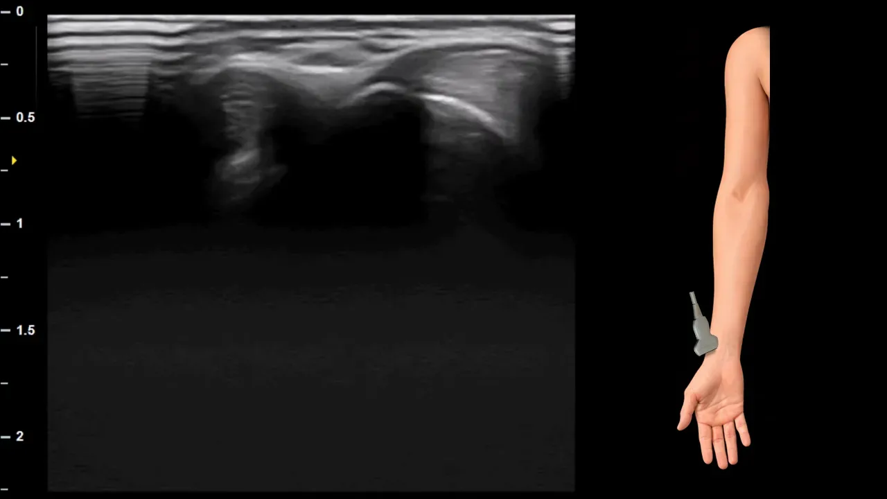
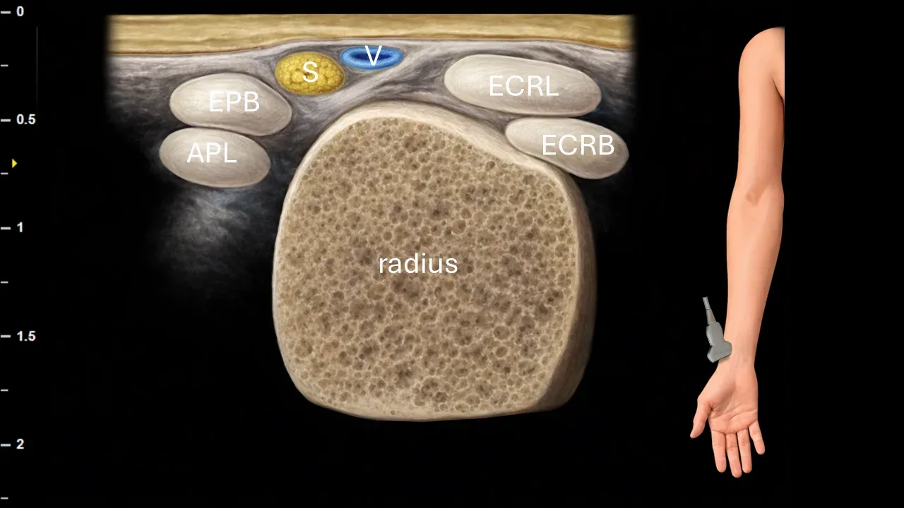
Figure 10. Carpus, transverse plane. S: superficial branch of radial nerve, V: cephalic vein, EPB: extensor pollicis brevis muscle, APL: abductor pollicis longus muscle, ECRL: extensor carpi radialis longus muscle, ECRB: extensor carpi radialis brevis muscle.
Transverse ultrasound section in the wrist region. The superficial branch of the radial nerve (S) is located in this projection between the 1st and 2nd dorsal compartments, i.e., between the tendons of the abductor pollicis longus muscle (APL) and extensor pollicis brevis muscle (EPB) on one side and the tendons of the extensor carpi radialis longus muscle (ECRL) and extensor carpi radialis brevis muscle (ECRB) on the other side. The cephalic vein (V) is also visible in close proximity to the nerve. The bony contour of the radius is also captured in the image.
Clinical Note
In this area, the superficial branch of the radialis nerve may be iatrogenically damaged during venous cannulation or catheterization of the vena cephalica, as the nerve runs in close anatomical proximity to it.
3. Deep branch of the radial nerve
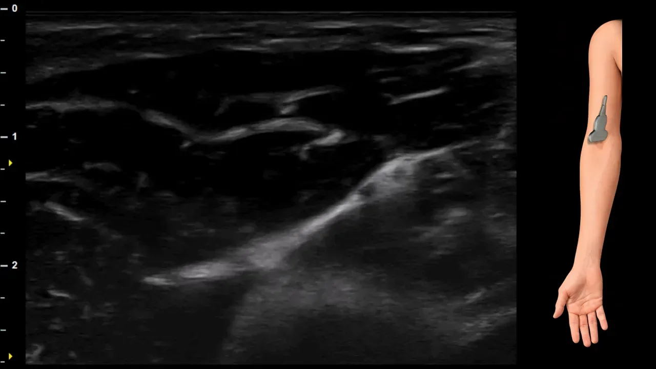
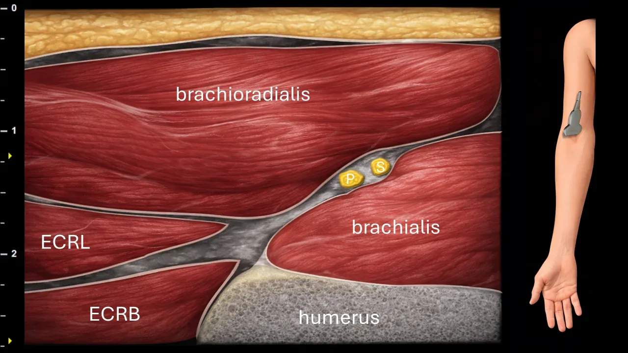
Figure 11. Elbow, transverse plane. P: superficial branch of n. radialis, S: deep branch of n. radialis, ECRL: m. extensor carpi radialis longus, ECRB: m. extensor carpi radialis brevis.
Transverse ultrasound section in the elbow region at the site of n. radialis division. In this projection, its two terminal branches are visible – the superficial branch (P) and the deep branch (S). The deep branch of n. radialis (S) extends deeper into the radial part of the elbow toward the supinator canal, while the superficial branch (P) continues more superficially. The image also shows surrounding muscular structures, particularly m. brachioradialis, m. brachialis, m. extensor carpi radialis longus (ECRL), m. extensor carpi radialis brevis (ECRB) and the bony contour of the humerus.
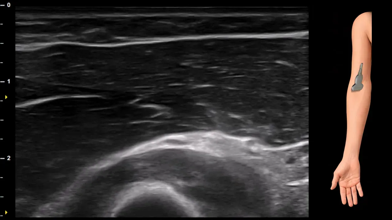
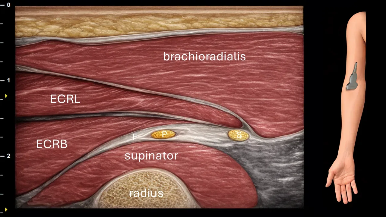
Figure 12. Proximal antebrachium, transverse plane. S: deep branch of radial nerve, P: posterior interosseous nerve, F: Frohse's arcade, ECRL: extensor carpi radialis longus muscle, ECRB: extensor carpi radialis brevis muscle.
Transverse ultrasound section of the proximal forearm. The deep branch of the radial nerve (S) enters the supinator canal and passes beneath Frohse's arcade (F), i.e., beneath the proximal fibrous margin of the supinator muscle. Distally, it continues as the posterior interosseous nerve (P). The image also shows surrounding muscle structures, particularly the brachioradialis muscle, extensor carpi radialis longus muscle (ECRL), extensor carpi radialis brevis muscle (ECRB), supinator muscle, and the bony contour of the radius.
Clinical Note
Frohse's arcade represents the most common site of compression of the deep branch of the radial nerve / posterior interosseous nerve, and is therefore a key area during sonographic examination of suspected posterior interosseous nerve syndrome or radial tunnel syndrome.
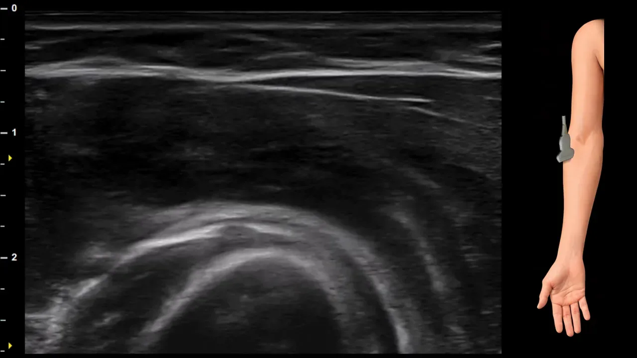
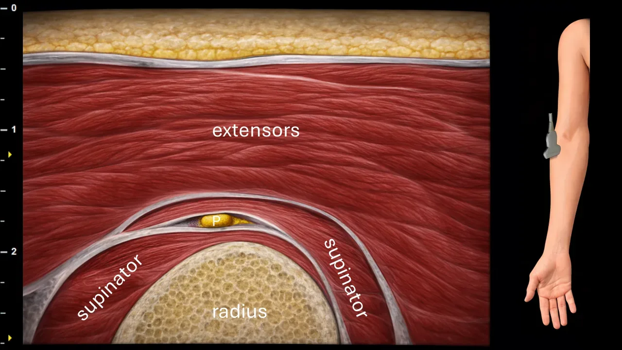
Figure 13. Proximal antebrachium, transverse plane. P: posterior interosseous nerve.
Transverse ultrasound section of the proximal forearm. The deep branch of the radial nerve enters the supinator canal here, where after passing through the supinator muscle it continues as the posterior interosseous nerve (P). In this projection, the nerve is positioned close to the radius, surrounded by muscle structures of the supinator muscle and the superficially located extensor musculature of the forearm.
Clinical Note
Supinator canal represents a possible site of compression of the deep branch of the radial nerve / posterior interosseous nerve, and is therefore an important area in sonographic examination of suspected compressive involvement in the radial tunnel.
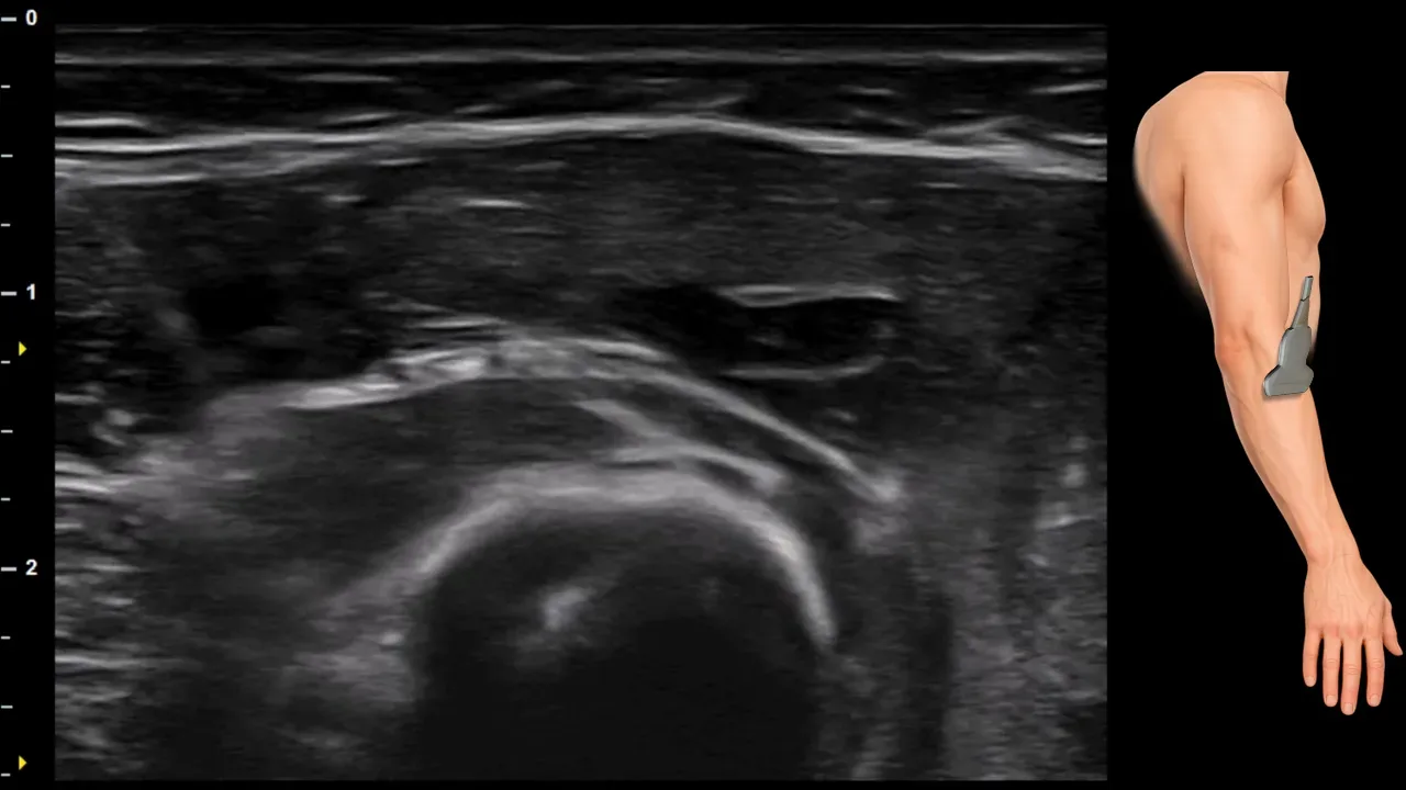
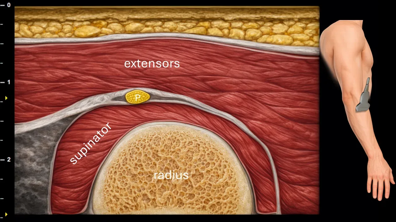
Figure 14. Proximal antebrachium, transverse plane. P: posterior interosseous nerve.
Transverse ultrasound section of the proximal forearm. The posterior interosseous nerve (P) at this level emerges from the supinator canal and continues further in the deep extensor compartment of the forearm. The nerve is located adjacent to the radius, between the supinator muscle and the more superficially located extensor muscles. The image also shows muscle structures of the extensor compartment and the bony contour of the radius.
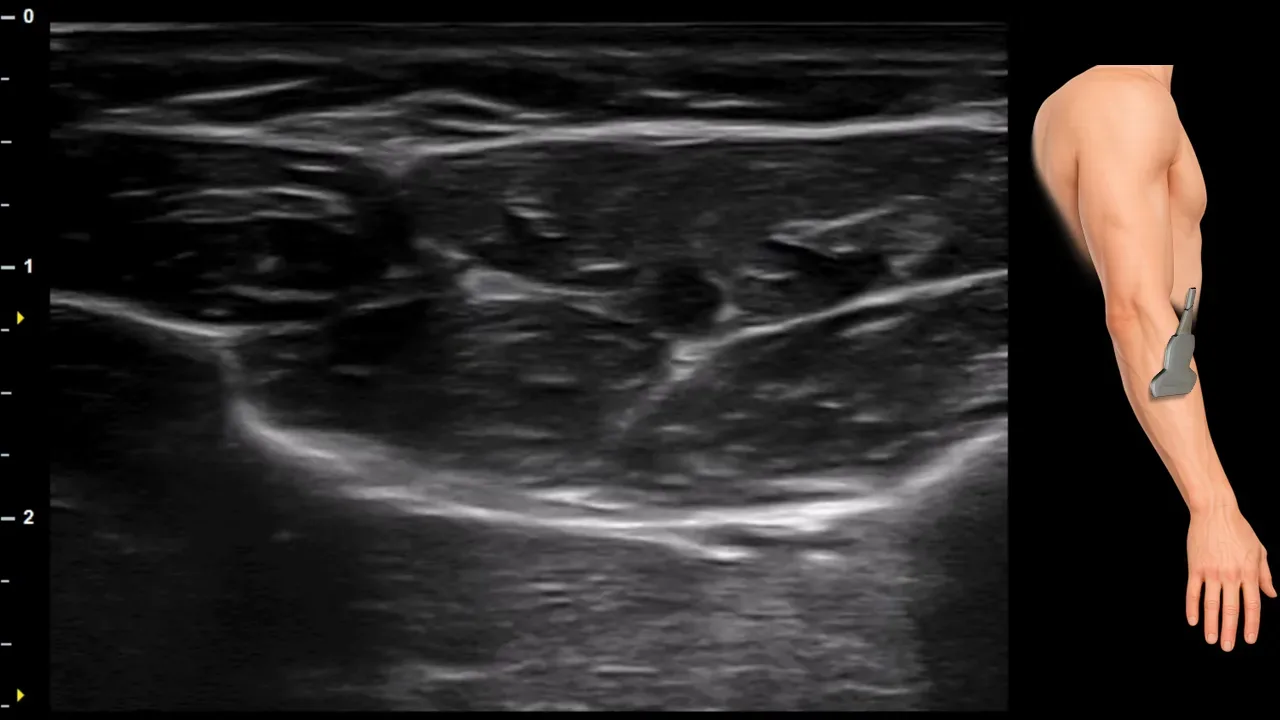
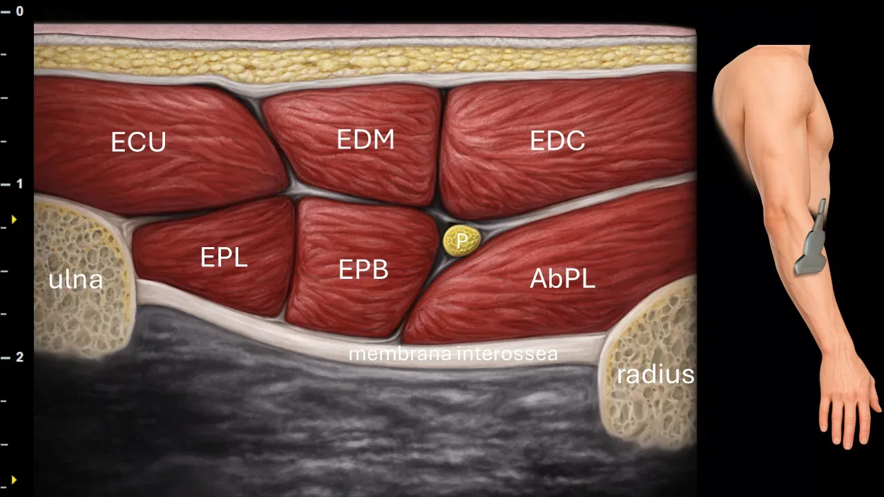
Figure 15. Mid-antebrachium, transverse plane. P: n. interosseus posterior, ECU: m. extensor carpi ulnaris, EDM: m. extensor digiti minimi, EDC: m. extensor digitorum communis, EPL: m. extensor pollicis longus, EPB: m. extensor pollicis brevis, AbPL: m. abductor pollicis longus.
Transverse ultrasound section of the middle portion of the forearm. N. interosseus posterior (P) runs here in the deep extensor compartment between individual muscle layers, in close proximity to the membrana interossea. The image shows superficially located extensor muscles, particularly m. extensor carpi ulnaris (ECU), m. extensor digiti minimi (EDM) and m. extensor digitorum communis (EDC), and deeper muscles m. extensor pollicis longus (EPL), m. extensor pollicis brevis (EPB) and m. abductor pollicis longus (AbPL). The image also shows the bony contours of the ulna and radius.
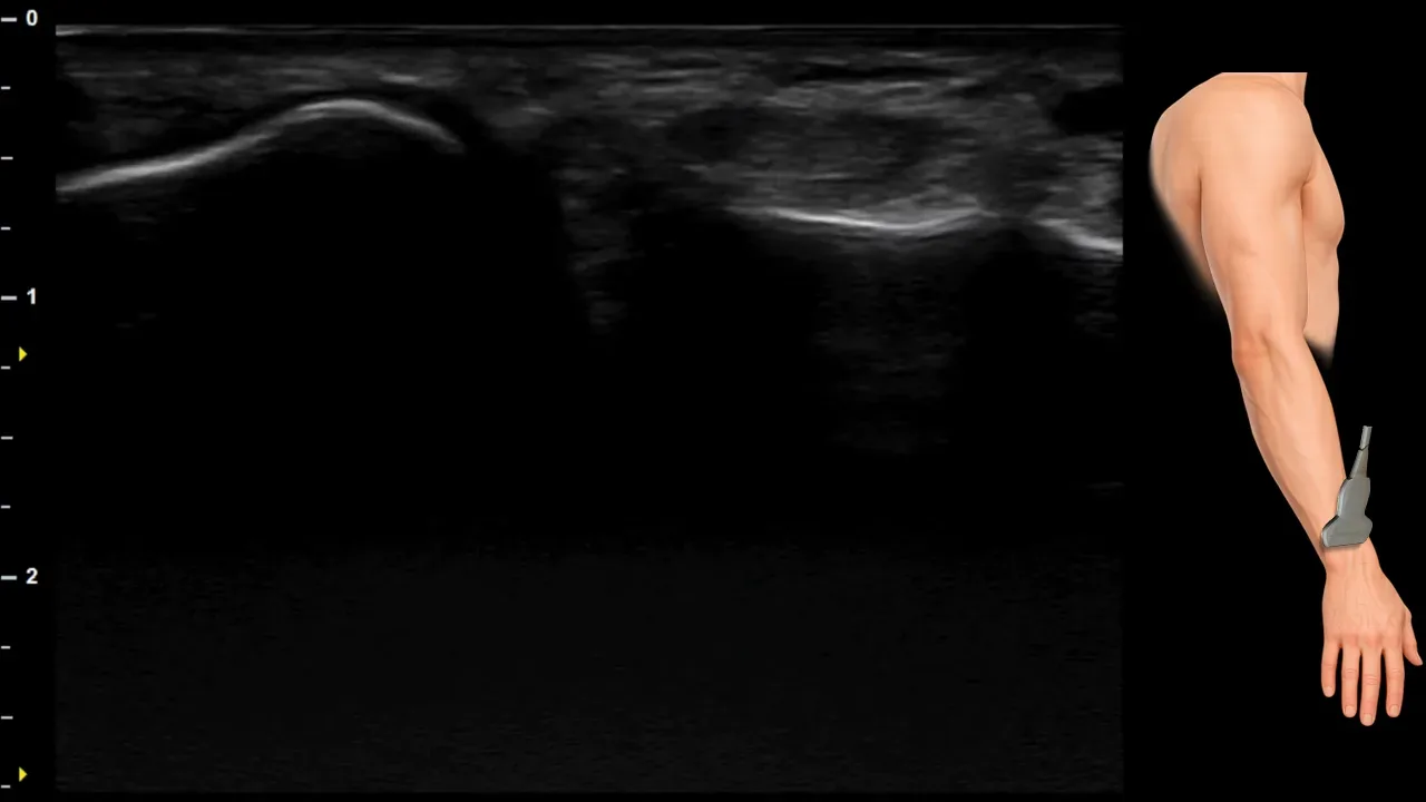
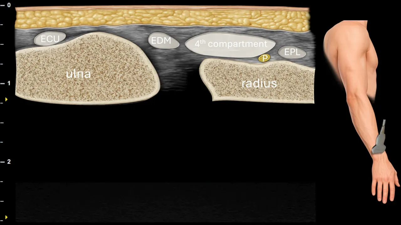
Figure 16. Carpus, transverse plane. P: n. interosseus posterior, ECU: m. extensor carpi ulnaris, EDM: m. extensor digiti minimi, EPL: m. extensor pollicis longus.
Transverse ultrasound section in the wrist area. N. interosseus posterior (P) is captured at this level at the dorsal side of the distal forearm, where it terminates in the dorsal wrist capsule. The nerve runs in close proximity to the tendon structures of the dorsal compartment, particularly adjacent to the 4th dorsal compartment, m. extensor digiti minimi (EDM) and m. extensor pollicis longus (EPL). Ulnarly, the m. extensor carpi ulnaris (ECU) is visible. The image also shows the bony contours of the ulna and radius.
Unlock the full Health Library
Full access to scanning protocols, anatomy, and clinical references. Cancel anytime.
- Every protocol and anatomy reference
- Original ultrasound illustrations and video demonstrations
- Sync across mobile and web