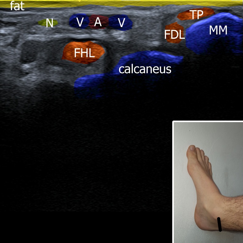
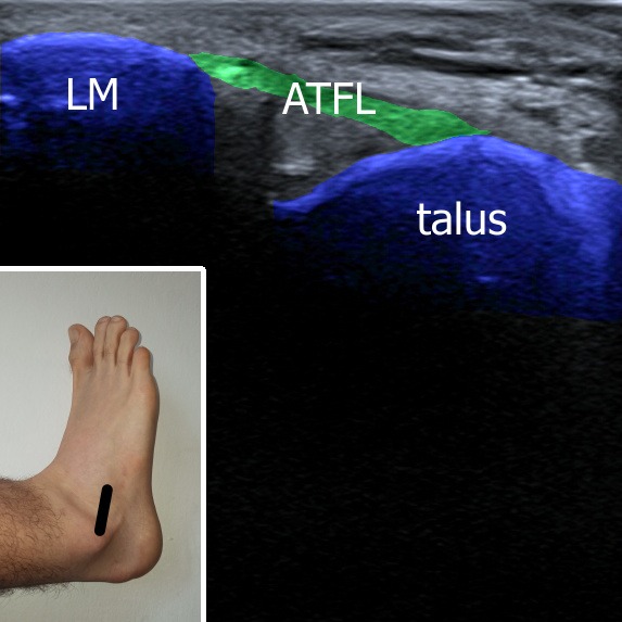
ANKLE

Ultrasound allows immediate evaluation of periarticular soft tissues, both statically and dynamically. Following structures can be evaluated: joint effusions, soft periarticular tissue (muscles, tendons and ligaments) and bone abnormalities.

Position of the patient:

The patient is lying supine and the knee flexed 90°. To perform examination of the dorsal part (Achilles tendon) the patient is lying prone with the knee extended and with the ankle fully dorsally flexed.

1. Anterior view
     A. Longitudinal plane
     B. Transversal plane
2. Medial view
     A. Longitudinal plane – deltoid ligament
     B. Transversal plane
3. Lateral view
     A. Longitudinal plane – ATFL
     B. Transversal plane
4. Posterior view
     A. Longitudinal plane
     B. Transversal plane
5. Plantar view
     A. Longitudinal plane

1. Anterior view

The probe is placed above the tibiotalar joint. Joint effusion can be present between tibia and talus. Muscles/tendons of extensor muscles and tibialis anterior can be evaluated.

Figure 1.

Musculoskeletal ultrasonography

1. Anterior view

The probe is placed above the tibiotalar joint. Joint effusion can be present between tibia and talus. Muscles/tendons of extensor muscles and tibialis anterior can be evaluated.

Figure 1.

Further content is locked

Login/Register and Subscribe To Unlock The Content