KNEE
Ultrasound allows immediate evaluation of periarticular soft tissues, both statically and dynamically. Following structures can be evaluated: intraarticular fluid collection, bursitis, evaluation of the tendons, meniscus, bone abnormalities and ligaments.
Position of the patient:
For examination of the anterior, medial and lateral compartment the patient is lying supine with the knee slightly flexed 20 – 30°. For imaging the posterior part of the knee the patient is lying prone on the bed.
1. Anterior view
A. Suprapatellar view, longitudinal plane
B. Suprapatellar view, transversal plane
C. Infrapatellar view, longitudinal plane
D. Infrapatellar view, transversal plane
E. Infrapetallar view, oblique plane
2. Medial view
A. Longitudinal plane
B. Longitudinal oblique plane
3. Lateral view
A. Longitudinal plane – ITB
B. Longitudinal plane – LCL
4. Posterior view
A. Longitudinal oblique plane
1. Anterior view
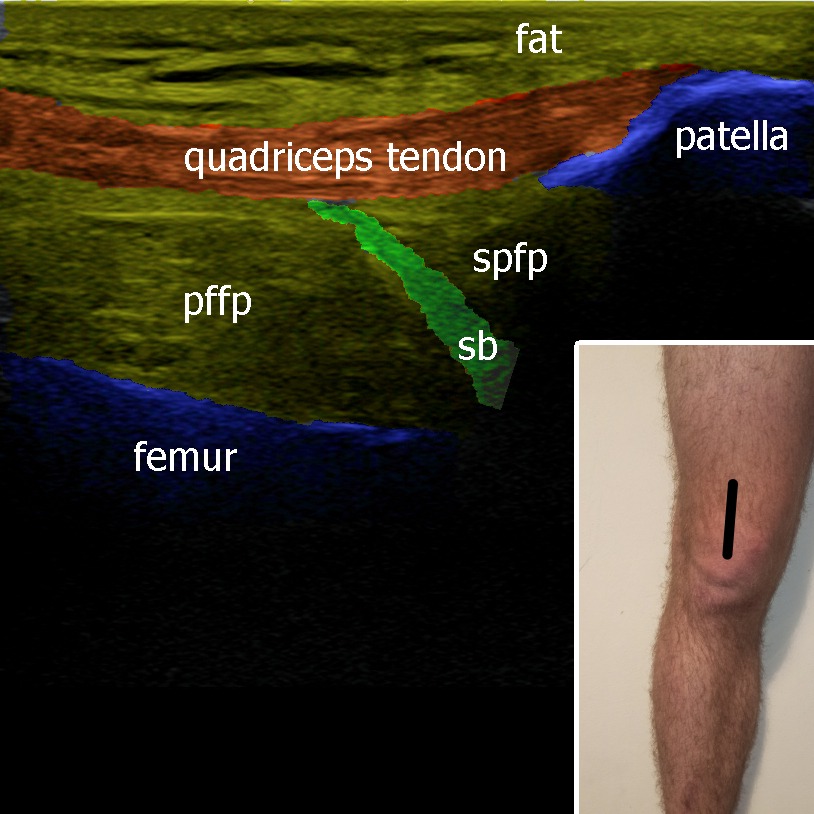
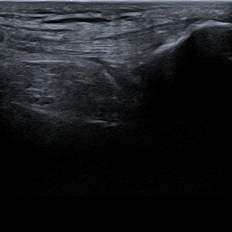
The probe is placed above the proximal part of the patella parallel with the long axis of the lower limb. Quadriceps tendon insertion onto patella can be evaluated. Underneath the tendon suprapatellar recessus of the knee joint can be visualized, especially when there is some fluid collection inside the joint.
Figure 1. pffp – prefemoral fat pad, spfp – suprapatellar fat pad, sb – recessus suprapatellaris.
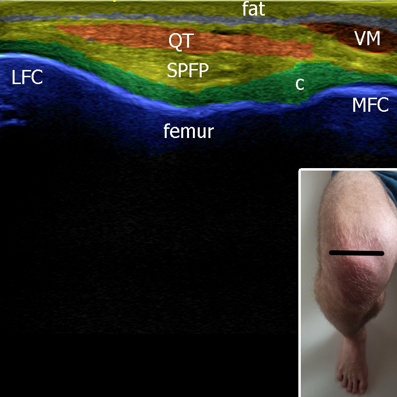
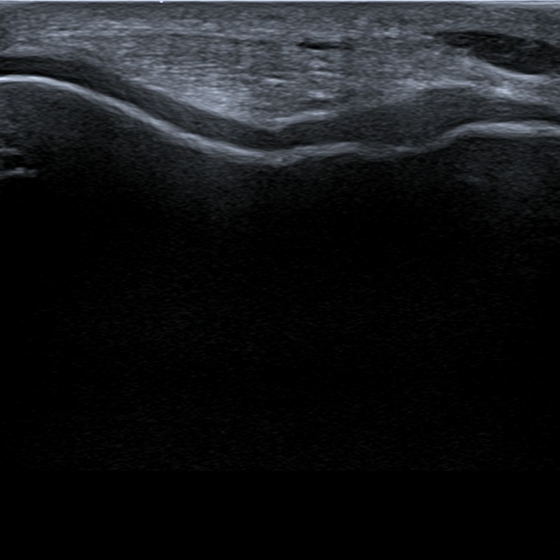
Positioning the knee into maximal flexion, we can evaluate the condyles of femur and the hyaline cartilage.
Figure 2. QT – quadriceps tendon, c – cartilage, VM – vastus medialis, spfp – suprpatellar fat pad, MFC – medial femoral condyle, LFC – lateral femoral condyle.
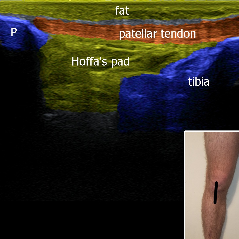
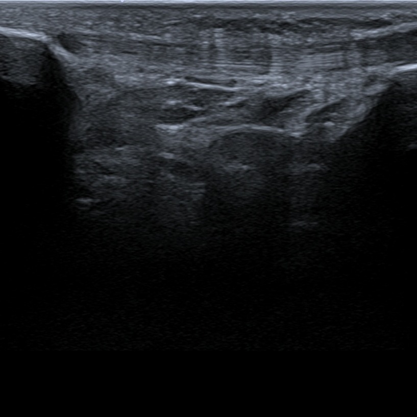
The probe is placed between the distal pole of the patella and tibia. Patellar ligament in it’s long axis can be evaluated. Underneath the patellar ligament can be found fat tissue called Hoffa’s fat pad.
Figure 3. P – patella.
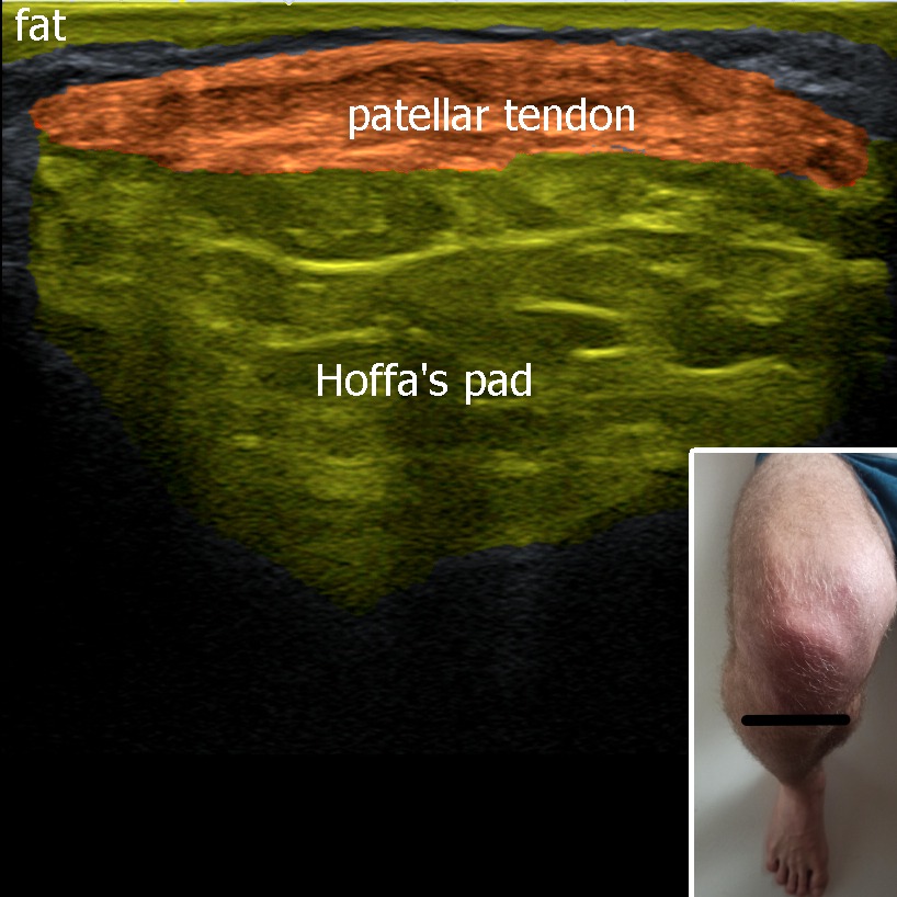
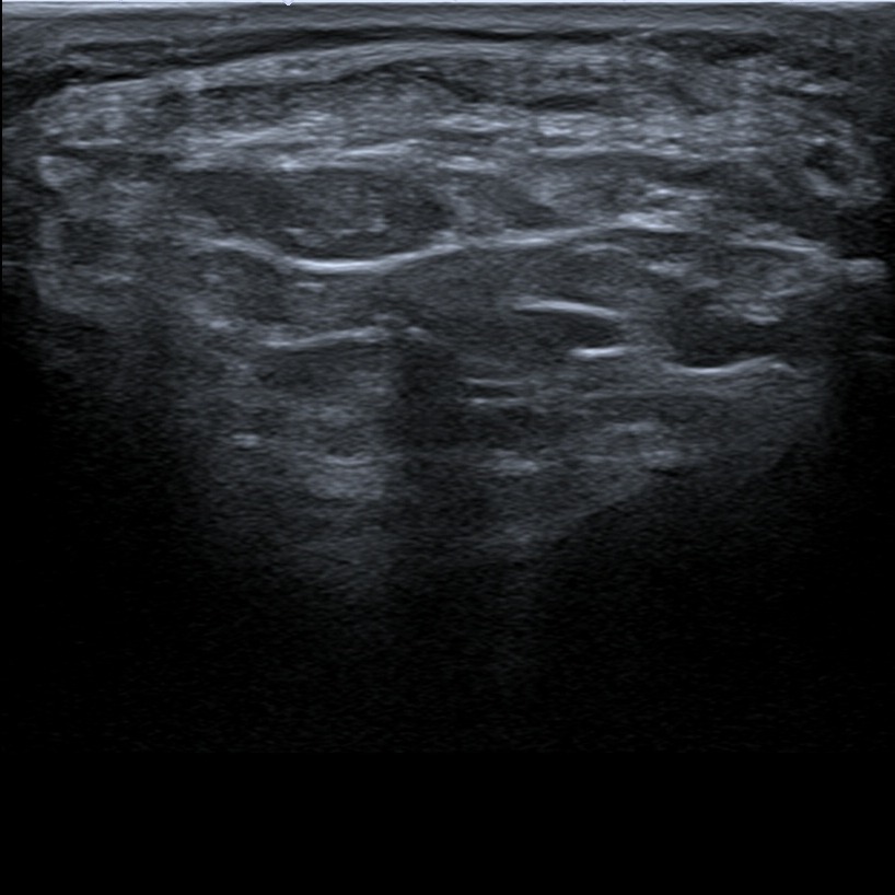
The patellar tendon can be visualized and evaluated also in the short axis.
Figure 4.
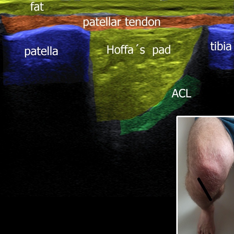
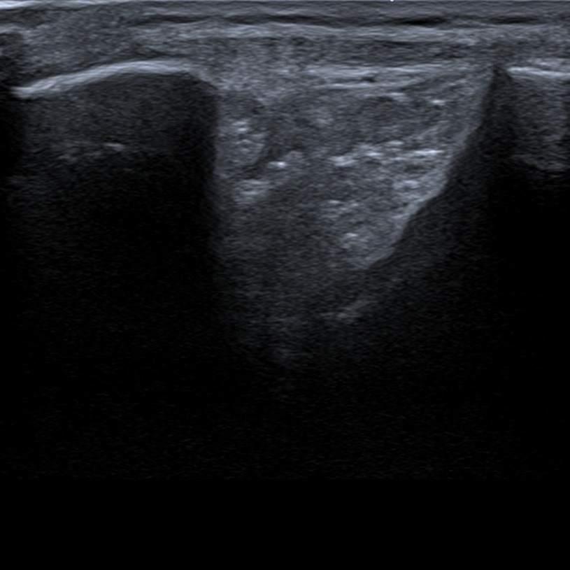
When the probe is placed oblique (in maximal knee flexion), parallel with the course of anterior cruciate ligament, the distal part of the ligament with it’s insertion onto tibia can be evaluated. Anterior drawer test can be performed in this position.
Figure 5. ACL – anterior cruciate ligament.
2. Medial view
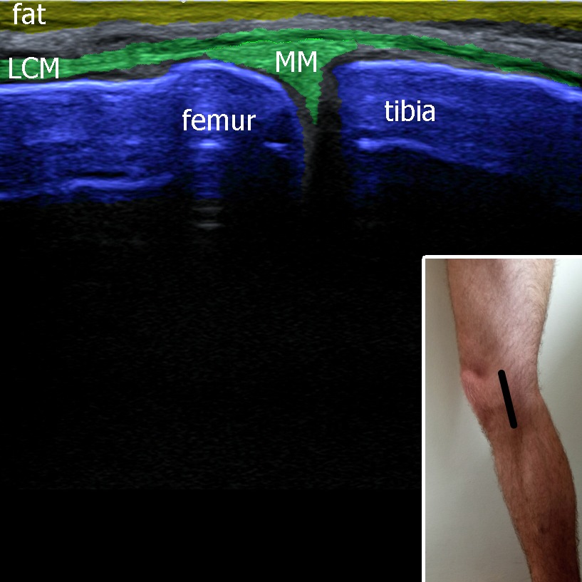
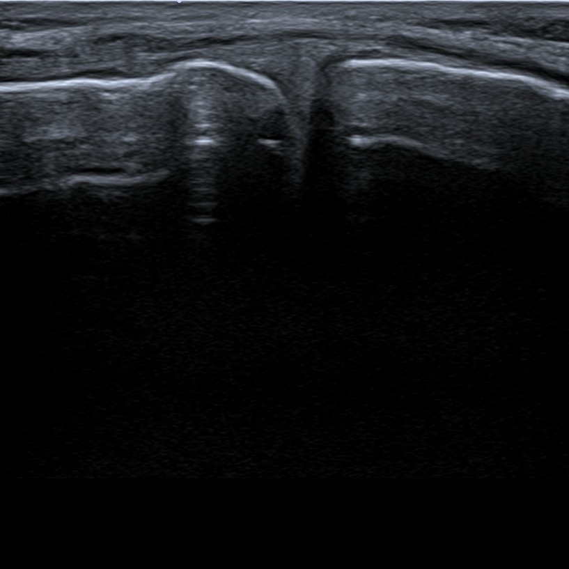
The probe is placed above the medial compartment of the knee. Medial collateral ligament can be evaluated as well as the medial meniscus.
Figure 6. MM – medial meniscus, LCM – medial collateral ligament.
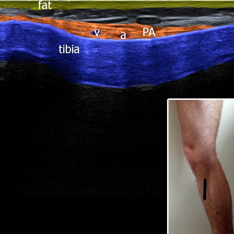
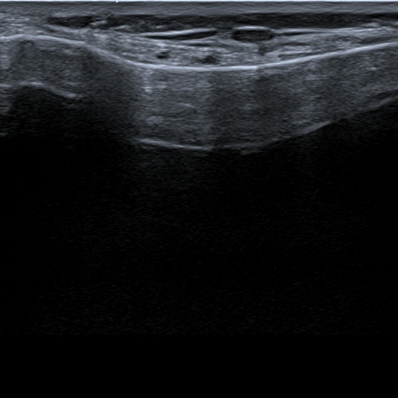
Sliding more distally with the probe, the pes anserinus (common insrerion of sartorius, gracilis and semitendinosus). Underneath the pes anserinus inferior medial genicular artery can be visualized.
Figure 7. PA – pes anserinus, a – inferior medial genicular artery, v – vein.
3. Lateral view
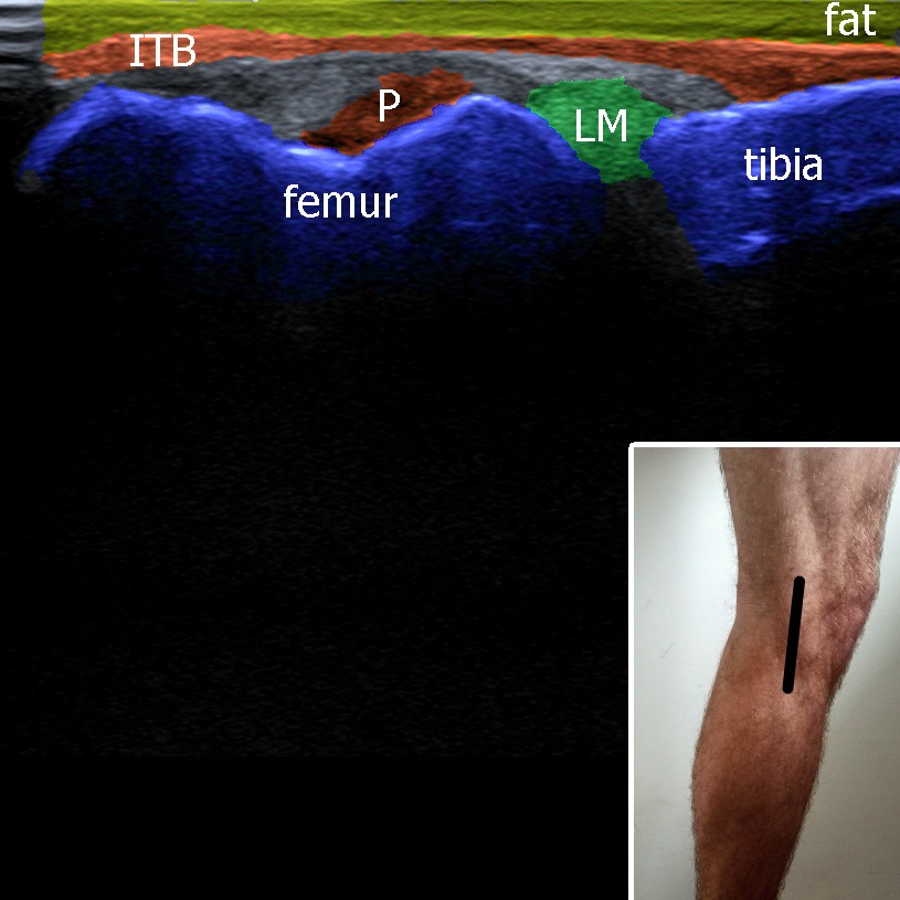
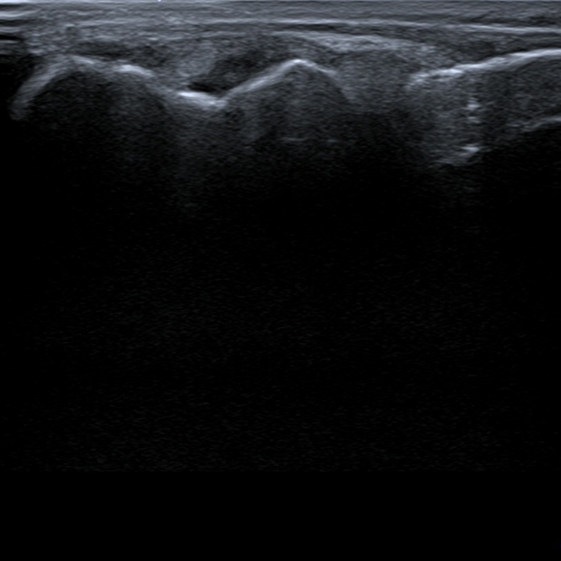
Placing the probe above femur and tibia the iliotibial band with it’s insertion onto tibia can be visualized. In this plane lateral meniscus of the knee can be also evaluated.
Figure 8. ITB – iliotibial band, LM – lateral meniscus, P – popliteus muscle.
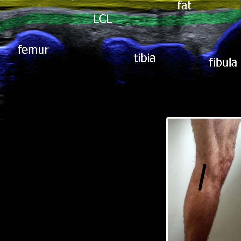
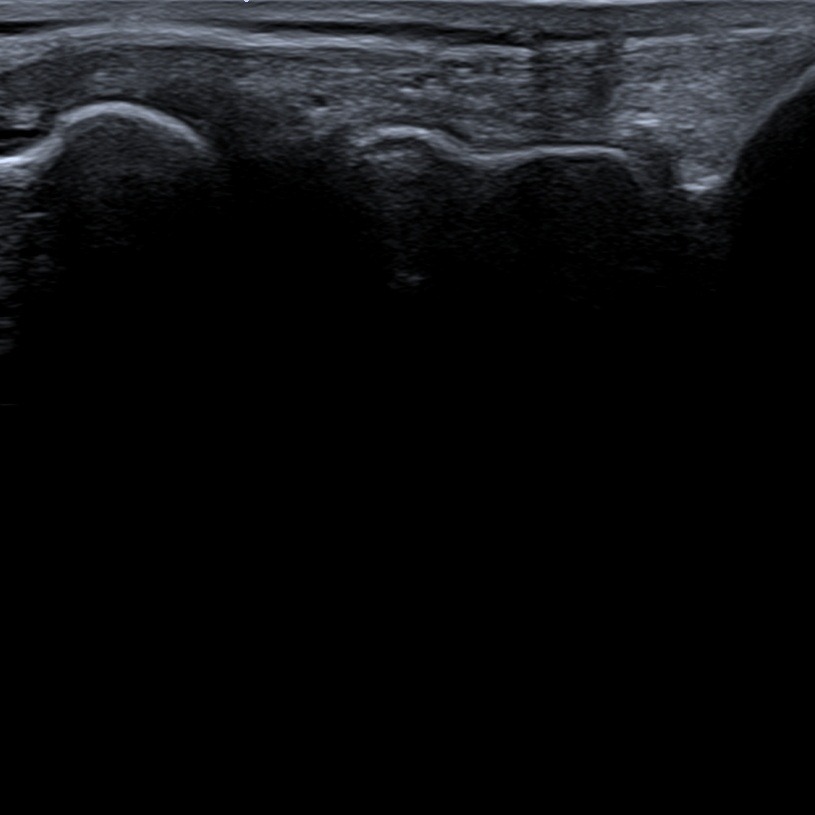
Sliding the probe more dorsal, lateral collateral ligament (that runs from femur to fibula) can be evaluated.
Figure 9. LCL – lateral collateral ligament.
4. Posterior view
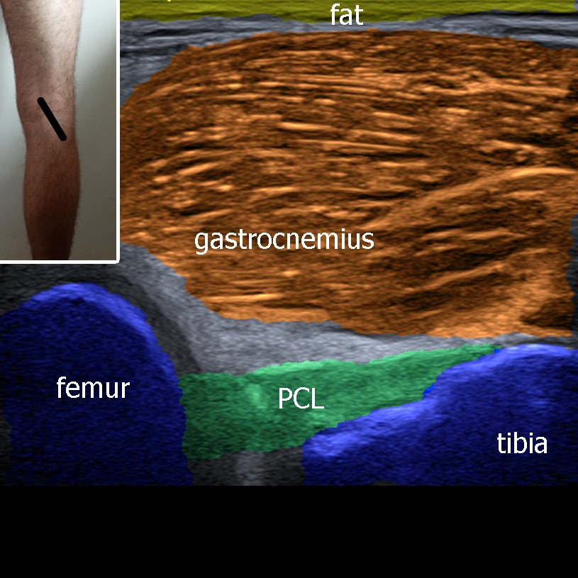
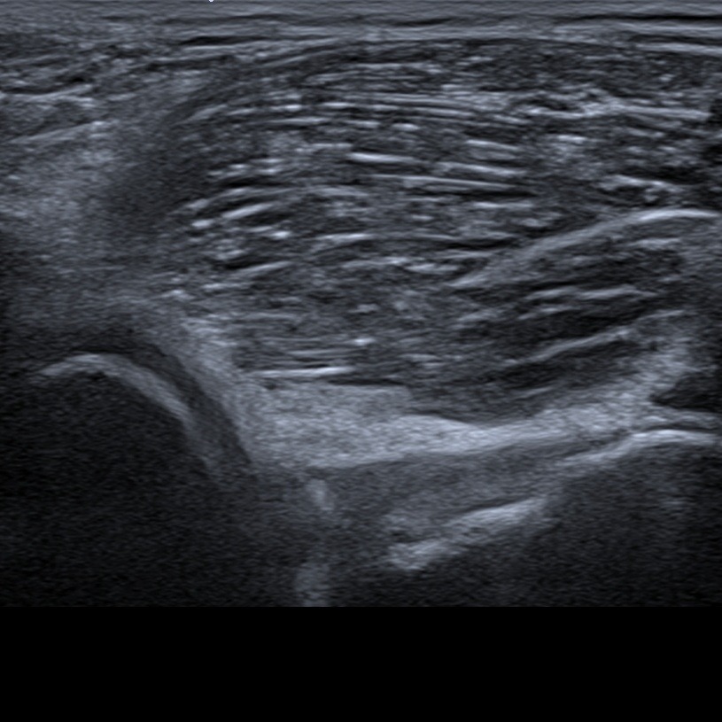
Putting the probe slightly oblique onto posterior compartment of the knee part of posterior cruciate ligament underneath the gastrocnemius muscle can be visualized.
Figure 10. PCL – posterior collateral ligament.
1. Anterior view
The probe is placed above the proximal part of the patella parallel with the long axis of the lower limb. Quadriceps tendon insertion onto patella can be evaluated. Underneath the tendon suprapatellar recessus of the knee joint can be visualized, especially when there is some fluid collection inside the joint.
Figure 1. pffp – prefemoral fat pad, spfp – suprapatellar fat pad, sb – recessus suprapatellaris.

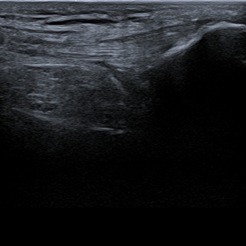
Positioning the knee into maximal flexion, we can evaluate the condyles of femur and the hyaline cartilage.
Figure 2. QT – quadriceps tendon, c – cartilage, VM – vastus medialis, spfp – suprpatellar fat pad, MFC – medial femoral condyle, LFC – lateral femoral condyle.


The probe is placed between the distal pole of the patella and tibia. Patellar ligament in it’s long axis can be evaluated. Underneath the patellar ligament can be found fat tissue called Hoffa’s fat pad.
Figure 3. P – patella.


The patellar tendon can be visualized and evaluated also in the short axis.
Figure 4.


When the probe is placed oblique (in maximal knee flexion), parallel with the course of anterior cruciate ligament, the distal part of the ligament with it’s insertion onto tibia can be evaluated. Anterior drawer test can be performed in this position.
Figure 5. ACL – anterior cruciate ligament.


2. Medial view
The probe is placed above the medial compartment of the knee. Medial collateral ligament can be evaluated as well as the medial meniscus.
Figure 6. MM – medial meniscus, LCM – medial collateral ligament.


Sliding more distally with the probe, the pes anserinus (common insrerion of sartorius, gracilis and semitendinosus). Underneath the pes anserinus inferior medial genicular artery can be visualized.
Figure 7. PA – pes anserinus, a – inferior medial genicular artery, v – vein.


3. Lateral view
Placing the probe above femur and tibia the iliotibial band with it’s insertion onto tibia can be visualized. In this plane lateral meniscus of the knee can be also evaluated.
Figure 8. ITB – iliotibial band, LM – lateral meniscus, P – popliteus muscle.


Sliding the probe more dorsal, lateral collateral ligament (that runs from femur to fibula) can be evaluated.
Figure 9. LCL – lateral collateral ligament.


4. Posterior view
Putting the probe slightly oblique onto posterior compartment of the knee part of posterior cruciate ligament underneath the gastrocnemius muscle can be visualized.
Figure 10. PCL – posterior collateral ligament.


1. Özçakar L, Kara M, Chang KV, Tekin L, Hung CY, Ulaülı AM, Wu CH, Tok F, Hsiao MY, Akkaya N, Wang TG, Çarli AB, Chen WS, De Muynck M. EURO-MUSCULUS/USPRM Basic Scanning Protocols for shoulder. Eur J Phys Rehabil Med. 2015 Aug;51(4):491-6. Epub 2015 Jul 9. PMID: 26158915.


