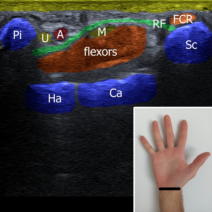
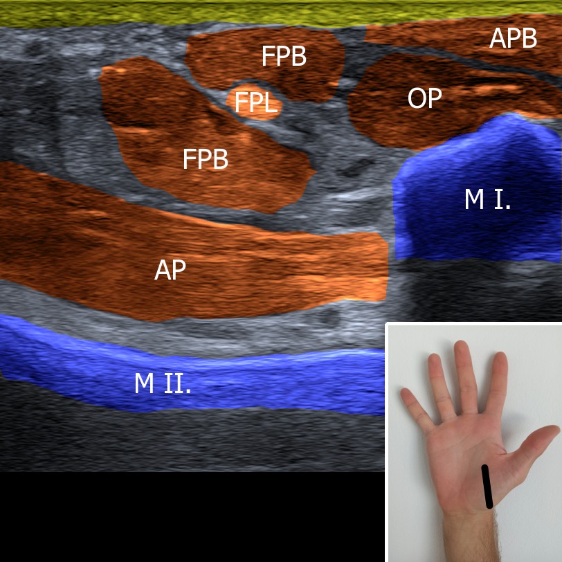
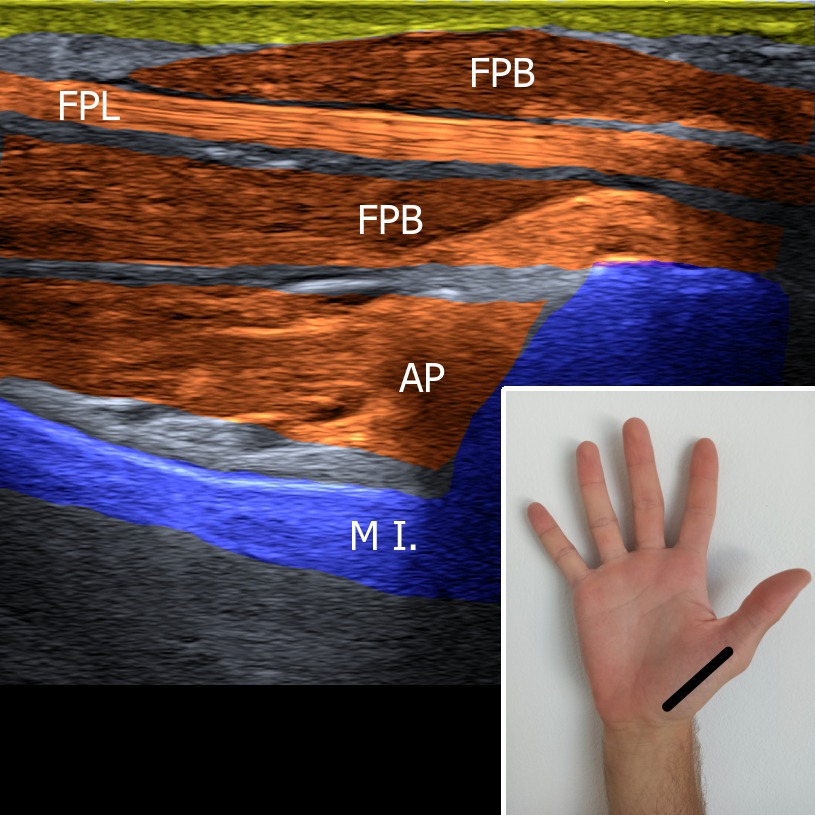
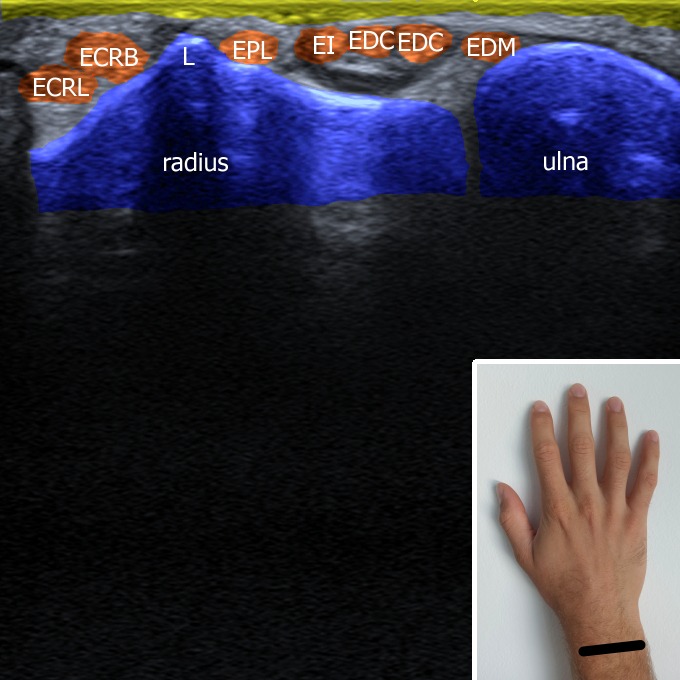
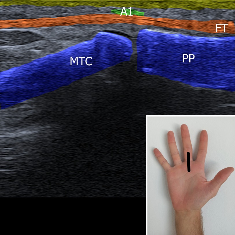
First of all carpal tunnel is located – the bony structures surrounding the carpal tunnel are pisiform, scaphoid, hamate and capitate bone. Tendons of the hand flexors and the median nerve are located in the carpal tunnel underneath the flexor retinaculum. At the ulnar side the Guyon’s canal can be seen with ulnar nerve and artery passing through.
Figure 1. U – ulnar nerve, M – median nerve, A – ulnar artery, Pi – os pisiforme, Ha – os hamatum, Ca – os capitatum, Sc – os scaphoideum, RF – retinaculum flexorum, FCR – flexor carpi ulnaris.