ELBOW
Patient is seated in front of the examiner. The position of the upper extremity varies from the scanning area (see figures below).
1. Anterior view
A. Longitudinal plane – ulnar side
B. Longitudinal plane – radial side
C. Longitudinal oblique plane
D. Transversal plane
2. Medial view
A. Lonitudinal plane
3. Lateral view
A. Longitudinal plane
4. Posterior view
A. Longitudinal plane
B. Transversal oblique plane
1. Anterior view
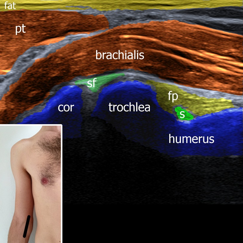
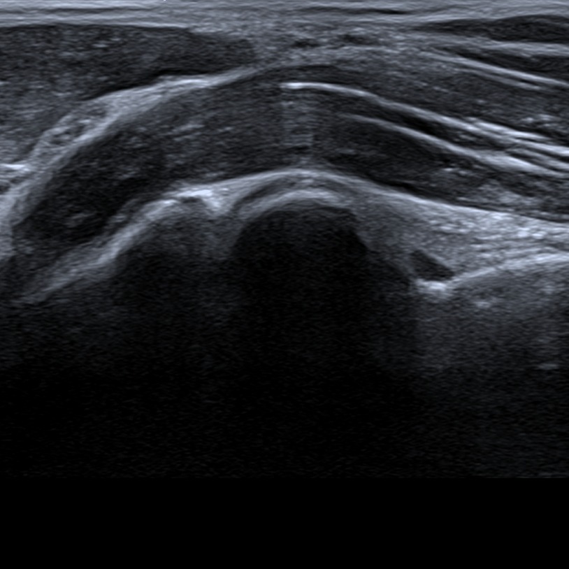
While examining the anterior compartment of the elbow, the patient is asked to extend his upper extremity and supine his forearm. In this plane humeroulnar joint (trochlea-coronoid process) can be evaluated. The joint space and the proximal coronoid fossa with anterior synovial recessus are places where the fluid can accumulate. Brachialis and pronator teres muscles can be assessed.
Figure 1. ulnar side. s – synovium in coronoid fossa, fp – fat pad, sf – synovial fringe, cor – processus coronoideus ulnae.
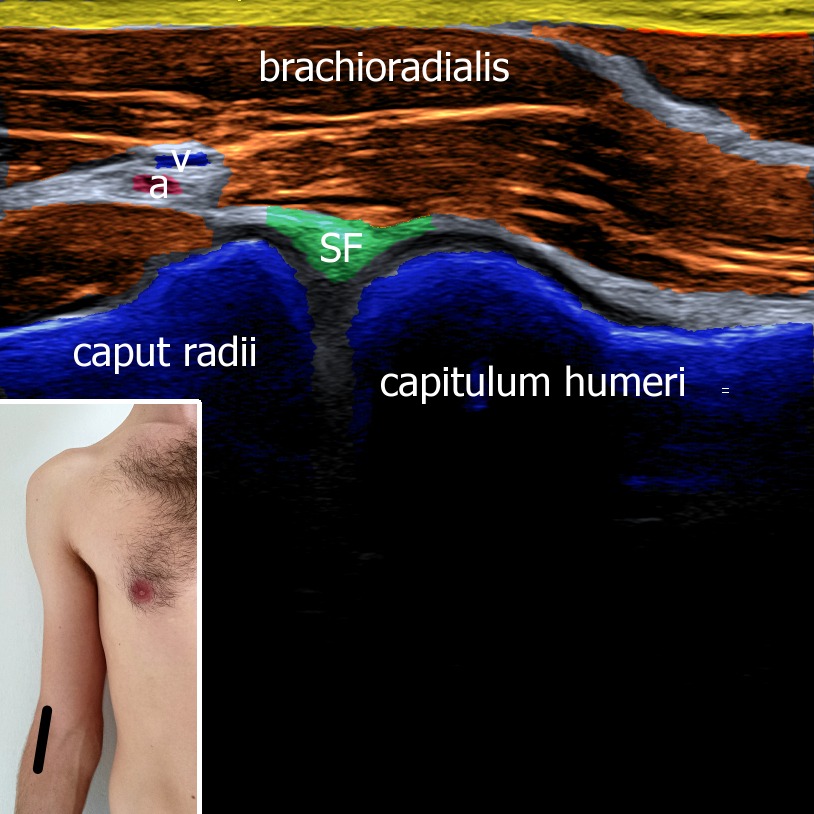
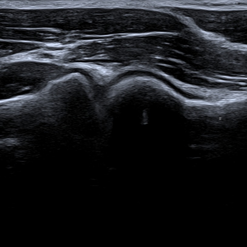
With the same position of the upper limb, we move the probe laterally to visualize the radiohumeral joint. On this plane we can differenciate radial head, capitulum humeri, radial fossa, brachioradialis muscle and vessels. The joint space itself and proximal radial fossa are the places where the fluid tends to accumulate.
Figure 2. sf – synovial fringe, a – artery, v – vein.
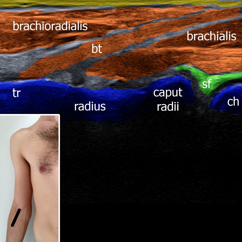
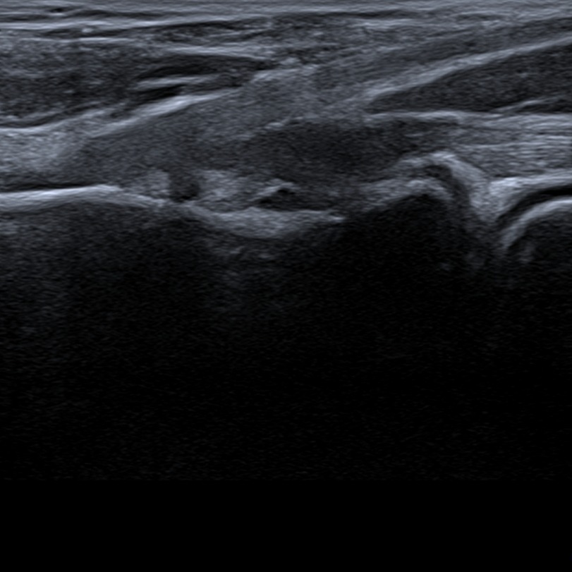
Shifting the probe in an oblique direction the distal biceps tendon and its attechment to the tuberositas radii can be visualized. The distal biceps tendon in long axis is quite diffucult for visalization because of its oblique course. Patient’s hand should be in maximal supination to get the tuberositas radii as superficial as possible.
Figure 3. tr – tuberositas radii, bt – biceps tendon, ch – capitullum humeri, sf – synovial fringe.
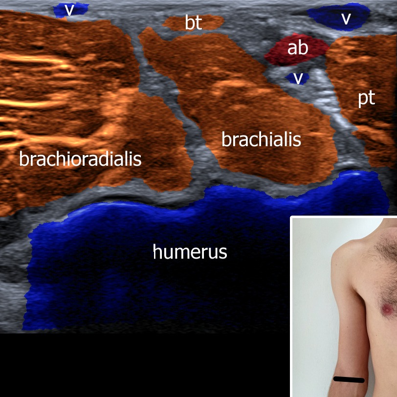
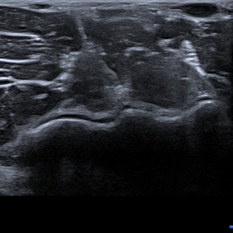
The transverse scan shows distal humeral epiphysis as a hyperechoic line covered by articular cartilage which appears hypoechoic. Regarding soft tissue pronator teres, brachialis and brachioradialis muscle as well as biceps tendon can be visualized. There are also vessels such as brachial artery and vein and superficial veins to be seen.
Figure 4. bt – biceps tendon, pt – pronator teres, ab – arteria brachialis, v – veins.
2. Medial view
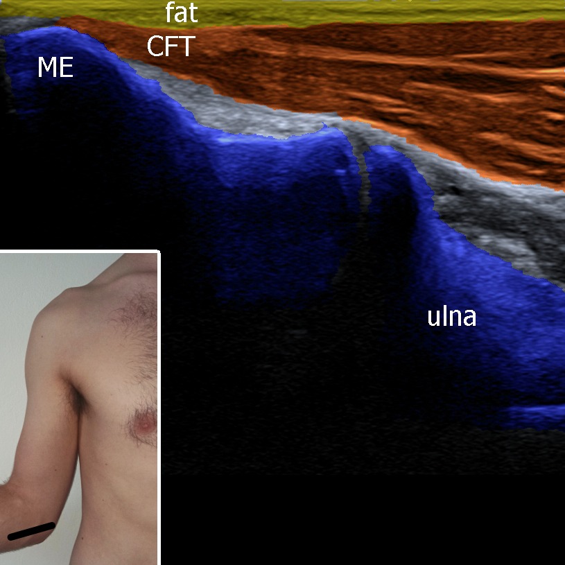
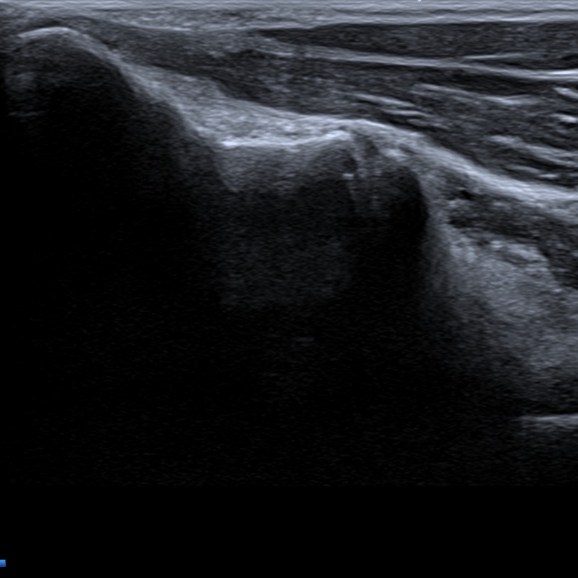
The upper extremity is extended or slightly flexed. The probe is attached to the medial epicondyle and aligned to the long axis of flexor muscles. Deep to the muscles the medial collateral ligament can be checked. The bones and attachments of the soft tissue should be checked for effusion, spurs, calcificaions, tears, erosions etc. Medial epicondyle tendinopathy known also as “golf elbow” can be visualised in this plan if present.
Figure 5. ME – medial epicondyle, CFT – common flexor tendon.
3. Lateral view
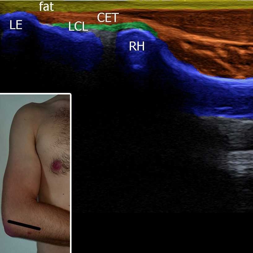
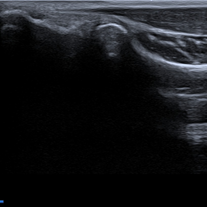
The elbow is in 90° flexion, the probe is attachet to the lateral epicondyle, visualization extensor muscles in the longitudinal axis. Lateral collateral ligament lies under the extensor muscles, binding lateral epicondyle of the humerus and radial head. Lateral epicondyle tendinopathy known also as “tennis elbow” can be visualised in this plane if present.
Figure 6. LE – lateral epicondyle, LCL – ligamentum collaterale laterale, CET – common extensor tendon, RH – radial head.
4. Posterior view
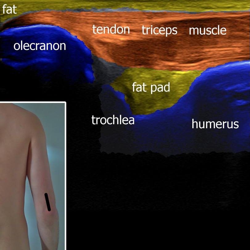
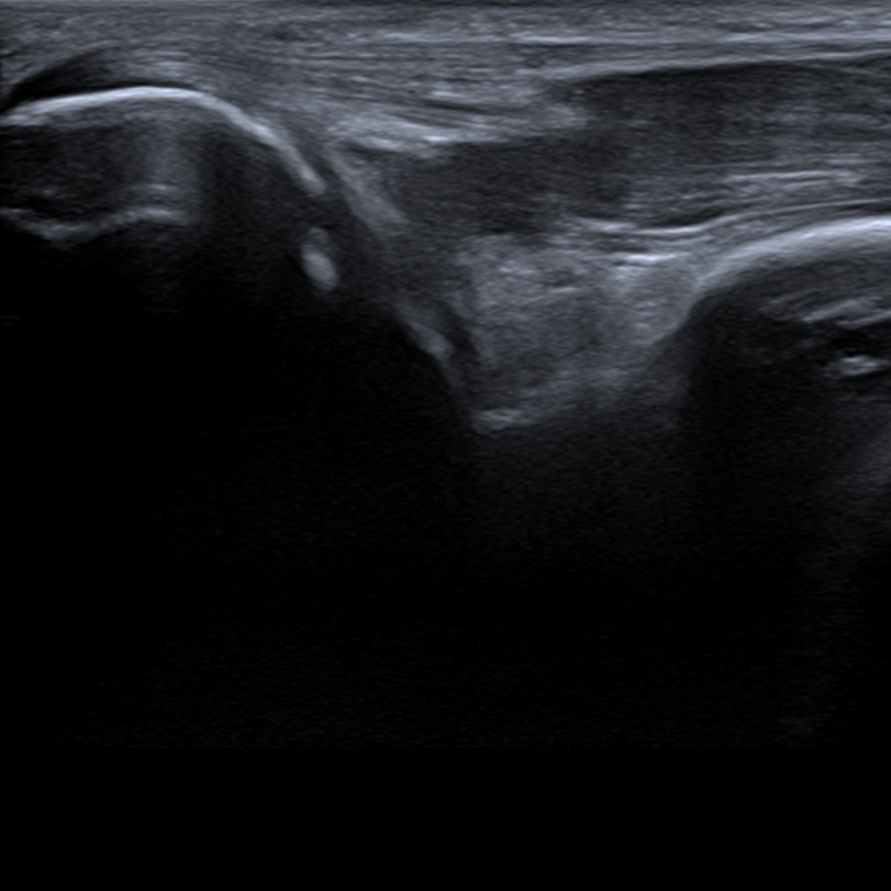
The probe is placed oved olecranon and triceps tendon, which is depict in the long axis. The triceps muscle with its muscletendineous junction and tendon inserting on the olecranon can be visualized. A small amount of fluid can be present between the fat pad and humerus in posterior joint recessus.
Figure 7. The elbow is flexed to 90°.
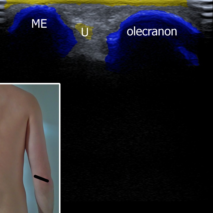
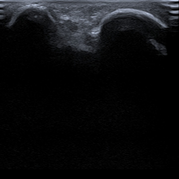
The ulnar nerve can be visualised between medial epicondyle of the humerus and olecranon. Dynamic assessment of the nerve can be performed doing flexion and extension, checking if the nerve subluxate.
Figure 8. U – ulnar nerve.
1. Anterior view
While examining the anterior compartment of the elbow, the patient is asked to extend his upper extremity and supine his forearm. In this plane humeroulnar joint (trochlea-coronoid process) can be evaluated. The joint space and the proximal coronoid fossa with anterior synovial recessus are places where the fluid can accumulate. Brachialis and pronator teres muscles can be assessed.
Figure 1. ulnar side. s – synovium in coronoid fossa, fp – fat pad, sf – synovial fringe, cor – processus coronoideus ulnae.


With the same position of the upper limb, we move the probe laterally to visualize the radiohumeral joint. On this plane we can differenciate radial head, capitulum humeri, radial fossa, brachioradialis muscle and vessels. The joint space itself and proximal radial fossa are the places where the fluid tends to accumulate.
Figure 2. sf – synovial fringe, a – artery, v – vein.


Shifting the probe in an oblique direction the distal biceps tendon and its attechment to the tuberositas radii can be visualized. The distal biceps tendon in long axis is quite diffucult for visalization because of its oblique course. Patient’s hand should be in maximal supination to get the tuberositas radii as superficial as possible.
Figure 3. tr – tuberositas radii, bt – biceps tendon, ch – capitullum humeri, sf – synovial fringe.


The transverse scan shows distal humeral epiphysis as a hyperechoic line covered by articular cartilage which appears hypoechoic. Regarding soft tissue pronator teres, brachialis and brachioradialis muscle as well as biceps tendon can be visualized. There are also vessels such as brachial artery and vein and superficial veins to be seen.
Figure 4. bt – biceps tendon, pt – pronator teres, ab – arteria brachialis, v – veins.


2. Medial view
The upper extremity is extended or slightly flexed. The probe is attached to the medial epicondyle and aligned to the long axis of flexor muscles. Deep to the muscles the medial collateral ligament can be checked. The bones and attachments of the soft tissue should be checked for effusion, spurs, calcificaions, tears, erosions etc. Medial epicondyle tendinopathy known also as “golf elbow” can be visualised in this plan if present.
Figure 5. ME – medial epicondyle, CFT – common flexor tendon.


3. Lateral view
The elbow is in 90° flexion, the probe is attachet to the lateral epicondyle, visualization extensor muscles in the longitudinal axis. Lateral collateral ligament lies under the extensor muscles, binding lateral epicondyle of the humerus and radial head. Lateral epicondyle tendinopathy known also as “tennis elbow” can be visualised in this plane if present.
Figure 6. LE – lateral epicondyle, LCL – ligamentum collaterale laterale, CET – common extensor tendon, RH – radial head.


4. Posterior view
The probe is placed oved olecranon and triceps tendon, which is depict in the long axis. The triceps muscle with its muscletendineous junction and tendon inserting on the olecranon can be visualized. A small amount of fluid can be present between the fat pad and humerus in posterior joint recessus.
Figure 7. The elbow is flexed to 90°.


The ulnar nerve can be visualised between medial epicondyle of the humerus and olecranon. Dynamic assessment of the nerve can be performed doing flexion and extension, checking if the nerve subluxate.
Figure 8. U – ulnar nerve.


1. Özçakar L, Kara M, Chang KV, Hung CY, Tekın L, Ulaşlı AM, Wu CH, Tok F, Hsıao MY, Akkaya N, Wang TG, Çarli AB, Chen WS, De Muynck M. EURO-MUSCULUS/USPRM Basic Scanning Protocols for elbow. Eur J Phys Rehabil Med. 2015 Aug;51(4):485-9. Epub 2015 Jul 9. PMID: 26158916.


