SHOULDER

Introduction

Ultrasound allows immediate evaluation of periarticular soft tissues, both statically and dynamically. Following structures can be evaluated: biceps tendon, rotator cuff muscles and tendons, subacromial bursa, AC joint, coracoacromial ligament, soft tissues mass, calcifications, some bony defects, etc.

Position of the patient:

Patient is seated in front of the examiner. The upper extremity is in the neutral position, but the position can be varied in order to perform dynamic testing or for better visualization of certain parts of the muscles.

Scanning protocol

1. Anterior view
     A. Horizontal plane – biceps brachii belly
     B. Horizontal plane – biceps brachii long and short head
     C. Horizontal plane – pectoralis major tendon
     D. Horizontal plane – subscapularis tendon, LHBB tendon
     E. Vertical plane – LHBB
     F. Vertical plane – biceps brachii myotendineous junctipn
     G. Oblique plane – coracoacromial ligament
2. Lateral view
     A. Horizontal plane – supraspinatus tendon
     B. Vertical plane – Rotator cuff
     C. Acromioclavicular joint
3. Posterior view
     A. Horizontal plane – labrum
     B. Horizontal plane – infraspinatus tendon
     C. Horizontal plane – suprascapular nerve 

1. Anterior view

A. Biceps brachii belly

Examination of the shoulder begins on the anterior side of the arm. The probe is placed in horizontal plane at the arm and biceps brachii muscle belly is visualized.

B. Biceps brachii long and short head

Moving the probe cranially the short (SHBB) and the long (LHBB) head of biceps brachii can be identified.

C. Pectoralis major tendon

Further cranially the pectoralis major tendon (P) between the deltoid and the short (S) and long (L) head of biceps brachii.

D. Subscapularis tendon, LHBB tendon short axis

Greater (TM) and lesser ™ tubercle is visualized moving the probe more cranially. In between (in bicipital sulcus) there is the long head of biceps brachii tendon (B). In this part, collection of the joint fluid can be seen around the biceps tendon. Subscapularis tendon (SSC) attaching the lesser tubercle can be also seen in this projection. 

E. LHBB tendon long axis

Rotating the probe 90° long head of biceps bracahii tendon (LHBB) can be visualized in long axis. Integrity or effusion of the tendon can be evaluated. Healthy tendon has fibralar appearence similar to spaghetti. 

F. Biceps brachii myotendineous junction

Moving the probe caudally the myotendineous junction (connection of the tendon – LHBBT and the muscle – LHBBM) of the biceps brachii can be visualized. 

G. Coracoacromial ligament

The probe is put into an oblique position for visualization of coracoacromial ligament (L) inbetween coracoid process (C) and acromion (A).

2. Lateral view

A. Supraspinatus tendon

The acromion (acr) and the greater tubercle on humerus are the bony landmarks to be looked for. In between these two structures supraspinatus tendon in long axis can be found. The width of supraspinatus tendon is approximately 4 cm, so the probe should be moved antero-posteriorly to scan all the parts of the tendon. This image is called a “birds-peak“. The subacromial bursa can be seen between supraspintus and deltoideus, when enlarged.


B. Rotator cuff

In this image the rotator cuff in short axis can be visualized. This image is called a “tire image”. The integrity of rotator cuff can be assessed by pushing the probe against the tendon. Normal tendon is not compresible, but when there is a rupture  the image of “flat tire” can be present – the ruptured tendon is compressible. For better visualization of the superior and posterior portions of the supraspinatus the hand is put behind patient’s back (Crass and modified Crass position). 

C. Acromioclavicular joint

The probe is placed on the top of the acromioclavicular joint. The acromion (A), the clavicle (C) and the joint (J) itself with joint capsule can be visualised. By shifting the probe posteriorly the status of the supraspinatus muscle can be assessed.

3. Posterior view

A. Labrum

The probe is placed underneath the spine of the scapula. The head of humerus and the glenoid (G) is the bony landmark. Triangular complex at the top of the glenoid represents the labrum. 


B. Infraspinatus tendon

Moving the probe more laterally, the tendon of infraspinatus is visualized in long axis. When the probe is moved caudaly, tendon of teres minor can be visualized.

C. Suprascapular nerve

Moving the probe more medially suprascapular nerve and artery can be visualized. 

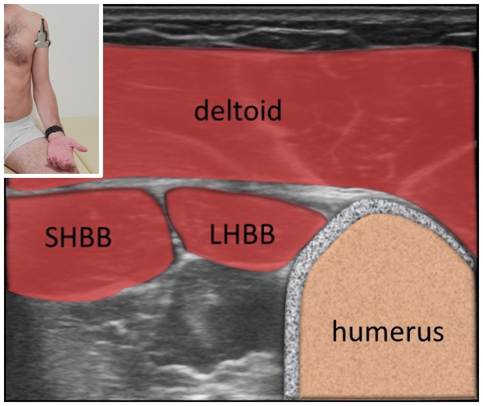
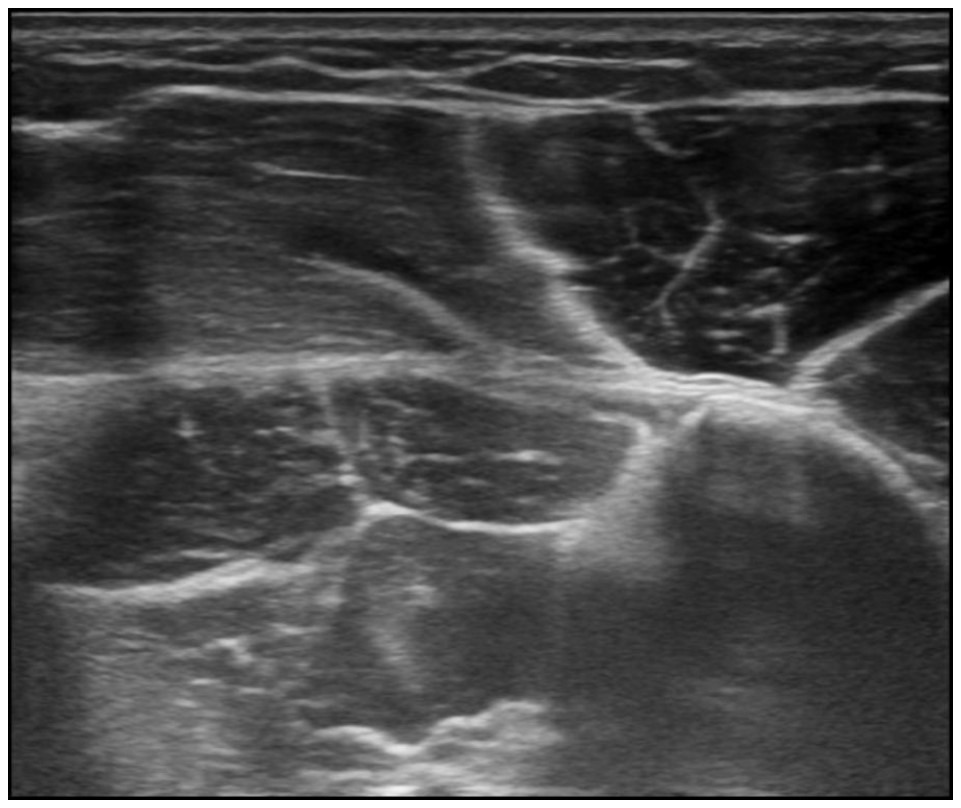
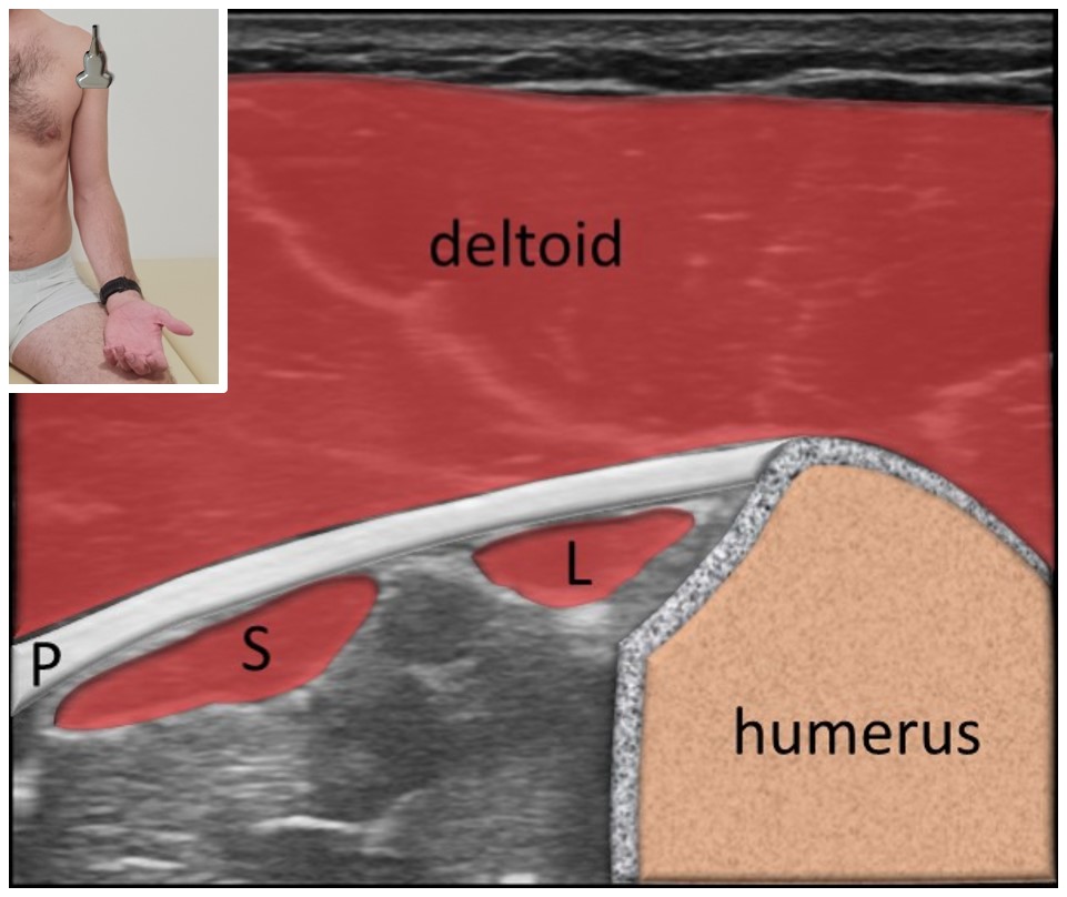
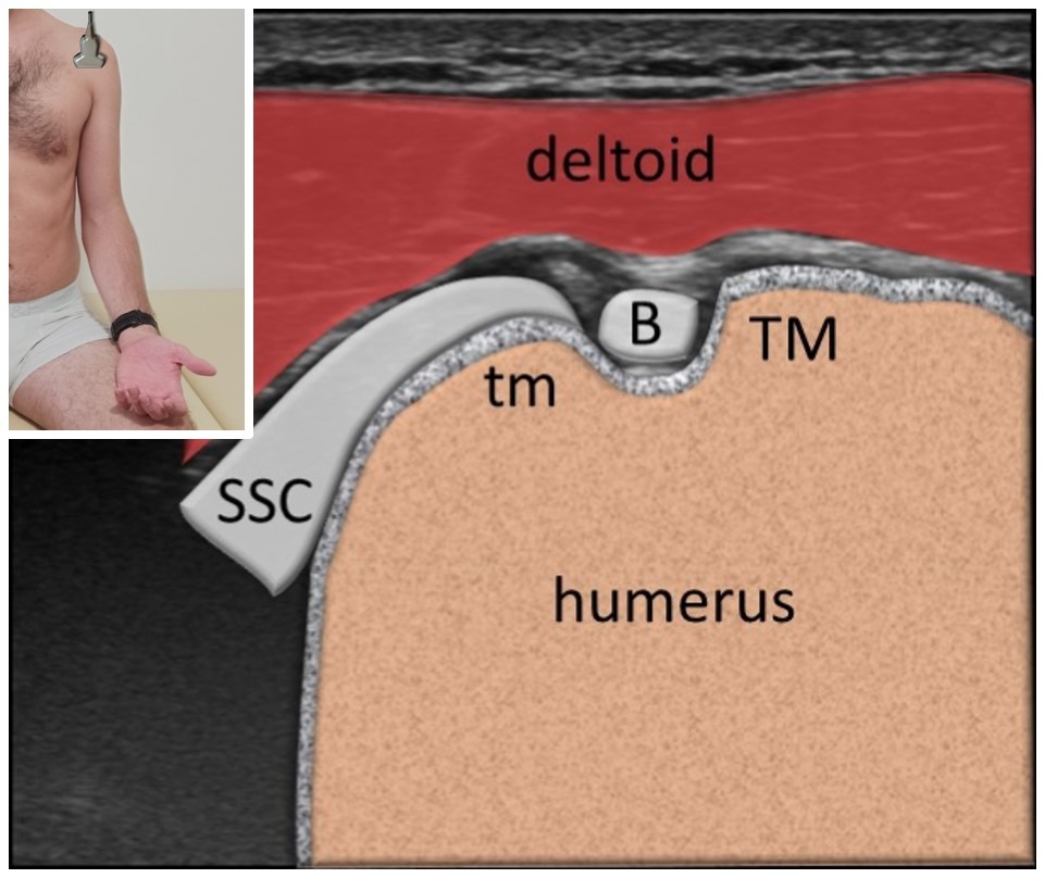
1. Özçakar L, Kara M, Chang KV, Tekin L, Hung CY, Ulaülı AM, Wu CH, Tok F, Hsiao MY, Akkaya N, Wang TG, Çarli AB, Chen WS, De Muynck M. EURO-MUSCULUS/USPRM Basic Scanning Protocols for shoulder. Eur J Phys Rehabil Med. 2015 Aug;51(4):491-6. Epub 2015 Jul 9. PMID: 26158915.