Ultrasound allows immediate evaluation of periarticular soft tissues, both statically and dynamically. Following structures can be evaluated: bursitis, periarticular soft tissues such as tendons, muscles and labrum, lymph nodes, vessels and nerves.
Position of the patient:
For examination of the anterior compartment the patient is lying down supine. For evaluation of the medial compartment is needed mild knee and hip flexion and external rotation. Lateral decubitus position is used for evaluation of the lateral compartment. Finally assessing the posterior part of the hip the patient is lying down in prone position.
Scanning protocol
1. Anterior view A. Longitudinal plane – labrum B. Longitudinal plane – joint recess 2. Medial view A. Longitudinal plane B. Transversal plane 3. Lateral view A. Longitudinal plane B. Transversal plane 4. Posterior view A. Longitudinal plane B. Transversal oblique plane
1. Anterior view
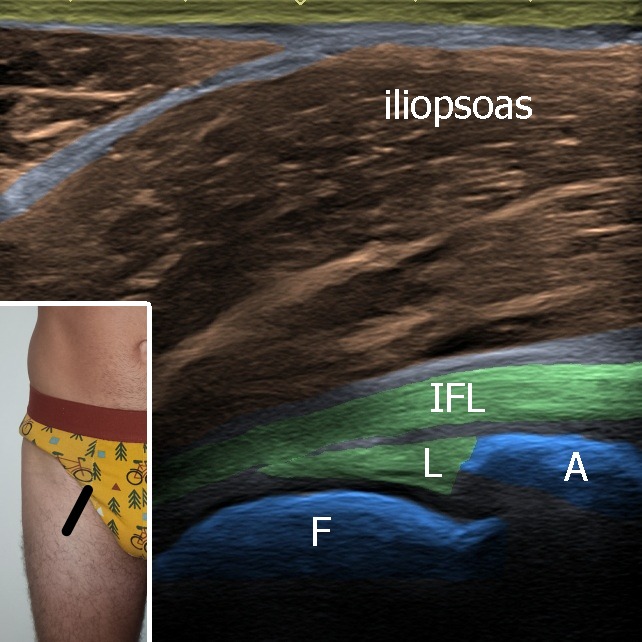
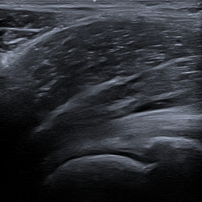
A. Longitudinal plane – labrum (Figure 1)
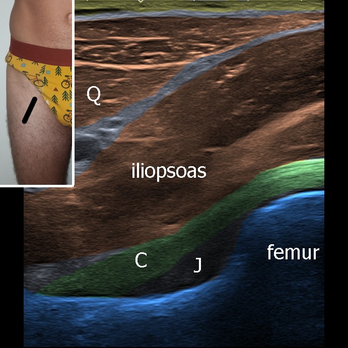
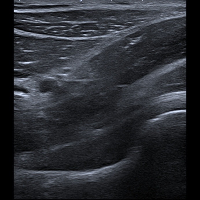
B. Longitudinal plane – joint recess (Figure 2)
The probe is placed above the hip joint parallel with the femur neck. In this sonogram we can evaluate the acetabular labrum, iliofemoral ligament and iliopsoas muscle.
With sliding the probe more distal anterior joint recess can be evaluated. Joint accumulation can be present.
Figure 1. IFL – iliofemoral ligament, L – labrum, F – femur, A – acetabulum.
Figure 2. Q – qudriceps muscle, C – joint capsule and iliofemorl ligament, J – anterior joint recess.
1. Anterior view
A. Longitudinal plane – labrum (Figure 1) – The probe is placed above the hip joint parallel with the femur neck. In this sonogram we can evaluate the acetabular labrum, iliofemoral ligament and iliopsoas muscle.
Figure 1. Anterior view, longitudinal plane – labrum. IFL – iliofemoral ligament, L – labrum, F – femur, A – acetabulum.
B. Longitudinal plane – anterior joint recess (Figure 2) – With sliding the probe more distal anterior joint recess can be evaluated. Joint accumulation can be present.