 MSK Academy
1. Ventrale Ansicht
1. Ventrale Ansicht
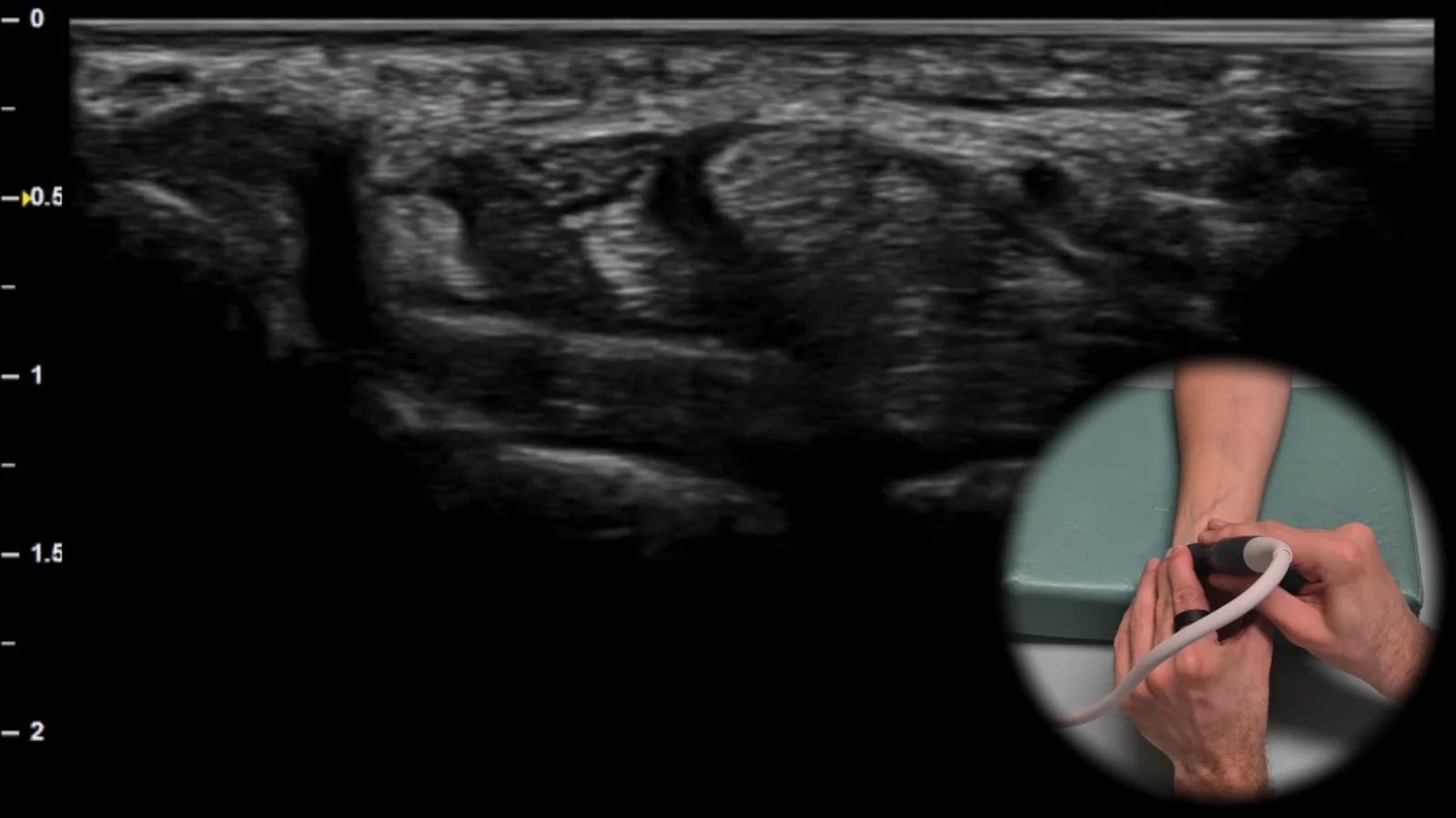
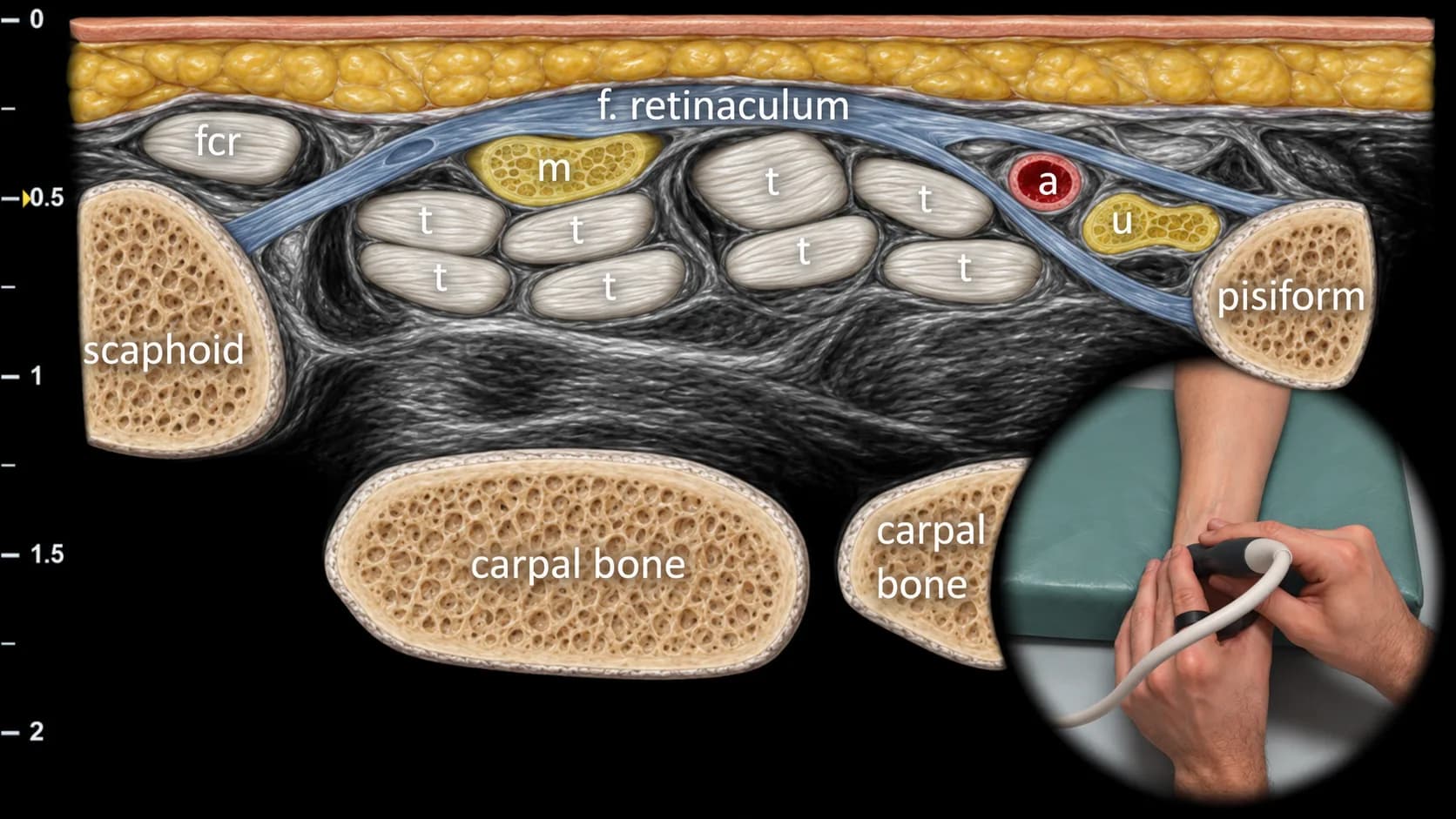
Abbildung 1. Volare Ansicht, Transversalebene. fcr: m. flexor carpi radialis, m: n. medianus, a: a. ulnaris, u: n. ulnaris, t: Flexorensehnen
Transversaler sonografischer Schnitt durch die volare Seite des Handgelenks auf Höhe des Karpaltunnels und des Guyon-Kanals. Zu den wichtigsten knöchernen Orientierungspunkten gehören das Os scaphoideum radial und das Os pisiforme ulnar, zwischen denen das Retinaculum flexorum über den Flexorensehnen (t) und dem n. medianus (m) im Bereich des Karpaltunnels erkennbar ist. Ulnar davon sind die a. ulnaris (a) und der n. ulnaris (u) bei ihrem Verlauf durch den Guyon-Kanal sichtbar, während radial die Sehne des m. flexor carpi radialis (fcr) dargestellt ist. Diese Projektion ist für die Beurteilung von Kompressionsneuropathien des n. medianus und des n. ulnaris sowie für den Nachweis einer Tenosynovitis der Flexoren und von Ganglionzysten im volaren Handgelenksbereich wesentlich.
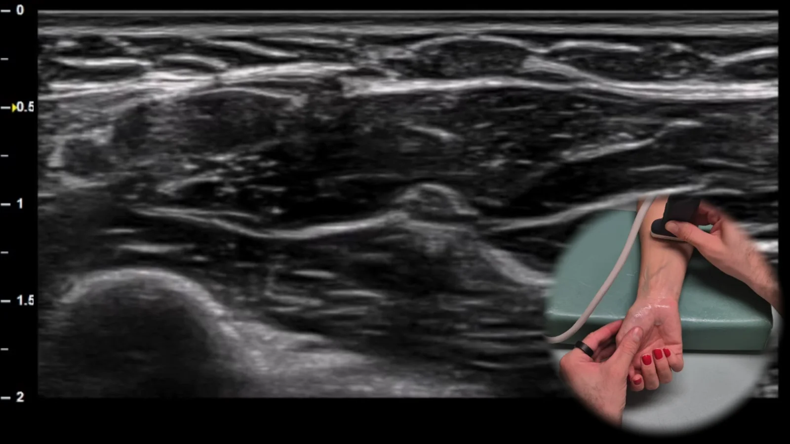
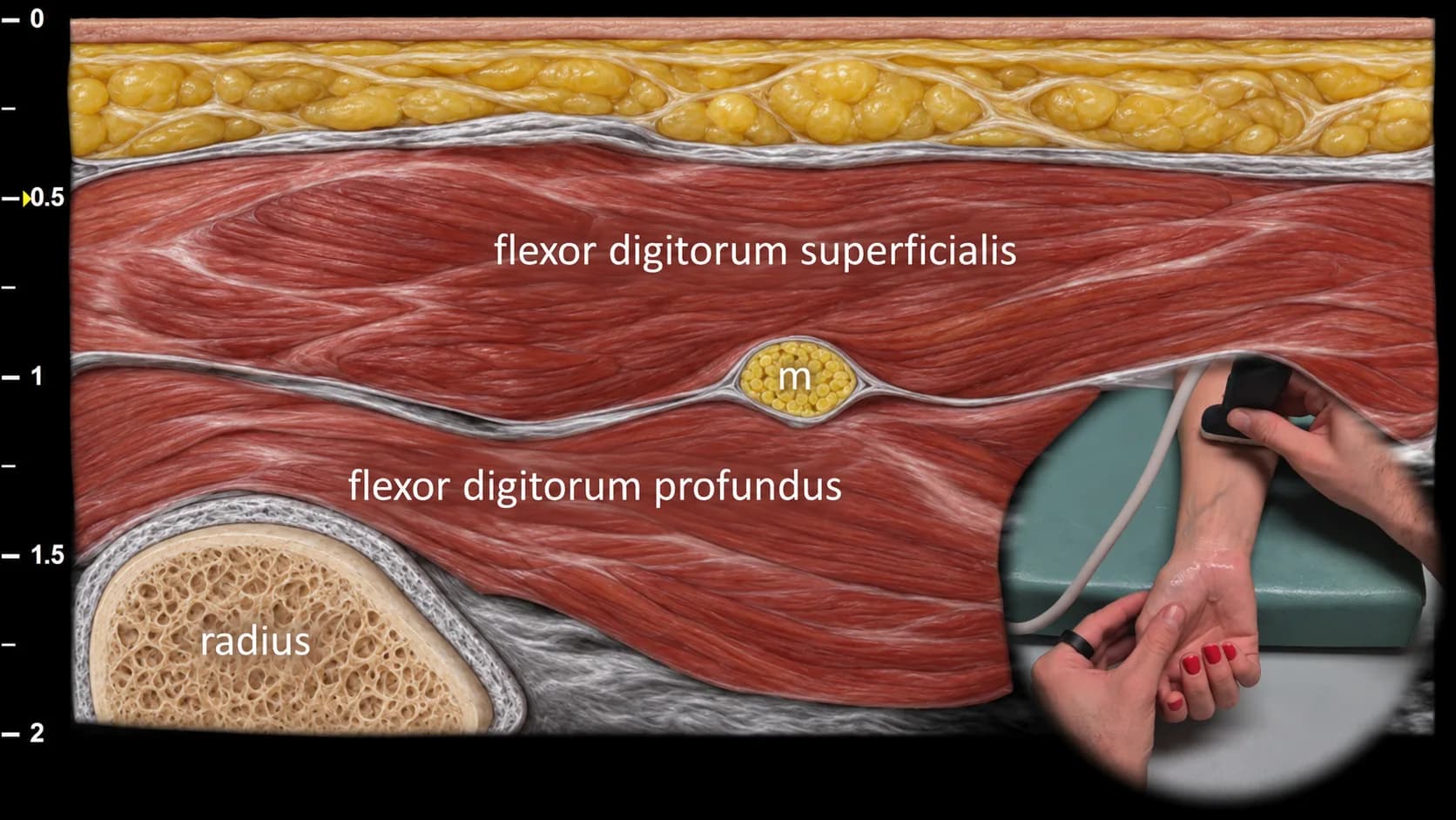
Abbildung 2. Ventrale Ansicht, Transversalebene. m: N. medianus
Sonografischer Querschnitt der volaren Seite des distalen Unterarms bei der proximalen Verfolgung des N. medianus ausgehend vom Bereich des Karpaltunnels. Der N. medianus (m) liegt in dieser Projektion zwischen dem M. flexor digitorum superficialis und dem M. flexor digitorum profundus, wobei der tiefer sichtbare Radius als knöcherne Orientierungsstruktur dient. Auf dieser Höhe weist der N. medianus ein typisches faszikuläres („wabenartiges“) Erscheinungsbild auf, was für die Beurteilung seiner Morphologie und die Erkennung pathologischer Veränderungen bei Kompressionsneuropathien oder einer anderweitigen Beteiligung des Nervs wichtig ist.
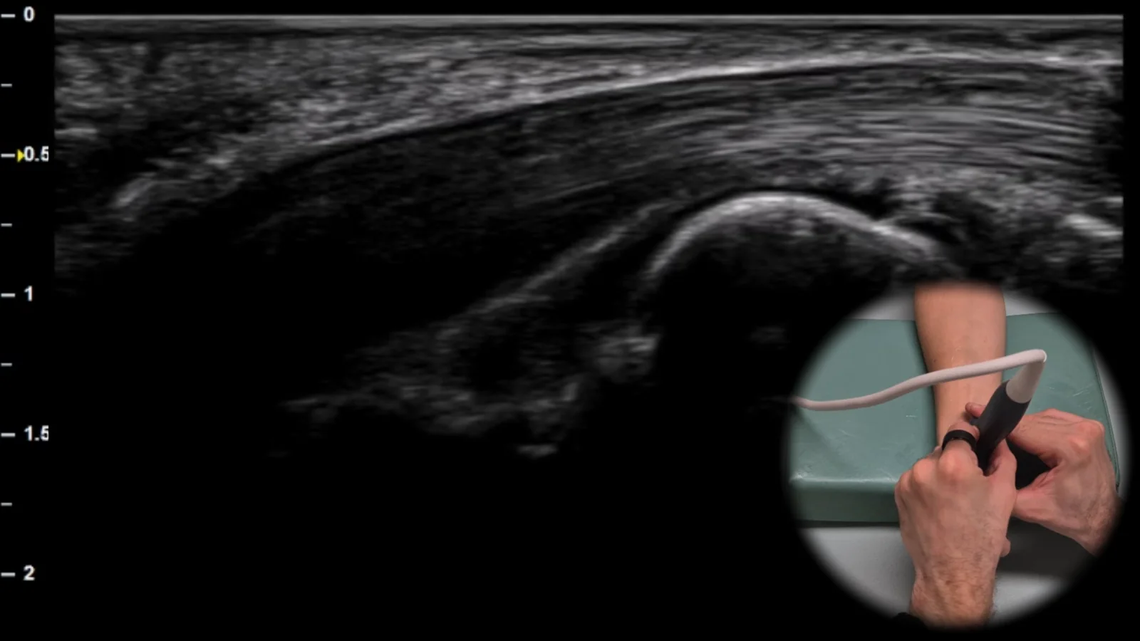
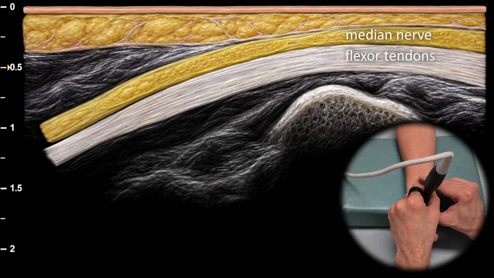
Abbildung 3. Ansicht von ventral, Sagittalebene. m: n. medianus
Longitudinaler sonografischer Schnitt der Volarseite des Handgelenks, der den n. medianus am Eingang des Karpaltunnels zeigt. Der Nervus medianus (m) ist in dieser Projektion als gestreifte Struktur mit parallelen echogenen Linien sichtbar und liegt oberflächlich über den Beugesehnen, was seiner typischen longitudinalen faszikulären Architektur entspricht. Diese Projektion ist wichtig für die Beurteilung der Nervenkontinuität, von Veränderungen seines Verlaufs und möglicher Kompressionszeichen am Eingang des Karpaltunnels.
Klinischer Hinweis
Bei einer Kompression des N. medianus kann an der Eintrittsstelle in den Karpaltunnel eine fokale Verengung des Nervs mit proximaler Erweiterung sichtbar sein, die in der Längsansicht einer „Flaschenhals“-Form ähnelt.
Zugang zur gesamten Bibliothek
Voller Zugriff auf Scan-Protokolle, Anatomie und klinische Referenzen. Jederzeit kündbar.
- Alle Protokolle und Anatomiereferenzen
- Originale Ultraschall-Illustrationen und Video-Demonstrationen
- Synchronisierung zwischen Mobilgerät und Web